

The DOI matches its Crossref record; 6 of the 6 references it cites were found in a registry or an index.
Membrane contact probability: An essential and predictive character for the structural and functional studies of membrane proteins
Open accessPublished version of a preprint
Abstract
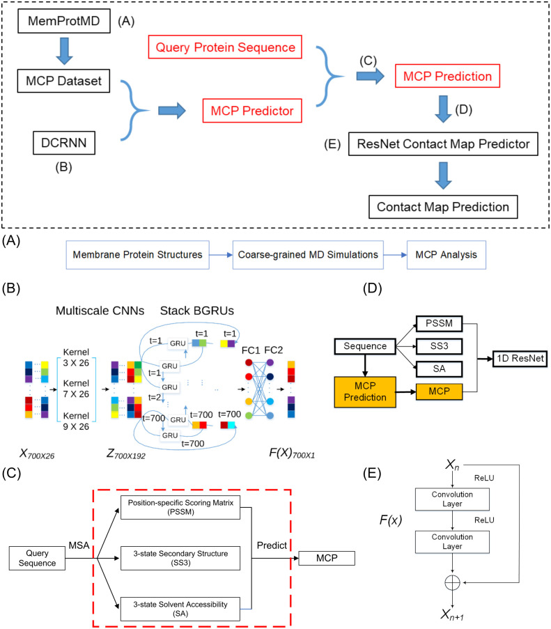
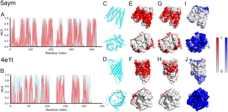
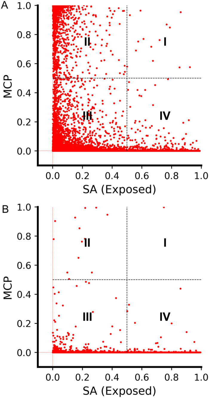
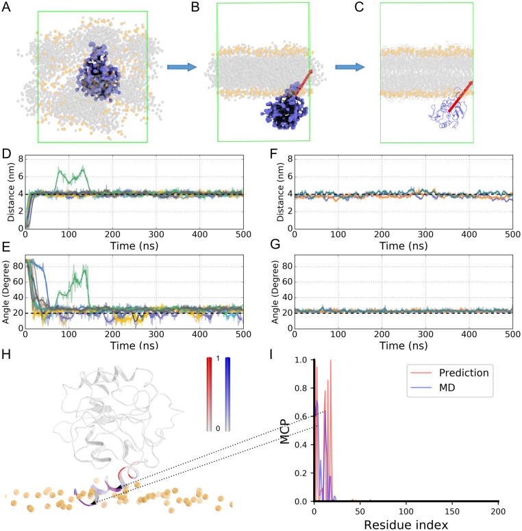
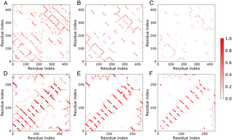
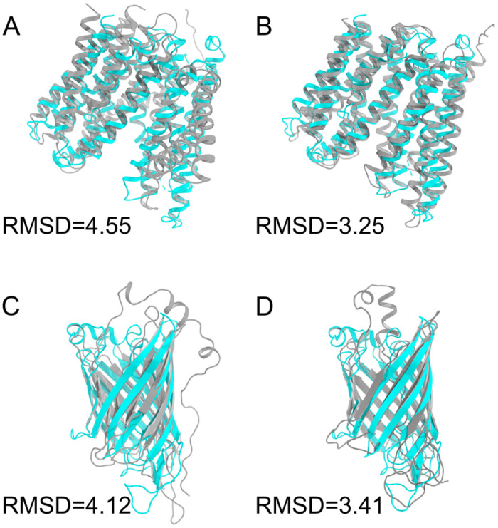
One of the unique traits of membrane proteins is that a significant fraction of their hydrophobic amino acids is exposed to the hydrophobic core of lipid bilayers rather than being embedded in the protein interior, which is often not explicitly considered in the protein structure and function predictions. Here, we propose a characteristic and predictive quantity, the membrane contact probability (MCP), to describe the likelihood of the amino acids of a given sequence being in direct contact with the acyl chains of lipid molecules. We show that MCP is complementary to solvent accessibility in characterizing the outer surface of membrane proteins, and it can be predicted for any given sequence with a machine learning-based method by utilizing a training dataset extracted from MemProtMD, a database generated from molecular dynamics simulations for the membrane proteins with a known structure. As the first of many potential applications, we demonstrate that MCP can be used to systematically improve the prediction precision of the protein contact maps and structures.
Overview
The claims and statistics of this paper haven’t been checked yet.
Run the check — free, no account needed. It reads the open-access text and checks the sources it cites.
Integrity signals
Registry
Cites
Statistics
Figures
Verification
Claims the paper makes
The claims and statistics of this paper haven’t been checked yet.
Run the check — free, no account needed. It reads the open-access text and checks the sources it cites.
Self-check
Percentages, sums and reported differences re-computed from the paper’s own numbers.
The self-check needs the paper’s full text.
Citations
Cites
Deep Residual Learning for Image Recognition
He, K., Zhang, X., Ren, S., & Sun, J. (2016) · 2016 IEEE Conference on Computer Vision and Pattern Recognition (CVPR) · doi:10.1109/cvpr.2016.90 · Cited by 235,474
Adam: A Method for Stochastic Optimization
Kingma, D. P., & Ba, J. (2014) · arXiv · doi:10.48550/arxiv.1412.6980 · Cited by 98,946
Highly accurate protein structure prediction with AlphaFold
Jumper, J., Evans, R., Pritzel, A., Green, T., Figurnov, M., Ronneberger, O., Tunyasuvunakool, K., Bates, R., Žídek, A., Potapenko, A., et al. (2021) · Nature · doi:10.1038/s41586-021-03819-2 · Cited by 43,764
Accurate prediction of protein structures and interactions using a 3-track network
Baek, M., DiMaio, F., Anishchenko, I., Dauparas, J., Ovchinnikov, S., Lee, G. R., Wang, J., Cong, Q., Kinch, L. N., Schaeffer, R. D., et al. (2021) · doi:10.1101/2021.06.14.448402 · Cited by 5,549
‘Improved parameters for the Martini coarse-grained protein force field’
de Jong, D. H., Singh, G., Bennett, W. F. D., Arnarez, C., Wassenaar, T. A., Schäfer, L. V., Periole, X., Tieleman, D. P., & Marrink, S. J. (2013) · Journal of Chemical Theory and Computation · doi:10.1021/ct300646g · Cited by 1,486
Accurate De Novo Prediction of Protein Contact Map by Ultra-Deep Learning Model
Wang, S., Sun, S., Li, Z., Zhang, R., & Xu, J. (2017) · PLOS Computational Biology · doi:10.1371/journal.pcbi.1005324 · Cited by 931
Research works printed but not linked: 83
- M Howarth, 2015, Say it with proteins: an alphabet of crystal structures, Nature Structural &Molecular Biology, 22, 349, 10.1038/nsmb.3011 · DOI
- Wang Z, Zhao F, Peng J, Xu J. Protein 8-class secondary structure prediction using Conditional Neural Fields. 2010 IEEE International Conference on Bioinformatics and Biomedicine (BIBM). 2010; p. 109–114. · DOI
- S Wang, 2016, Protein Secondary Structure Prediction Using Deep Convolutional Neural Fields, Scientific Reports, 6, 18962, 10.1038/srep18962 · DOI
- S Wang, 2016, RaptorX-Property: a web server for protein structure property prediction, Nucleic Acids Research, 44, W430, 10.1093/nar/gkw306 · DOI
- Y Zhou, 2017, Prediction of Protein Secondary Structure. vol. 1484 of Methods in Molecular Biology
- J Yang, 2020, Improved protein structure prediction using predicted interresidue orientations, Proceedings of the National Academy of Sciences, 117, 1496, 10.1073/pnas.1914677117 · DOI · Paper page
- A Senior, 2020, Improved protein structure prediction using potentials from deep learning, Nature, 577, 706, 10.1038/s41586-019-1923-7 · DOI · Paper page
- M Uhlén, 2015, Tissue-based map of the human proteome, Science, 347, 1260419, 10.1126/science.1260419 · DOI · Paper page
- Y Cheng, 2018, Membrane protein structural biology in the era of single particle cryo-EM, Current Opinion in Structural Biology, 52, 58, 10.1016/j.sbi.2018.08.008 · DOI
- B Rost, 1996, Bridging the protein sequence-structure gap by structure predictions, Annual Review of Biophysics and Biomolecular Structure, 25, 113, 10.1146/annurev.bb.25.060196.000553 · DOI
- T Flock, 2012, Deciphering membrane protein structures from protein sequences, Genome Biology, 13, 160, 10.1186/gb-2012-13-6-160 · DOI
- A Singh, 2020, Deep learning 3D structures, Nature Methods, 17, 249, 10.1038/s41592-020-0779-y · DOI
- B Adhikari, 2018, DNCON2: improved protein contact prediction using two-level deep convolutional neural networks, Bioinformatics, 34, 1466, 10.1093/bioinformatics/btx781 · DOI
- J Hanson, 2018, Accurate prediction of protein contact maps by coupling residual two-dimensional bidirectional long short-term memory with convolutional neural networks, Bioinformatics, 34, 4039, 10.1093/bioinformatics/bty481 · DOI
- B Zhang, 2018, Protein Solvent-Accessibility Prediction by a Stacked Deep Bidirectional Recurrent Neural Network, Biomolecules, 8, 33, 10.3390/biom8020033 · DOI
- W Kabsch, 1983, Dictionary of protein secondary structure: Pattern recognition of hydrogen-bonded and geometrical features, Biopolymers, 22, 2577, 10.1002/bip.360221211 · DOI · Paper page
- J Ma, 2015, AcconPred: Predicting Solvent Accessibility and Contact Number Simultaneously by a Multitask Learning Framework under the Conditional Neural Fields Model, BioMed Research International, 2015, 678764
- T Beuming, 2004, A knowledge-based scale for the analysis and prediction of buried and exposed faces of transmembrane domain proteins, Bioinformatics, 20, 1822, 10.1093/bioinformatics/bth143 · DOI
- Z Yuan, 2006, Predicting the solvent accessibility of transmembrane residues from protein sequence, Journal of Proteome Research, 5, 1063, 10.1021/pr050397b · DOI
- K Illergård, 2009, MPRAP: An accessibility predictor for a-helical transmem-brane proteins that performs well inside and outside the membrane, BMC Bioinformatics, 11, 333, 10.1186/1471-2105-11-333 · DOI
- C Lu, 2019, TMP-SSurface: A Deep Learning-Based Predictor for Surface Accessibility of Transmembrane Protein Residues, Crystals, 9, 640, 10.3390/cryst9120640 · DOI
- JK Leman, 2017, Computing structure-based lipid accessibility of membrane proteins with mp_lipid_acc in RosettaMP, BMC Bioinformatics, 18, 115, 10.1186/s12859-017-1541-z · DOI
- L Adamian, 2006, Prediction of transmembrane helix orientation in polytopic membrane proteins, BMC Structural Biology, 6, 13, 10.1186/1472-6807-6-13 · DOI
- M Phatak, 2011, Solvent and lipid accessibility prediction as a basis for model quality assessment in soluble and membrane proteins, Current Protein & Peptide Science, 12, 563, 10.2174/138920311796957603 · DOI
- JS Lai, 2013, Lipid exposure prediction enhances the inference of rotational angles of transmembrane helices, BMC Bioinformatics, 14, 304, 10.1186/1471-2105-14-304 · DOI
- T Nugent, 2010, Predicting Transmembrane Helix Packing Arrangements using Residue Contacts and a Force-Directed Algorithm, PLoS Computational Biology, 6, 10.1371/journal.pcbi.1000714 · DOI
- A Krogh, 2001, Predicting transmembrane protein topology with a hidden Markov model: application to complete genomes, Journal of Molecular Biology, 305, 567, 10.1006/jmbi.2000.4315 · DOI · Paper page
- DT Jones, 2007, Improving the accuracy of transmembrane protein topology prediction using evolutionary information, Bioinformatics, 23, 538, 10.1093/bioinformatics/btl677 · DOI
- H Viklund, 2008, OCTOPUS: improving topology prediction by two-track ANN-based preference scores and an extended topological grammar, Bioinformatics, 24, 1662, 10.1093/bioinformatics/btn221 · DOI
- SM Reynolds, 2008, Transmembrane topology and signal peptide prediction using dynamic bayesian networks, PLoS Computational Biology, 4, e1000213, 10.1371/journal.pcbi.1000213 · DOI
- SH Feng, 2020, Topology prediction improvement of α-helical transmembrane proteins through helix-tail modeling and multiscale deep learning fusion, Journal of Molecular Biology, 432, 1279, 10.1016/j.jmb.2019.12.007 · DOI
- Li Z, Yu Y. Protein Secondary Structure Prediction Using Cascaded Convolutional and Recurrent Neural Networks. In: Proceedings of the Twenty-Fifth International Joint Conference on Artificial Intelligence (IJCAI); 2016.
- PJ Stansfeld, 2015, MemProtMD: Automated Insertion of Membrane Protein Structures into Explicit Lipid Membranes, Structure(London, England:1993), 23, 1350
- TD Newport, 2019, The MemProtMD database: a resource for membrane-embedded protein structures and their lipid interactions, Nucleic Acids Research, 47, D390, 10.1093/nar/gky1047 · DOI
- Y Li, 2019, Ensembling multiple raw coevolutionary features with deep residual neural networks for contact-map prediction in CASP13, Proteins: Structure, 87, 1082, 10.1002/prot.25798 · DOI · Paper page
- R Taniguchi, 2015, Outward- and inward-facing structures of a putative bacterial transition-metal transporter with homology to ferroportin, Nature Communications, 6, 8545, 10.1038/ncomms9545 · DOI · Paper page
- JW Fairman, 2012, Crystal structures of the outer membrane domain of intimin and invasin from enterohemorrhagic E. coli and enteropathogenic Y. pseudotuberculosis, Structure, 20, 1233, 10.1016/j.str.2012.04.011 · DOI
- MA Lomize, 2012, OPM database and PPM web server: resources for positioning of proteins in membranes, Nucleic Acids Research, 40, D370, 10.1093/nar/gkr703 · DOI
- M Huang, 2001, Crystal structure of Sar1-GDP at 1.7 Å resolution and the role of the NH2 terminus in ER export, The Journal of Cell Biology, 155, 937, 10.1083/jcb.200106039 · DOI
- MCS Lee, 2005, Sar1p N-Terminal Helix Initiates Membrane Curvature and Completes the Fission of a COPII Vesicle, Cell, 122, 605, 10.1016/j.cell.2005.07.025 · DOI
- PJ Stansfeld, 2013, Multiscale Simulations Reveal Conserved Patterns of Lipid Interactions with Aquaporins, Structure(London, England:1993), 21, 810
- JK Leman, 2013, Simultaneous prediction of protein secondary structure and transmembrane spans, Proteins: Structure, Function, and Bioinformatics, 81, 1127, 10.1002/prot.24258 · DOI
- S Wang, 2017, Folding Membrane Proteins by Deep Transfer Learning, Cell Systems, 5, 202, 10.1016/j.cels.2017.09.001 · DOI
- G Wang, 2003, PISCES: a protein sequence culling server, Bioinformatics, 19, 1589, 10.1093/bioinformatics/btg224 · DOI
- S Seemayer, 2014, CCMpred—fast and precise prediction of protein residue–residue contacts from correlated mutations, Bioinformatics, 30, 3128, 10.1093/bioinformatics/btu500 · DOI
- D Jones, 2012, PSICOV: precise structural contact prediction using sparse inverse covariance estimation on large multiple sequence alignments, Bioinformatics, 28, 184, 10.1093/bioinformatics/btr638 · DOI
- D Jones, 2015, MetaPSICOV: combining coevolution methods for accurate prediction of contacts and long range hydrogen bonding in proteins, Bioinformatics, 31, 999, 10.1093/bioinformatics/btu791 · DOI
- DWA Buchan, 2018, Improved protein contact predictions with the MetaPSICOV2 server in CASP12, Proteins, 86, 78, 10.1002/prot.25379 · DOI
- B Adhikari, 2018, CONFOLD2: improved contact-driven ab initio protein structure modeling, BMC Bioinformatics, 19, 1, 10.1186/s12859-018-2032-6 · DOI
- A Lesk, 1980, Solvent accessibility, protein surfaces, and protein folding, Biophysical Journal, 32, 35, 10.1016/S0006-3495(80)84914-9 · DOI
- M Zhang, 2018, Structure of the mechanosensitive OSCA channels, Nature Structural & Molecular Biology, 25, 850, 10.1038/s41594-018-0117-6 · DOI
- WC Wimley, 1996, Experimentally determined hydrophobicity scale for proteins at membrane interfaces, Nature Structural & Molecular Biology, 3, 842, 10.1038/nsb1096-842 · DOI
- MA Lomize, 2006, OPM: Orientations of Proteins in Membranes database, Bioinformatics, 22, 623, 10.1093/bioinformatics/btk023 · DOI
- T Nugent, 2013, Membrane protein orientation and refinement using a knowledge-based statistical potential, BMC Bioinformatics, 14, 1, 10.1186/1471-2105-14-276 · DOI
- P Bond, 2006, Insertion and assembly of membrane proteins via simulation, Journal of the American Chemical Society, 128, 2697, 10.1021/ja0569104 · DOI
- KA Scott, 2008, Coarse-grained MD simulations of membrane protein-bilayer self-assembly, Structure, 16, 621, 10.1016/j.str.2008.01.014 · DOI · Paper page
- C Arnarez, 2013, Evidence for cardiolipin binding sites on the membrane-exposed surface of the cytochrome bc1, Journal of the American Chemical Society, 135, 3112, 10.1021/ja310577u · DOI
- AP Chetwynd, 2008, CGDB: a database of membrane protein/lipid interactions by coarse-grained molecular dynamics simulations, Molecular Membrane Biology, 25, 662, 10.1080/09687680802446534 · DOI
- Z Liu, 2021, TMP- SSurface2: A Novel Deep Learning-Based Surface Accessibility Predictor for Transmembrane Protein Sequence, Frontiers in Genetics, 12
- W Jeon, 2019, FP2VEC: a new molecular featurizer for learning molecular properties, Bioinformatics, 35, 4979, 10.1093/bioinformatics/btz307 · DOI
- GB Kim, 2021, DeepTFactor: A deep learning-based tool for the prediction of transcription factors, Proceedings of the National Academy of Sciences of the United States of America, 118, 1
- Józefowicz R, Zaremba W, Sutskever I. An Empirical Exploration of Recurrent Network Architectures. In: In Proceedings of the 32nd International Conference on Machine Learning (ICML-15); 2015.
- M Remmert, 2012, HHblits: lightning-fast iterative protein sequence searching by HMM-HMM alignment, Nature Methods, 9, 173, 10.1038/nmeth.1818 · DOI · Paper page
- NK Fox, 2014, SCOPe: Structural Classification of Proteins—extended, integrating SCOP and ASTRAL data and classification of new structures, Nucleic Acids Research, 42, D304, 10.1093/nar/gkt1240 · DOI · Paper page
- JM Chandonia, 2019, SCOPe: classification of large macromolecular structures in the structural classification of proteins—extended database, Nucleic Acids Research, 47, D475, 10.1093/nar/gky1134 · DOI
- M Betancourt, 1999, Pair potentials for protein folding: Choice of reference states and sensitivity of predicted native states to variations in the interaction schemes, Protein Science, 8, 361, 10.1110/ps.8.2.361 · DOI
- S Miyazawa, 1985, Estimation of effective interresidue contact energies from protein crystal structures: quasi-chemical approximation, Macromolecules, 18, 534, 10.1021/ma00145a039 · DOI
- MR Betancourt, 1999, Pair potentials for protein folding: choice of reference states and sensitivity of predicted native states to variations in the interaction schemes, Protein Science, 8, 361, 10.1110/ps.8.2.361 · DOI
- SD Dunn, 2008, Mutual information without the influence of phylogeny or entropy dramatically improves residue contact prediction, Bioinformatics, 24, 333, 10.1093/bioinformatics/btm604 · DOI
- LJ McGuffin, 2000, The PSIPRED protein structure prediction server, Bioinformatics, 16, 404, 10.1093/bioinformatics/16.4.404 · DOI
- AT Brunger, 1998, Crystallography & NMR system: A new software suite for macromolecular structure determination, Acta Crystallographica-Section D-Biological Crystallography, 54, 905, 10.1107/S0907444998003254 · DOI
- N Siew, 2000, MaxSub: an automated measure for the assessment of protein structure prediction quality, Bioinformatics, 16, 776, 10.1093/bioinformatics/16.9.776 · DOI
- A Šali, 1993, Comparative protein modelling by satisfaction of spatial restraints, Journal of Molecular Biology, 234, 779, 10.1006/jmbi.1993.1626 · DOI · Paper page
- S Marrink, 2007, The MARTINI force field: coarse grained model for biomolecular simulations, The Journal of Physical Chemistry B, 111, 7812, 10.1021/jp071097f · DOI
- L Monticelli, 2008, The MARTINI Coarse-Grained Force Field: Extension to Proteins, Journal of Chemical Theory and Computation, 4, 819, 10.1021/ct700324x · DOI
- G Bussi, 2007, Canonical sampling through velocity rescaling, The Journal of Chemical Physics, 126, 014101, 10.1063/1.2408420 · DOI
- H Berendsen, 1984, Molecular dynamics with coupling to an external bath, Journal of Chemical Physics, 81, 3684, 10.1063/1.448118 · DOI
- T Wassenaar, 2014, Going Backward: A Flexible Geometric Approach to Reverse Transformation from Coarse Grained to Atomistic Models, Journal of Chemical Theory and Computation, 10, 676, 10.1021/ct400617g · DOI
- T Darden, 1993, Particle mesh Ewald: An Nlog(N) method for Ewald sums in large systems, Journal of Chemical Physics, 98, 10089, 10.1063/1.464397 · DOI
- M Parrinello, 1981, Polymorphic transitions in single crystals: A new molecular dynamics method, Journal of Applied Physics, 52, 7182, 10.1063/1.328693 · DOI
- K Lindorff-Larsen, 2010, Improved side-chain torsion potentials for the Amber ff99SB protein force field, Proteins, 78, 1950, 10.1002/prot.22711 · DOI
- JPM Jämbeck, 2012, Derivation and Systematic Validation of a Refined All-Atom Force Field for Phosphatidylcholine Lipids, The Journal of Physical Chemistry B, 116, 3164, 10.1021/jp212503e · DOI
- JPM Jämbeck, 2012, An Extension and Further Validation of an All-Atomistic Force Field for Biological Membranes, Journal of Chemical Theory and Computation, 8, 2938, 10.1021/ct300342n · DOI
Metadata
- Authors
- Wang, L., Zhang, J., Wang, D., & Song, C.
- Affiliations
- Peking University (CN)
- Corresponding author
- Song, Chen
- Venue
- PLOS Computational Biology
- Publisher
- Public Library of Science (PLoS)
- Published
- 2022-03-30
- Volume
- 18
- Issue
- 3
- Pages
- e1009972
- ISSN
- 1553-734X (ISSN-L)
- 1553-7358
- PMC
- PMC9000120
- OpenAlex
- W4220830943
- Cited by
- 18
- References
- 6
- Citations by year
- 2026: 6 · 2025: 2 · 2024: 3 · 2023: 5 · 2022: 2
- FWCI
- 0.98
- Type
- journal article
- Language
- en
- Update notices
- new_version (2022-04-11)
- Open access
- gold
- doi.org
- Licence
- Locations
- PLoS Computational Biology (publishedVersion, cc-by)
- bioRxiv (Cold Spring Harbor Laboratory) (acceptedVersion, cc-by-nc-nd)
- PubMed Central (publishedVersion, cc-by)
- PubMed (publishedVersion)
- RePEc: Research Papers in Economics (submittedVersion)
- DOAJ (DOAJ: Directory of Open Access Journals) (submittedVersion)
- Funders
- Ministry of Science and Technology of the People’s Republic of China (2021YFE0108100)
- National Natural Science Foundation of China (21873006, 32071251)
- Topics
- Life Sciences › Biochemistry, Genetics and Molecular Biology › Molecular Biology › Protein Structure and Dynamics
- Life Sciences › Biochemistry, Genetics and Molecular Biology › Molecular Biology › Machine Learning in Bioinformatics
- Life Sciences › Biochemistry, Genetics and Molecular Biology › Molecular Biology › Lipid Membrane Structure and Behavior
- Keywords
- membrane proteins, lipid bilayers, hydrophobic amino acids, solvent accessibility, protein structure prediction, machine learning prediction, molecular dynamics simulation, protein contact maps
- MeSH terms
- Amino Acids, Lipid Bilayers, Membrane Proteins, Probability, Molecular Dynamics Simulation
About these checks
Registry, retraction, Cites and statistics results are computed, not judged, with one exception: a printed reference that no index lists is read once by a model before it counts as a citation error. Claims are extracted by a model and kept only when their quote appears verbatim in the text; each cited source is then checked, with a model’s help, for whether it says what the claim relies on. Those verdicts can be wrong — read the claim’s quote and the cited source before relying on one.
Statistics and arithmetic are recomputed, not judged.
Confidence bands are shown only for fields calibrated on our annotated test set. None is calibrated yet, so no band is shown.
What counts as a claim ›How we measure confidence ›How we check this ›
Are you an author, or do you see a problem with this page? Request a review
