

The DOI matches its Crossref record; 5 of the 5 references it cites were found in a registry or an index.
Exploring a diverse world of effector domains and amyloid signaling motifs in fungal NLR proteins
Open accessPublished version of a preprint
Abstract
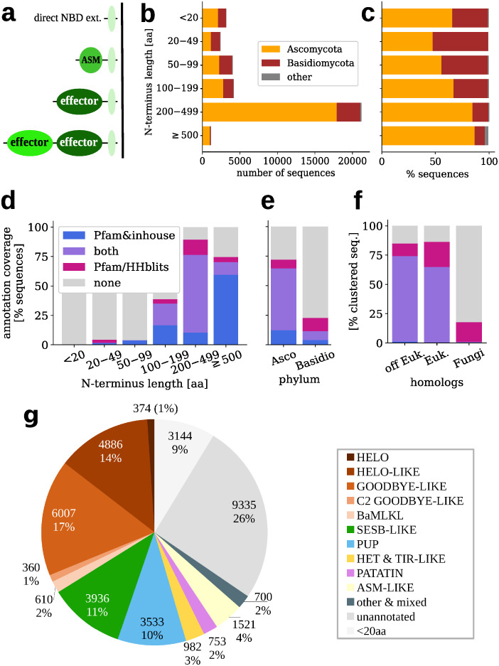
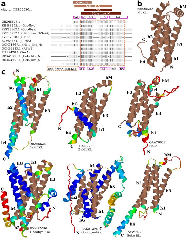
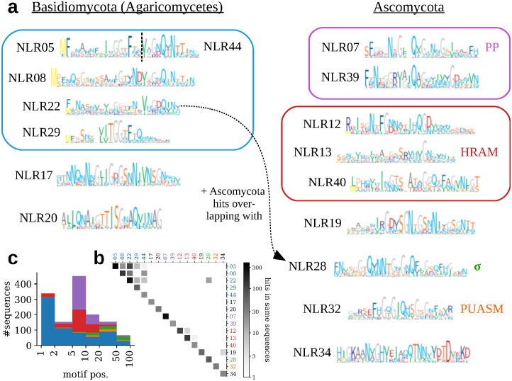
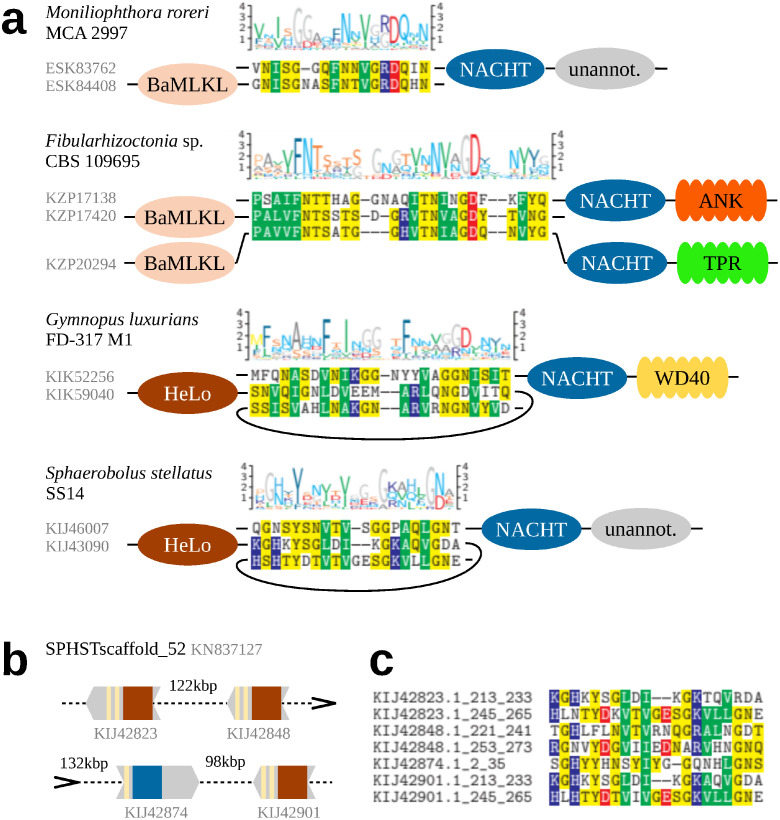
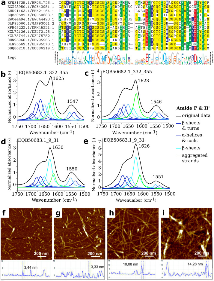
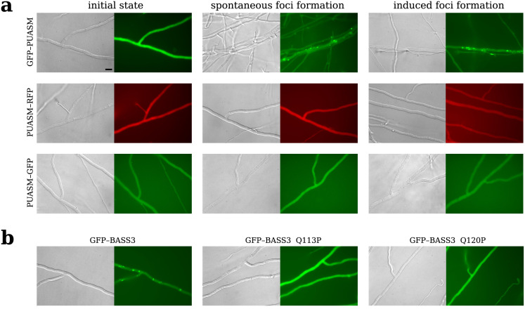
NLR proteins are intracellular receptors constituting a conserved component of the innate immune system of cellular organisms. In fungi, NLRs are characterized by high diversity of architectures and presence of amyloid signaling. Here, we explore the diverse world of effector and signaling domains of fungal NLRs using state-of-the-art bioinformatic methods including MMseqs2 for fast clustering, probabilistic context-free grammars for sequence analysis, and AlphaFold2 deep neural networks for structure prediction. In addition to substantially improving the overall annotation, especially in basidiomycetes, the study identifies novel domains and reveals the structural similarity of MLKL-related HeLo- and Goodbye-like domains forming the most abundant superfamily of fungal NLR effectors. Moreover, compared to previous studies, we found several times more amyloid motif instances, including novel families, and validated aggregating and prion-forming properties of the most abundant of them in vitro and in vivo . Also, through an extensive in silico search, the NLR-associated amyloid signaling was identified in basidiomycetes. The emerging picture highlights similarities and differences in the NLR architectures and amyloid signaling in ascomycetes, basidiomycetes and other branches of life.
Overview
The claims and statistics of this paper haven’t been checked yet.
Run the check — free, no account needed. It reads the open-access text and checks the sources it cites.
Integrity signals
Registry
Cites
Statistics
Figures
Verification
Claims the paper makes
The claims and statistics of this paper haven’t been checked yet.
Run the check — free, no account needed. It reads the open-access text and checks the sources it cites.
Self-check
Percentages, sums and reported differences re-computed from the paper’s own numbers.
The self-check needs the paper’s full text.
Citations
Cites
Highly accurate protein structure prediction with AlphaFold
Jumper, J., Evans, R., Pritzel, A., Green, T., Figurnov, M., Ronneberger, O., Tunyasuvunakool, K., Bates, R., Žídek, A., Potapenko, A., et al. (2021) · Nature · doi:10.1038/s41586-021-03819-2 · Cited by 43,764
Potter, S. C., Luciani, A., Eddy, S. R., Park, Y., Lopez, R., & Finn, R. D. (2018) · Nucleic Acids Research · doi:10.1093/nar/gky448 · Cited by 2,607
UniRef clusters: a comprehensive and scalable alternative for improving sequence similarity searches
Suzek, B. E., Wang, Y., Huang, H., McGarvey, P. B., Wu, C. H., & the UniProt Consortium (2015) · Bioinformatics · doi:10.1093/bioinformatics/btu739 · Cited by 1,928
Distinct β-sheet structure in protein aggregates determined by ATR-FTIR spectroscopy
Shivu, B., Seshadri, S., Li, J., Oberg, K. A., Uversky, V. N., & Fink, A. L. (2013) · Biochemistry · doi:10.1021/bi400625v · Cited by 289
The STIR-domain superfamily in signal transduction, development and immunity
Novatchkova, M., Leibbrandt, A., Werzowa, J., Neubüser, A., & Eisenhaber, F. (2003) · Trends in Biochemical Sciences · doi:10.1016/s0968-0004(03)00067-7 · Cited by 255
Research works printed but not linked: 134
- JDG Jones, 2016, Intracellular innate immune surveillance devices in plants and animals, Science, 354, aaf6395, 10.1126/science.aaf6395 · DOI
- J Uehling, 2017, Do fungi have an innate immune response? An NLR-based comparison to plant and animal immune systems, PLoS Pathogens, 13, e1006578, 10.1371/journal.ppat.1006578 · DOI
- Z Duxbury, 2021, A Comparative Overview of the Intracellular Guardians of Plants and Animals: NLRs in Innate Immunity and Beyond, Annual Review of Plant Biology, 72, 155, 10.1146/annurev-arplant-080620-104948 · DOI
- EV Koonin, 2002, Origin and evolution of eukaryotic apoptosis: the bacterial connection, Cell Death & Differentiation, 9, 394, 10.1038/sj.cdd.4400991 · DOI
- A Daskalov, 2020, NLR Function in Fungi as Revealed by the Study of Self/Non-self Recognition Systems, 123
- Y Xiong, 2020, Resistosome and inflammasome: platforms mediating innate immunity, Current opinion in plant biology, 56, 47, 10.1016/j.pbi.2020.03.010 · DOI
- G Bi, 2021, Regulation of Cell Death and Signaling by Pore-Forming Resistosomes, Annual review of phytopathology, 59, 239, 10.1146/annurev-phyto-020620-095952 · DOI
- IML Saur, 2021, NOD-like receptor-mediated plant immunity: from structure to cell death, Nat Rev Immunol, 21, 305, 10.1038/s41577-020-00473-z · DOI
- EA van der Biezen, 1998, The NB-ARC domain: a novel signalling motif shared by plant resistance gene products and regulators of cell death in animals, Current Biology, 8, R226, 10.1016/S0960-9822(98)70145-9 · DOI
- E Koonin, 2000, The NACHT family—a new group of predicted NTPases implicated in apoptosis and MHC transcription activation, Trends in Biochemical Sciences, 25, 223, 10.1016/S0968-0004(00)01577-2 · DOI
- W Dyrka, 2014, Diversity and Variability of NOD-Like Receptors in Fungi, Genome Biology and Evolution, 6, 3137, 10.1093/gbe/evu251 · DOI
- W Dyrka, 2020, Identification of NLR-associated amyloid signaling motifs in bacterial genomes, Journal of Molecular Biology, 432, 6005, 10.1016/j.jmb.2020.10.004 · DOI
- LA Gao, 2022, Prokaryotic innate immunity through pattern recognition of conserved viral proteins, Science, 377, eabm4096, 10.1126/science.abm4096 · DOI
- S Saupe, 1995, A gene responsible for vegetative incompatibility in the fungus Podospora anserina encodes a protein with a GTP-binding motif and G beta homologous domain, Gene, 162, 135, 10.1016/0378-1119(95)00272-8 · DOI
- M Paoletti, 2016, Vegetative incompatibility in fungi: From recognition to cell death, whatever does the trick, Fungal Biol Rev, 30, 152, 10.1016/j.fbr.2016.08.002 · DOI
- AP Gonçalves, 2020, Conflict, Competition, and Cooperation Regulate Social Interactions in Filamentous Fungi, Annual Review of Microbiology, 74, 693, 10.1146/annurev-micro-012420-080905 · DOI
- JM Urbach, 2017, The NBS-LRR architectures of plant R-proteins and metazoan NLRs evolved in independent events, Proceedings of the National Academy of Sciences, 114, 1063, 10.1073/pnas.1619730114 · DOI
- E Espagne, 2002, HET-E and HET-D belong to a new subfamily of WD40 proteins involved in vegetative incompatibility specificity in the fungus Podospora anserina, Genetics, 161, 71, 10.1093/genetics/161.1.71 · DOI
- E Bastiaans, 2014, Natural variation of heterokaryon incompatibility gene het-c in Podospora anserina reveals diversifying selection, Molecular Biology and Evolution, 31, 962, 10.1093/molbev/msu047 · DOI
- MA Armant, 2002, Toll-like receptors: a family of pattern-recognition receptors in mammals, Genome biology, 3, REVIEWS3011, 10.1186/gb-2002-3-8-reviews3011 · DOI
- VY Toshchakov, 2020, A survey of TIR domain sequence and structure divergence, Immunogenetics, 72, 181, 10.1007/s00251-020-01157-7 · DOI
- D Lapin, 2022, Molecular innovations in plant TIR-based immunity signaling, The Plant Cell, 34, 1479, 10.1093/plcell/koac035 · DOI
- J Heller, 2018, NLR surveillance of essential SEC-9 SNARE proteins induces programmed cell death upon allorecognition in filamentous fungi, Proc Natl Acad Sci U S A, 115, E2292, 10.1073/pnas.1719705115 · DOI
- M Paoletti, 2009, Fungal incompatibility: Evolutionary origin in pathogen defense?, BioEssays, 31, 1201, 10.1002/bies.200900085 · DOI
- M Paoletti, 2007, Genesis of a Fungal Non-Self Recognition Repertoire, PLoS ONE, 2, e283, 10.1371/journal.pone.0000283 · DOI
- D Chevanne, 2010, WD-repeat instability and diversification of the Podospora anserina hnwd non-self recognition gene family, BMC Evolutionary Biology, 10, 134, 10.1186/1471-2148-10-134 · DOI
- OJ Rando, 2007, Timescales of Genetic and Epigenetic Inheritance, Cell, 128, 655, 10.1016/j.cell.2007.01.023 · DOI
- M Iotti, 2012, Self/nonself recognition in Tuber melanosporum is not mediated by a heterokaryon incompatibility system, Fungal Biology, 116, 261, 10.1016/j.funbio.2011.11.009 · DOI
- RD Finn, 2016, The Pfam protein families database: towards a more sustainable future, Nucleic Acids Research
- SJ Saupe, 2020, Amyloid Signaling in Filamentous Fungi and Bacteria, Annual Review of Microbiology, 74, 673, 10.1146/annurev-micro-011320-013555 · DOI
- DL Ollis, 1992, The alpha/beta hydrolase fold, Protein Engineering, Design and Selection, 5, 197, 10.1093/protein/5.3.197 · DOI
- A Mushegian, 1994, Unexpected sequence similarity between nucleosidases and phosphoribosyltransferases of different specificity, Protein science: a publication of the Protein Society, 3, 1081, 10.1002/pro.5560030711 · DOI
- C Mao, 1997, The crystal structure of Escherichia coli purine nucleoside phosphorylase: a comparison with the human enzyme reveals a conserved topology, Structure (London, England: 1993), 5, 1373, 10.1016/S0969-2126(97)00287-6 · DOI
- A Balguerie, 2003, Domain organization and structure-function relationship of the HET-s prion protein of Podospora anserina, The EMBO Journal, 22, 2071, 10.1093/emboj/cdg213 · DOI
- ND Fedorova, 2005, Comparative analysis of programmed cell death pathways in filamentous fungi, BMC Genomics, 6, 177, 10.1186/1471-2164-6-177 · DOI
- J Greenwald, 2010, The mechanism of prion inhibition by HET-S, Molecular Cell, 38, 889, 10.1016/j.molcel.2010.05.019 · DOI
- A Daskalov, 2012, Genomic Clustering and Homology between HET-S and the NWD2 STAND Protein in Various Fungal Genomes, PLoS ONE, 7, e34854, 10.1371/journal.pone.0034854 · DOI
- A Daskalov, 2016, Identification of a novel cell death-inducing domain reveals that fungal amyloid-controlled programmed cell death is related to necroptosis, Proceedings of the National Academy of Sciences of the United States of America, 113, 2720, 10.1073/pnas.1522361113 · DOI
- J Wang, 2019, Reconstitution and structure of a plant NLR resistosome conferring immunity, Science, 364, eaav5870, 10.1126/science.aav5870 · DOI
- G Bi, 2021, The ZAR1 resistosome is a calcium-permeable channel triggering plant immune signaling, Cell, 184, 3528, 10.1016/j.cell.2021.05.003 · DOI
- A Daskalov, 2015, Signal transduction by a fungal NOD-like receptor based on propagation of a prion amyloid fold, PLoS Biology, 13, e1002059, 10.1371/journal.pbio.1002059 · DOI
- C Wasmer, 2008, Amyloid Fibrils of the HET-s(218–289) Prion Form a Beta Solenoid with a Triangular Hydrophobic Core, Science, 319, 1523, 10.1126/science.1151839 · DOI
- H van Melckebeke, 2010, Atomic-Resolution Three-Dimensional Structure of HET-s(218–289) Amyloid Fibrils by Solid-State NMR Spectroscopy, Journal of the American Chemical Society, 132, 13765, 10.1021/ja104213j · DOI
- C Seuring, 2012, The mechanism of toxicity in HET-S/HET-s prion incompatibility, PLoS Biology, 10, e1001451, 10.1371/journal.pbio.1001451 · DOI
- A Daskalov, 2015, Theme and variations: evolutionary diversification of the HET-s functional amyloid motif, Scientific Reports, 5, 12494, 10.1038/srep12494 · DOI
- A Daskalov, 2021, Structural and molecular basis of cross-seeding barriers in amyloids, Proceedings of the National Academy of Sciences, 118, 10.1073/pnas.2014085118 · DOI
- S Graziani, 2004, Bistability and hysteresis of the ‘Secteur’ differentiation are controlled by a two-gene locus in Nectria haematococca, BMC Biology, 2, 18, 10.1186/1741-7007-2-18 · DOI
- X Sun, 2002, Identification of a novel homotypic interaction motif required for the phosphorylation of receptor-interacting protein (RIP) by RIP3, The Journal of Biological Chemistry, 277, 9505, 10.1074/jbc.M109488200 · DOI
- M Rebsamen, 2009, DAI/ZBP1 recruits RIP1 and RIP3 through RIP homotypic interaction motifs to activate NF-kB, EMBO reports, 10, 916, 10.1038/embor.2009.109 · DOI
- AV Kajava, 2014, Evolutionary link between metazoan RHIM motif and prion-forming domain of fungal heterokaryon incompatibility factor HET-s/HET-s, Scientific Reports, 4, 1, 10.1038/srep07436 · DOI
- AB Ahmed, 2015, A structure-based approach to predict predisposition to amyloidosis, Alzheimer’s & Dementia, 11, 681, 10.1016/j.jalz.2014.06.007 · DOI
- W Dyrka, 2021, Searching for universal model of amyloid signaling motifs using probabilistic context-free grammars, BMC Bioinformatics, 22, 222, 10.1186/s12859-021-04139-y · DOI
- C Clavé, 2022, Fungal gasdermin-like proteins are controlled by proteolytic cleavage, Proceedings of the National Academy of Sciences, 119, e2109418119, 10.1073/pnas.2109418119 · DOI
- AG Johnson, 2022, Bacterial gasdermins reveal an ancient mechanism of cell death, Science, 375, 221, 10.1126/science.abj8432 · DOI
- E Gluck-Thaler, 2022, Giant Starship Elements Mobilize Accessory Genes in Fungal Genomes, Molecular Biology and Evolution, 39, 10.1093/molbev/msac109 · DOI
- J Li, 2012, The RIP1/RIP3 necrosome forms a functional amyloid signaling complex required for programmed necrosis, Cell, 150, 339, 10.1016/j.cell.2012.06.019 · DOI · Paper page
- A Kleino, 2017, Peptidoglycan-Sensing Receptors Trigger the Formation of Functional Amyloids of the Adaptor Protein Imd to Initiate Drosophila NF-kB Signaling, Immunity, 47, 635, 10.1016/j.immuni.2017.09.011 · DOI
- M Steinegger, 2017, MMseqs2 enables sensitive protein sequence searching for the analysis of massive data sets, Nature Biotechnology, 35, 1026, 10.1038/nbt.3988 · DOI · Paper page
- M Mirdita, 2016, Uniclust databases of clustered and deeply annotated protein sequences and alignments, Nucleic Acids Research, 45, D170, 10.1093/nar/gkw1081 · DOI
- M Remmert, 2012, HHblits: lightning-fast iterative protein sequence searching by HMM-HMM alignment, Nature Methods, 9, 173, 10.1038/nmeth.1818 · DOI · Paper page
- R Oliva, 2010, Recent developments in effector biology of filamentous plant pathogens, Cellular Microbiology, 12, 705, 10.1111/j.1462-5822.2010.01471.x · DOI
- T Liu, 2011, Two host cytoplasmic effectors are required for pathogenesis of Phytophthora sojae by suppression of host defenses, Plant physiology, 155, 490, 10.1104/pp.110.166470 · DOI
- D Zhang, 2016, Transposons to toxins: the provenance, architecture and diversification of a widespread class of eukaryotic effectors, Nucleic Acids Research, 44, 3513, 10.1093/nar/gkw221 · DOI
- S Voss, 2018, RiCRN1, a Crinkler Effector From the Arbuscular Mycorrhizal Fungus Rhizophagus irregularis, Functions in Arbuscule Development, Frontiers in microbiology, 9, 2068, 10.3389/fmicb.2018.02068 · DOI
- A Hetmann, 2019, Supresja odporności podstawowej typu PTI przez syntetyzowane w fitopatogenach białka efektorowe wprowadzane do wnętrza komórek infekowanej rośliny, Postępy Biochemii, 65, 58, 10.18388/pb.2019_257 · DOI
- SJ Grimshaw, 2004, Structure of the sterile alpha motif (SAM) domain of the Saccharomyces cerevisiae mitogen-activated protein kinase pathway-modulating protein STE50 and analysis of its interaction with the STE11 SAM, The Journal of Biological Chemistry, 279, 2192, 10.1074/jbc.M305605200 · DOI
- S Ray, 2020, Sterile α-motif domain requirement for cellular signaling and survival, Journal of Biological Chemistry, 295, 7113, 10.1074/jbc.RA119.011895 · DOI
- LAJ O’Neill, 2007, The family of five: TIR-domain-containing adaptors in Toll-like receptor signalling, Nature reviews Immunology, 7, 353, 10.1038/nri2079 · DOI · Paper page
- Q Zhang, 2011, TIR domain-containing adaptor SARM is a late addition to the ongoing microbe-host dialog, Developmental and comparative immunology, 35, 461, 10.1016/j.dci.2010.11.013 · DOI
- ND Peterson, 2022, Pathogen infection and cholesterol deficiency activate the C. elegans p38 immune pathway through a TIR-1/SARM1 phase transition, eLife, 11, e74206, 10.7554/eLife.74206 · DOI
- S Detke, 1998, Cloning of the Candida albicans nucleoside transporter by complementation of nucleoside transport-deficient Saccharomyces, Yeast, 14, 1257, 10.1002/(SICI)1097-0061(1998100)14:14<1257::AID-YEA326>3.0.CO;2-6 · DOI
- S Lu, 2019, CDD/SPARCLE: the conserved domain database in 2020, Nucleic Acids Research, 48, D265, 10.1093/nar/gkz991 · DOI
- J Soeding, 2005, Protein homology detection by HMM-HMM comparison, Bioinformatics, 21, 951, 10.1093/bioinformatics/bti125 · DOI
- L Zimmermann, 2018, A Completely Reimplemented MPI Bioinformatics Toolkit with a New HHpred Server at its Core, Journal of Molecular Biology, 430, 2237, 10.1016/j.jmb.2017.12.007 · DOI · Paper page
- M Rübbelke, 2020, Locking mixed-lineage kinase domain-like protein in its auto-inhibited state prevents necroptosis, Proceedings of the National Academy of Sciences, 117, 33272, 10.1073/pnas.2017406117 · DOI
- M Mirdita, 2022, ColabFold: making protein folding accessible to all, Nature Methods, 19, 679, 10.1038/s41592-022-01488-1 · DOI · Paper page
- Y Zhang, 2005, TM-align: a protein structure alignment algorithm based on the TM-score, Nucleic Acids Research, 33, 2302, 10.1093/nar/gki524 · DOI · Paper page
- M Landau, 2005, ConSurf 2005: the projection of evolutionary conservation scores of residues on protein structures, Nucleic Acids Research, 33, W299, 10.1093/nar/gki370 · DOI
- H Ashkenazy, 2016, ConSurf 2016: an improved methodology to estimate and visualize evolutionary conservation in macromolecules, Nucleic Acids Research, 44, W344, 10.1093/nar/gkw408 · DOI · Paper page
- H Adachi, 2019, An N-terminal motif in NLR immune receptors is functionally conserved across distantly related plant species, eLife, 8, e49956, 10.7554/eLife.49956 · DOI
- DE McNamara, 2019, Direct Activation of Human MLKL by a Select Repertoire of Inositol Phosphate Metabolites, Cell Chemical Biology, 26, 863, 10.1016/j.chembiol.2019.03.010 · DOI
- B Wu, 2012, Evolution of prokaryotic homologues of the eukaryotic SEFIR protein domain, Gene, 492, 160, 10.1016/j.gene.2011.10.033 · DOI
- F Jacob, 2013, Evolution and Conservation of Plant NLR Functions, Frontiers in Immunology, 4, 297, 10.3389/fimmu.2013.00297 · DOI
- Y Guo, 2018, Fungus-Bacterium Symbionts Promote Plant Health and Performance, Microbes and Environments, 33, 239, 10.1264/jsme2.ME3303rh · DOI
- TL Bailey, 2009, MEME Suite: tools for motif discovery and searching, Nucleic Acids Research, 37, W202, 10.1093/nar/gkp335 · DOI · Paper page
- N Alkan, 2013, Global Aspects of pacC Regulation of Pathogenicity Genes in Colletotrichum gloeosporioides as Revealed by Transcriptome Analysis, Molecular Plant-Microbe Interactions, 26, 1345, 10.1094/MPMI-03-13-0080-R · DOI · Paper page
- R Sarroukh, 2013, ATR-FTIR: a “rejuvenated” tool to investigate amyloid proteins, Biochimica et biophysica acta, 1828, 2328, 10.1016/j.bbamem.2013.04.012 · DOI
- JM Ruysschaert, 2018, ATR-FTIR analysis of amyloid proteins, Methods Mol Biol, 1777, 69, 10.1007/978-1-4939-7811-3_3 · DOI
- FS Ruggeri, 2019, Atomic force microscopy for single molecule characterisation of protein aggregation, Archives of Biochemistry and Biophysics, 664, 134, 10.1016/j.abb.2019.02.001 · DOI
- M Biancalana, 2010, Molecular mechanism of Thioflavin-T binding to amyloid fibrils, Biochimica et Biophysica Acta (BBA)—Proteins and Proteomics, 1804, 1405, 10.1016/j.bbapap.2010.04.001 · DOI
- C Xue, 2017, Thioflavin T as an amyloid dye: fibril quantification, optimal concentration and effect on aggregation, Royal Society Open Science, 4, 160696, 10.1098/rsos.160696 · DOI
- MR Nilsson, 2004, Techniques to study amyloid fibril formation in vitro, Methods, 34, 151, 10.1016/j.ymeth.2004.03.012 · DOI
- H Li, 2009, Encyclopedia of Analytical Chemistry
- PM Martins, 2020, MIRRAGGE—Minimum Information Required for Reproducible AGGregation Experiments, Frontiers in Molecular Neuroscience, 13, 222, 10.3389/fnmol.2020.582488 · DOI
- N Wilkosz, 2020, Molecular Spectroscopic Markers of Abnormal Protein Aggregation, Molecules, 25, 10.3390/molecules25112498 · DOI
- R Khurana, 2000, Do Parallel β-Helix Proteins Have a Unique Fourier Transform Infrared Spectrum?, Biophysical Journal, 78, 994, 10.1016/S0006-3495(00)76657-4 · DOI
- Y Zou, 2013, Parallel β-Sheet Fibril and Antiparallel β-Sheet Oligomer: New Insights into Amyloid Formation of Hen Egg White Lysozyme under Heat and Acidic Condition from FTIR Spectroscopy, The Journal of Physical Chemistry B, 117, 4003, 10.1021/jp4003559 · DOI
- M Grelich-Mucha, 2021, Autofluorescence of Amyloids Determined by Enantiomeric Composition of Peptides, The Journal of Physical Chemistry B, 125, 5502, 10.1021/acs.jpcb.1c00808 · DOI
- K Berthelot, 2011, In Vivo and In Vitro Analyses of Toxic Mutants of HET-s: FTIR Antiparallel Signature Correlates with Amyloid Toxicity, Journal of Molecular Biology, 412, 137, 10.1016/j.jmb.2011.07.009 · DOI
- JR Requena, 2014, The structure of the infectious prion protein, Prion, 8, 60, 10.4161/pri.28368 · DOI
- SD Moran, 2014, How to Get Insight into Amyloid Structure and Formation from Infrared Spectroscopy, The journal of physical chemistry letters, 5, 1984, 10.1021/jz500794d · DOI
- T Bardin, 2021, Partial Prion Cross-Seeding between Fungal and Mammalian Amyloid Signaling Motifs, mBio, 12, e02782, 10.1128/mBio.02782-20 · DOI
- M Paoletti, 2007, The Fungus-Specific HET Domain Mediates Programmed Cell Death in Podospora anserina, Eukaryotic Cell, 6, 2001, 10.1128/EC.00129-07 · DOI
- W Dyrka, 2009, A Stochastic Context Free Grammar based Framework for Analysis of Protein Sequences, BMC Bioinformatics, 10, 323, 10.1186/1471-2105-10-323 · DOI
- GW Wilburn, 2020, Remote homology search with hidden Potts models, PLOS Computational Biology, 16, 1, 10.1371/journal.pcbi.1008085 · DOI
- AP Muntoni, 2020, Aligning biological sequences by exploiting residue conservation and coevolution, Phys Rev E, 102, 062409, 10.1103/PhysRevE.102.062409 · DOI
- H Talibart, 2021, PPalign: optimal alignment of Potts models representing proteins with direct coupling information, BMC Bioinformatics, 22, 317, 10.1186/s12859-021-04222-4 · DOI
- Merrill W, Weiss G, Goldberg Y, Schwartz R, Smith NA, Yahav E. A Formal Hierarchy of RNN Architectures. In: Proceedings of the 58th Annual Meeting of the Association for Computational Linguistics. Online: Association for Computational Linguistics; 2020. p. 443–459. · DOI
- Bhattamishra S, Patel A, Goyal N. On the Computational Power of Transformers and Its Implications in Sequence Modeling. In: Proceedings of the 24th Conference on Computational Natural Language Learning. Online: Association for Computational Linguistics; 2020. p. 455–475. · DOI
- Merrill W. Formal Language Theory Meets Modern NLP. CoRR. 2021;abs/2102.10094.
- Nambiar A, Heflin M, Liu S, Maslov S, Hopkins M, Ritz A. Transforming the Language of Life: Transformer Neural Networks for Protein Prediction Tasks. In: Proceedings of the 11th ACM International Conference on Bioinformatics, Computational Biology and Health Informatics. BCB’20. New York, NY, USA: Association for Computing Machinery; 2020. · DOI
- N Brandes, 2022, ProteinBERT: A universal deep-learning model of protein sequence and function, Bioinformatics, 38, 2102, 10.1093/bioinformatics/btac020 · DOI · Paper page
- SF Altschul, 1997, Gapped BLAST and PSI-BLAST: a new generation of protein database search programs, Nucleic Acids Research, 25, 3389, 10.1093/nar/25.17.3389 · DOI · Paper page
- F Sievers, 2011, Fast, scalable generation of high-quality protein multiple sequence alignments using Clustal Omega, Molecular Systems Biology, 7, 539, 10.1038/msb.2011.75 · DOI
- TU Consortium, 2020, UniProt: the universal protein knowledgebase in 2021, Nucleic Acids Research, 49, D480, 10.1093/nar/gkaa1100 · DOI · Paper page
- W Dyrka, 2019, Estimating probabilistic context-free grammars for proteins using contact map constraints, PeerJ, 7, e6559, 10.7717/peerj.6559 · DOI
- W Li, 2006, Cd-hit: a fast program for clustering and comparing large sets of protein or nucleotide sequences, Bioinformatics, 22, 1658, 10.1093/bioinformatics/btl158 · DOI
- L Fu, 2012, CD-HIT: accelerated for clustering the next-generation sequencing data, Bioinformatics, 28, 3150, 10.1093/bioinformatics/bts565 · DOI · Paper page
- Bailey TL, Elkan C. Fitting a mixture model by expectation maximization to discover motifs in biopolymers. In: Proceedings of the Second International Conference on Intelligent Systems for Molecular Biology. AAAI Press, Menlo Park, California; 1994. p. 28–36.
- SR Eddy, 2011, Accelerated Profile HMM Searches, PLoS Computational Biology, 7, e1002195, 10.1371/journal.pcbi.1002195 · DOI · Paper page
- K Katoh, 2013, MAFFT multiple sequence alignment software version 7: improvements in performance and usability, Molecular Biology and Evolution, 30, 772, 10.1093/molbev/mst010 · DOI · Paper page
- S Capella-Gutierrez, 2009, trimAl: a tool for automated alignment trimming in large-scale phylogenetic analyses, Bioinformatics, 25, 1972, 10.1093/bioinformatics/btp348 · DOI
- NR Coordinators, 2017, Database resources of the National Center for Biotechnology Information, Nucleic Acids Research, 46, D8, 10.1093/nar/gkx1095 · DOI
- PW Harrison, 2020, The European Nucleotide Archive in 2020, Nucleic Acids Research, 49, D82, 10.1093/nar/gkaa1028 · DOI
- McKinney W. Data Structures for Statistical Computing in Python. In: St fan van der Walt, Jarrod Millman, editors. Proceedings of the 9th Python in Science Conference; 2010. p. 56–61. · DOI
- JD Hunter, 2007, Matplotlib: A 2D graphics environment, Computing in Science & Engineering, 9, 90, 10.1109/MCSE.2007.55 · DOI · Paper page
- ML Waskom, 2021, seaborn: statistical data visualization, Journal of Open Source Software, 6, 3021, 10.21105/joss.03021 · DOI · Paper page
- E Beitz, 2000, TeXshade: shading and labeling of multiple sequence alignments using LaTeX2e, Bioinformatics, 16, 135, 10.1093/bioinformatics/16.2.135 · DOI
- ER Gansner, 2000, An open graph visualization system and its applications to software engineering, Software Practice and Experience, 30, 1203, 10.1002/1097-024X(200009)30:11<1203::AID-SPE338>3.0.CO;2-N · DOI
- RA Sayle, 1995, RASMOL: biomolecular graphics for all, Trends in Biochemical Sciences, 20, 374, 10.1016/S0968-0004(00)89080-5 · DOI
- A Savitzky, 1964, Smoothing and differentiation of data by simplified least squares procedures, Analytical Chemistry, 36, 1627, 10.1021/ac60214a047 · DOI · Paper page
- A Balguerie, 2004, The sequences appended to the amyloid core region of the HET-s prion protein determine higher-order aggregate organization in vivo, Journal of Cell Science, 117, 2599, 10.1242/jcs.01116 · DOI
- T Bergès, 1989, Heat Shock at an Elevated Temperature Improves Transformation Efficiency of Protoplasts from Podospora anserina, Microbiology, 135, 601, 10.1099/00221287-135-3-601 · DOI
- L Benkemoun, 2006, Methods for the in vivo and in vitro analysis of [Het-s] prion infectivity, Methods, 39, 61, 10.1016/j.ymeth.2006.04.006 · DOI
Metadata
- Authors
- Wojciechowski, J. W., Tekoglu, E., Gąsior-Głogowska, M., Coustou, V., Szulc, N., Szefczyk, M., Kopaczyńska, M., Saupe, S. J., & Dyrka, W.
- Affiliations
- Wrocław University of Science and Technology (PL)
- Centre National de la Recherche Scientifique (FR)
- Corresponding author
- Saupe, Sven J.
- Dyrka, Witold
- ORCID
- Venue
- PLOS Computational Biology
- Publisher
- Public Library of Science (PLoS)
- Published
- 2022-12-21
- Volume
- 18
- Issue
- 12
- Pages
- e1010787
- ISSN
- 1553-734X (ISSN-L)
- 1553-7358
- PMC
- PMC9815663
- OpenAlex
- W4312064942
- Cited by
- 19
- References
- 5
- Citations by year
- 2026: 2 · 2025: 10 · 2024: 1 · 2023: 6
- FWCI
- 2.34
- Type
- journal article
- Language
- en
- Update notices
- new_version (2023-01-05)
- Open access
- gold
- doi.org
- Licence
- Locations
- PLoS Computational Biology (publishedVersion, cc-by)
- bioRxiv (Cold Spring Harbor Laboratory) (acceptedVersion)
- PubMed Central (publishedVersion, cc-by)
- PubMed (publishedVersion)
- HAL (submittedVersion, other-oa)
- RePEc: Research Papers in Economics (submittedVersion)
- Digital Collections portal (Koç University) (submittedVersion)
- DOAJ (DOAJ: Directory of Open Access Journals) (submittedVersion)
- Funders
- Narodowe Centrum Nauki (2019/35/B/NZ2/03997)
- Wroclawskie Centrum Sieciowo-Superkomputerowe, Politechnika Wroclawska (98)
- Narodowe Centrum Badań i Rozwoju (POWR.03.02.00-00-I003/16)
- Agence Nationale de la Recherche (SFAS, R-17-CE11-0035)
- Topics
- Life Sciences › Agricultural and Biological Sciences › Plant Science › Plant-Microbe Interactions and Immunity
- Life Sciences › Biochemistry, Genetics and Molecular Biology › Molecular Biology › Fungal and yeast genetics research
- Life Sciences › Immunology and Microbiology › Immunology › Immune Response and Inflammation
- Keywords
- NLR proteins, effector domain, Basidiomycota, Ascomycota, prion formation, protein aggregation, domain architecture, protein structure prediction, sequence clustering
- MeSH terms
- NLR Proteins, Amyloid, Fungal Proteins, Amyloidogenic Proteins
About these checks
Registry, retraction, Cites and statistics results are computed, not judged, with one exception: a printed reference that no index lists is read once by a model before it counts as a citation error. Claims are extracted by a model and kept only when their quote appears verbatim in the text; each cited source is then checked, with a model’s help, for whether it says what the claim relies on. Those verdicts can be wrong — read the claim’s quote and the cited source before relying on one.
Statistics and arithmetic are recomputed, not judged.
Confidence bands are shown only for fields calibrated on our annotated test set. None is calibrated yet, so no band is shown.
What counts as a claim ›How we measure confidence ›How we check this ›
Are you an author, or do you see a problem with this page? Request a review
