

The DOI matches its Crossref record; 96 of the 96 references it cites were found in a registry or an index.
Deep learning for lung cancer prognostication: A retrospective multi-cohort radiomics study
Open accessAbstract
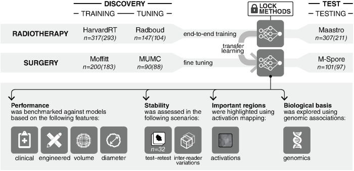
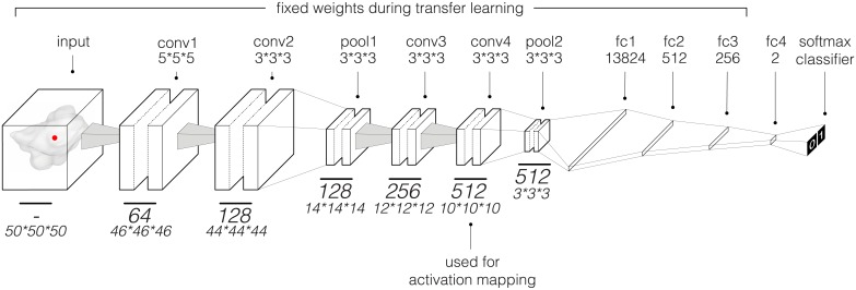
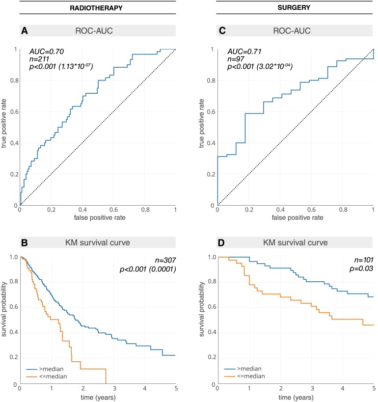
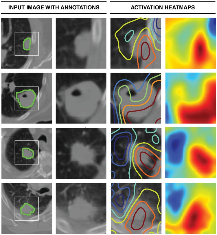
BACKGROUND: Non-small-cell lung cancer (NSCLC) patients often demonstrate varying clinical courses and outcomes, even within the same tumor stage. This study explores deep learning applications in medical imaging allowing for the automated quantification of radiographic characteristics and potentially improving patient stratification. METHODS AND FINDINGS: We performed an integrative analysis on 7 independent datasets across 5 institutions totaling 1,194 NSCLC patients (age median = 68.3 years [range 32.5-93.3], survival median = 1.7 years [range 0.0-11.7]). Using external validation in computed tomography (CT) data, we identified prognostic signatures using a 3D convolutional neural network (CNN) for patients treated with radiotherapy (n = 771, age median = 68.0 years [range 32.5-93.3], survival median = 1.3 years [range 0.0-11.7]). We then employed a transfer learning approach to achieve the same for surgery patients (n = 391, age median = 69.1 years [range 37.2-88.0], survival median = 3.1 years [range 0.0-8.8]). We found that the CNN predictions were significantly associated with 2-year overall survival from the start of respective treatment for radiotherapy (area under the receiver operating characteristic curve [AUC] = 0.70 [95% CI 0.63-0.78], p < 0.001) and surgery (AUC = 0.71 [95% CI 0.60-0.82], p < 0.001) patients. The CNN was also able to significantly stratify patients into low and high mortality risk groups in both the radiotherapy (p < 0.001) and surgery (p = 0.03) datasets. Additionally, the CNN was found to significantly outperform random forest models built on clinical parameters-including age, sex, and tumor node metastasis stage-as well as demonstrate high robustness against test-retest (intraclass correlation coefficient = 0.91) and inter-reader (Spearman's rank-order correlation = 0.88) variations. To gain a better understanding of the characteristics captured by the CNN, we identified regions with the most contribution towards predictions and highlighted the importance of tumor-surrounding tissue in patient stratification. We also present preliminary findings on the biological basis of the captured phenotypes as being linked to cell cycle and transcriptional processes. Limitations include the retrospective nature of this study as well as the opaque black box nature of deep learning networks. CONCLUSIONS: Our results provide evidence that deep learning networks may be used for mortality risk stratification based on standard-of-care CT images from NSCLC patients. This evidence motivates future research into better deciphering the clinical and biological basis of deep learning networks as well as validation in prospective data.
Overview
The claims and statistics of this paper haven’t been checked yet.
Run the check — free, no account needed. It reads the open-access text and checks the sources it cites.
Integrity signals
Registry
Cites
Statistics
Figures
Verification
Claims the paper makes
The claims and statistics of this paper haven’t been checked yet.
Run the check — free, no account needed. It reads the open-access text and checks the sources it cites.
Claims other papers make about this one
Each is checked against this paper’s own text: does it say what the citing paper says it does?
The source backs part of the claim, not all of it — or backs it for a different population, condition or comparison.
In the citing paper
“Especially in medical imaging there is a large potential as deep learning can successfully be used for identifying and segmenting objects within the 3-dimensional (3D) image space13–16.”
From Deep convolutional neural networks to predict cardiovascular risk from computed tomography (2021) · cites this paper as [15]
Experimental — accuracy not yet measuredIn the citing paper
“Deep-learning methods, in comparison, are robust to differences in acquisition and segmentation variability and provide improved performance10.”
From Foundation Model for Cancer Imaging Biomarkers (2024) · cites this paper as [10]
In the citing paper
“While many studies investigating imaging-based biomarkers incorporate supervised deep-learning algorithms into their models9–11, they are typically applied in scenarios where large datasets are available for training and testing.”
From Foundation Model for Cancer Imaging Biomarkers (2024) · cites this paper as [10]
Self-check
Percentages, sums and reported differences re-computed from the paper’s own numbers.
The self-check needs the paper’s full text.
Citations
Cites
ImageNet classification with deep convolutional neural networks
Krizhevsky, A., Sutskever, I., & Hinton, G. E. (2017) · Communications of the ACM · doi:10.1145/3065386 · Cited by 112,155
Controlling the False Discovery Rate: A Practical and Powerful Approach to Multiple Testing
Benjamini, Y., & Hochberg, Y. (1995) · Journal of the Royal Statistical Society Series B: Statistical Methodology · doi:10.1111/j.2517-6161.1995.tb02031.x · Cited by 109,482
Adam: A Method for Stochastic Optimization
Kingma, D. P., & Ba, J. (2014) · arXiv · doi:10.48550/arxiv.1412.6980 · Cited by 98,946
Jemal, A., Bray, F., Ferlay, J., Ward, E., & Forman, D. (2011) · CA A Cancer Journal for Clinicians · doi:10.3322/caac.20107 · Cited by 55,077
Subramanian, A., Tamayo, P., Mootha, V. K., Mukherjee, S., Ebert, B. L., Gillette, M. A., Paulovich, A., Pomeroy, S. L., Golub, T. R., Lander, E. S., et al. (2005) · Proceedings of the National Academy of Sciences · doi:10.1073/pnas.0506580102 · Cited by 53,625
Fully convolutional networks for semantic segmentation
Long, J., Shelhamer, E., & Darrell, T. (2015) · 2015 IEEE Conference on Computer Vision and Pattern Recognition (CVPR) · doi:10.1109/cvpr.2015.7298965 · Cited by 38,834
(2014) · Cited by 33,419
Batch Normalization: Accelerating Deep Network Training by Reducing Internal Covariate Shift
Ioffe, S., & Szegedy, C. (2015) · arXiv · doi:10.48550/arxiv.1502.03167 · Cited by 27,818
Global cancer statistics, 2012
Torre, L. A., Bray, F., Siegel, R. L., Ferlay, J., Lortet‐Tieulent, J., & Jemal, A. (2015) · CA A Cancer Journal for Clinicians · doi:10.3322/caac.21262 · Cited by 27,235
Grad-CAM: Visual Explanations from Deep Networks via Gradient-Based Localization
Selvaraju, R. R., Cogswell, M., Das, A., Vedantam, R., Parikh, D., & Batra, D. (2017) · 2017 IEEE International Conference on Computer Vision (ICCV) · doi:10.1109/iccv.2017.74 · Cited by 24,513
Research works printed but not linked: 1
- Maas AL, Hannun AY, Ng AY. Rectifier nonlinearities improve neural network acoustic models. 30th International Conference on Machine Learning; 2013 Jun 16–21; Atlanta, GA, US.
Cited by
Sorted by citations
Explainable artificial intelligence (XAI) in deep learning-based medical image analysis
van der Velden, B. H. M., Kuijf, H. J., Gilhuijs, K. G. A., & Viergever, M. A. (2022) · Medical Image Analysis · doi:10.1016/j.media.2022.102470 · Cited by 1,328
Predicting cancer outcomes with radiomics and artificial intelligence in radiology
Bera, K., Braman, N., Gupta, A., Velcheti, V., & Madabhushi, A. (2022) · Nature Reviews Clinical Oncology · doi:10.1038/s41571-021-00560-7 · Cited by 819
Harnessing multimodal data integration to advance precision oncology
Boehm, K. M., Khosravi, P., Vanguri, R., Gao, J., & Shah, S. P. (2022) · Nature Reviews Cancer · doi:10.1038/s41568-021-00408-3 · Cited by 662
The Future of Sensitivity Analysis: An essential discipline for systems modeling and policy support
Razavi, S., Jakeman, A. J., Saltelli, A., Prieur, C., Iooss, B., Borgonovo, E., Plischke, E., Piano, S. L., Iwanaga, T., Becker, W., et al. (2020) · Environmental Modelling & Software · doi:10.1016/j.envsoft.2020.104954 · Cited by 703
Deep Learning Predicts Lung Cancer Treatment Response from Serial Medical Imaging
Xu, Y., Hosny, A., Zeleznik, R., Parmar, C., Coroller, T. P., Franco, I. “., Mak, R. H., & Aerts, H. J. W. L. (2019) · Clinical Cancer Research · doi:10.1158/1078-0432.ccr-18-2495 · Cited by 658
Self-citation
Metadata
- Authors
- Hosny, A., Parmar, C., Coroller, T. P., Grossmann, P., Zeleznik, R., Kumar, A., Bussink, J., Gillies, R. J., Mak, R. H., & Aerts, H. J. W. L.
- Affiliations
- Brigham and Women's Hospital (US)
- Radboud University Medical Center (NL)
- Moffitt Cancer Center (US)
- Corresponding author
- Aerts, Hugo J. W. L.
- ORCID
- Venue
- PLOS Medicine
- Publisher
- Public Library of Science (PLoS)
- Published
- 2018-11-30
- Volume
- 15
- Issue
- 11
- Pages
- e1002711
- ISSN
- 1549-1277 (ISSN-L)
- 1549-1676
- PMC
- PMC6269088
- OpenAlex
- W2903150666
- Cited by
- 579
- References
- 95
- Citations by year
- 2026: 30 · 2025: 61 · 2024: 78 · 2023: 93 · 2022: 95 · 2021: 103 · 2020: 84 · 2019: 33 · 2018: 1
- FWCI
- 38.12
- Type
- journal article
- Language
- en
- Open access
- gold
- doi.org
- Licence
- Locations
- PLoS Medicine (publishedVersion, cc-by)
- Europe PMC (PubMed Central) (publishedVersion, cc-by)
- PubMed Central (publishedVersion, cc-by)
- PubMed (publishedVersion)
- DOAJ (DOAJ: Directory of Open Access Journals) (submittedVersion, cc-by-sa)
- Figshare (submittedVersion, cc-by)
- Radboud Repository (Radboud University) (submittedVersion)
- Radboud Repository (Radboud University) (submittedVersion, other-oa)
- Funders
- National Institutes of Health (U24CA194354, U01CA190234)
- Topics
- Health Sciences › Medicine › Radiology, Nuclear Medicine and Imaging › Radiomics and Machine Learning in Medical Imaging
- Physical Sciences › Computer Science › Artificial Intelligence › AI in cancer detection
- Health Sciences › Medicine › Pulmonary and Respiratory Medicine › Lung Cancer Diagnosis and Treatment
- Keywords
- non-small cell lung cancer, outcome prediction, radiomics, 3D convolutional neural networks, computed tomography, transfer learning, overall survival, radiotherapy, surgical resection, patient stratification, deep learning interpretability, random forest comparison, prognostic signature
- MeSH terms
- Clinical Decision-Making, Preliminary Data, Deep Learning, Adult, Aged, Aged, 80 and over, Carcinoma, Non-Small-Cell Lung, Diagnosis, Computer-Assisted, Female, Humans, Lung Neoplasms, Male, Middle Aged, Neoplasm Staging, Predictive Value of Tests, Radiographic Image Interpretation, Computer-Assisted, Retrospective Studies, Risk Factors, Tomography, X-Ray Computed, Reproducibility of Results, Risk Assessment
About these checks
Registry, retraction, Cites and statistics results are computed, not judged, with one exception: a printed reference that no index lists is read once by a model before it counts as a citation error. Claims are extracted by a model and kept only when their quote appears verbatim in the text; each cited source is then checked, with a model’s help, for whether it says what the claim relies on. Those verdicts can be wrong — read the claim’s quote and the cited source before relying on one.
Statistics and arithmetic are recomputed, not judged.
Confidence bands are shown only for fields calibrated on our annotated test set. None is calibrated yet, so no band is shown.
What counts as a claim ›How we measure confidence ›How we check this ›
Are you an author, or do you see a problem with this page? Request a review
