Cited by 70

The DOI matches its Crossref record; 3 of the 3 references it cites were found in a registry or an index.

Dynamics of Th17 Cells and Their Role in Schistosoma japonicum Infection in C57BL/6 Mice

Wen, Xiaoyun, He, Lei, Chi, Ying, Zhou, Sha, Hoellwarth, Jason, Zhang, Cui, Zhu, Jifeng, Wu, Calvin, Dhesi, Shawn, Wang, Xuefeng, Liu, Feng, Su, Chuan·2011·PLoS Neglected Tropical Diseases

Overview

The claims and statistics of this paper haven’t been checked yet.

Run the check — free, no account needed. It reads the open-access text and checks the sources it cites.

A few free checks a day · papers already verified open instantly · results are public

Integrity signals

Registry

Crossref · 10.1371/journal.pntd.0001399

Cites

3 of 3 references verified0 citation errors · 0 not checkable

Statistics

Figures

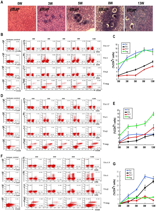
Figure 1 Kinetics of Th17, Th1, Th2 and Treg cell induction in S. japonicum infection. For each of three independent experiments, 20 female C57BL/6 mice were infected with 12 cercariae of S. japonicum per mouse. Four mice were randomly chosen and sacrificed at 0 (before infection), 3, 5, 8 or 13 weeks post-infection. Liver sections were stained with H&E for microscopic examination. Single cell suspensions of splenocytes, lymphocytes or liver cells were stimulated with PMA and ionomycin in the presence of Golgistop for 6 h. Cells were surface stained with anti-CD3-APC and anti-CD4-FITC and then intracellularly stained with PE-conjugated antibodies against IL-17A, IFN-γ, IL-4 or isotype IgG2a control antibody for FACS analysis of Th17, Th1 or Th2 cells. Tregs were detected in single cell suspensions of splenocytes, lymphocytes or liver cells using the Mouse Regulatory T Cell Staining Kit. A. Histopathology in the livers (magnification: 400×). Results are representative of three independent experiments. The kinetics of the percentages of Th17, Th1, Th2 and Treg cells in total CD4+ T cells from mouse spleens (B), mesenteric lymph nodes (D) and livers (F). Data are expressed as the mean ± SD of 12 mice from three independent experiments. Flow cytometric analysis of CD4+ T cell subsets in mouse splenocytes (C), mesenteric lymphocytes (E) and hepatocytes (G) from one representative experiment. Cells were gated on the CD3+ population for analysis of Th17, Th1, Th2 cells, or gated on the CD3+CD4+ population for analysis of Treg cells.
Figure 1 Kinetics of Th17, Th1, Th2 and Treg cell induction in S. japonicum infection. For each of three independent experiments, 20 female C57BL/6 mice were infected with 12 cercariae of S. japonicum per mouse. Four mice were randomly chosen and sacrificed at 0 (before infection), 3, 5, 8 or 13 weeks post-infection. Liver sections were stained with H&E for microscopic examination. Single cell suspensions of splenocytes, lymphocytes or liver cells were stimulated with PMA and ionomycin in the presence of Golgistop for 6 h. Cells were surface stained with anti-CD3-APC and anti-CD4-FITC and then intracellularly stained with PE-conjugated antibodies against IL-17A, IFN-γ, IL-4 or isotype IgG2a control antibody for FACS analysis of Th17, Th1 or Th2 cells. Tregs were detected in single cell suspensions of splenocytes, lymphocytes or liver cells using the Mouse Regulatory T Cell Staining Kit. A. Histopathology in the livers (magnification: 400×). Results are representative of three independent experiments. The kinetics of the percentages of Th17, Th1, Th2 and Treg cells in total CD4+ T cells from mouse spleens (B), mesenteric lymph nodes (D) and livers (F). Data are expressed as the mean ± SD of 12 mice from three independent experiments. Flow cytometric analysis of CD4+ T cell subsets in mouse splenocytes (C), mesenteric lymphocytes (E) and hepatocytes (G) from one representative experiment. Cells were gated on the CD3+ population for analysis of Th17, Th1, Th2 cells, or gated on the CD3+CD4+ population for analysis of Treg cells.CC BY · PubMed Central
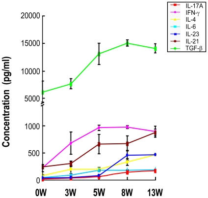
Figure 2 Kinetics of cytokines in the supernatant produced by splenocytes from S. japonicum infected mice. For each of three independent experiments, 20 female C57BL/6 mice were infected with 12 cercariae of S. japonicum per mouse. At 0 (before infection), 3, 5, 8 and 13 weeks post-infection, four mice at each time point were randomly chosen and sacrificed. Single cell suspensions of splenocytes were prepared and then cultured in the presence of PMA and ionomycin. The culture supernatants were collected after 72 h of incubation for detection of IL-17, IFN-γ, IL-4, TGF-β, IL-6, IL-23 and IL-21 by ELISA. Data are expressed as the mean ± SD of 12 mice from three independent experiments.
Figure 2 Kinetics of cytokines in the supernatant produced by splenocytes from S. japonicum infected mice. For each of three independent experiments, 20 female C57BL/6 mice were infected with 12 cercariae of S. japonicum per mouse. At 0 (before infection), 3, 5, 8 and 13 weeks post-infection, four mice at each time point were randomly chosen and sacrificed. Single cell suspensions of splenocytes were prepared and then cultured in the presence of PMA and ionomycin. The culture supernatants were collected after 72 h of incubation for detection of IL-17, IFN-γ, IL-4, TGF-β, IL-6, IL-23 and IL-21 by ELISA. Data are expressed as the mean ± SD of 12 mice from three independent experiments.CC BY · PubMed Central
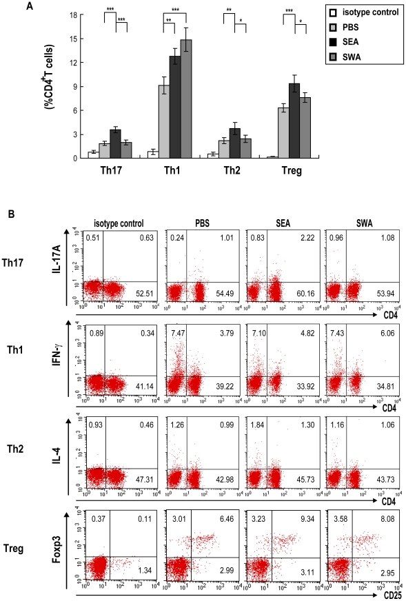
Figure 3 Proportions of Th17, Th1, Th2 and Treg cells in mice immunized with SEA or SWA. For each of three independent experiments, C57BL/6 mice (8 per group) were each injected subcutaneously in the back with 100 µl of an IFA emulsified solution containing 50 µg of SEA, 50 µg of SWA or PBS. The immunizations were repeated two times with a 14-day interval. Two weeks after the final vaccination, the mice were sacrificed, and single cell suspensions of splenocytes were prepared. The splenocytes were stimulated with PMA/ionomycin in the presence of Golgistop for 6 h, followed by surface staining with anti-CD3-APC and anti-CD8-FITC and then intracellular staining with PE-conjugated antibodies against IL-17A, IFN-γ, IL-4 or isotype IgG2a control antibody for FACS analysis of Th17, Th1 or Th2 cells. Splenocytes were stained with the Mouse Regulatory T Cell Staining Kit for detection of Treg cells. A. Proportions of Th17, Th1, Th2 and Treg cells in CD4+ T cells. Results are expressed as mean ± SD of 24 mice from 3 independent experiments. *P<0.05; **P<0.01; ***P<0.001, compared to control mice injected with PBS. B. Flow cytometric analysis from one representative experiment. Cells were gated on the CD3+ population for analysis of Th17, Th1 and Th2 cells or on CD3+CD4+ population for analysis of Treg cells.
Figure 3 Proportions of Th17, Th1, Th2 and Treg cells in mice immunized with SEA or SWA. For each of three independent experiments, C57BL/6 mice (8 per group) were each injected subcutaneously in the back with 100 µl of an IFA emulsified solution containing 50 µg of SEA, 50 µg of SWA or PBS. The immunizations were repeated two times with a 14-day interval. Two weeks after the final vaccination, the mice were sacrificed, and single cell suspensions of splenocytes were prepared. The splenocytes were stimulated with PMA/ionomycin in the presence of Golgistop for 6 h, followed by surface staining with anti-CD3-APC and anti-CD8-FITC and then intracellular staining with PE-conjugated antibodies against IL-17A, IFN-γ, IL-4 or isotype IgG2a control antibody for FACS analysis of Th17, Th1 or Th2 cells. Splenocytes were stained with the Mouse Regulatory T Cell Staining Kit for detection of Treg cells. A. Proportions of Th17, Th1, Th2 and Treg cells in CD4+ T cells. Results are expressed as mean ± SD of 24 mice from 3 independent experiments. *P<0.05; **P<0.01; ***P<0.001, compared to control mice injected with PBS. B. Flow cytometric analysis from one representative experiment. Cells were gated on the CD3+ population for analysis of Th17, Th1 and Th2 cells or on CD3+CD4+ population for analysis of Treg cells.CC BY · PubMed Central
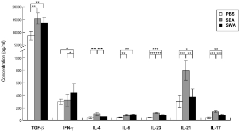
Figure 4 ELISA detection of cytokines in the supernatant produced by SEA or SWA immunized mice splenocytes. For each of three independent experiments, C57BL/6 mice (8 per group) were each injected subcutaneously in the back with 100 µl of an IFA emulsified solution containing 50 µg of SEA, 50 µg of SWA or PBS. The immunizations were repeated two times with a 14-day interval. Two weeks after the final vaccination, the mice were sacrificed, and single cell suspensions of splenocytes were prepared. The splenocytes (2×10⁵/well in 200 µl complete media) were cultured in 96 well plates with PMA/ionomycin. Culture supernatants were collected after 72 h, and cytokines were detected by ELISA. Results are expressed as mean ± SD of 24 mice from three independent experiments. *P<0.05; **P<0.01; ***P<0.001, compared to control groups as indicated.
Figure 4 ELISA detection of cytokines in the supernatant produced by SEA or SWA immunized mice splenocytes. For each of three independent experiments, C57BL/6 mice (8 per group) were each injected subcutaneously in the back with 100 µl of an IFA emulsified solution containing 50 µg of SEA, 50 µg of SWA or PBS. The immunizations were repeated two times with a 14-day interval. Two weeks after the final vaccination, the mice were sacrificed, and single cell suspensions of splenocytes were prepared. The splenocytes (2×10⁵/well in 200 µl complete media) were cultured in 96 well plates with PMA/ionomycin. Culture supernatants were collected after 72 h, and cytokines were detected by ELISA. Results are expressed as mean ± SD of 24 mice from three independent experiments. *P<0.05; **P<0.01; ***P<0.001, compared to control groups as indicated.CC BY · PubMed Central
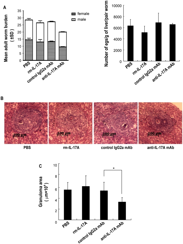
Figure 5 Protection against S. japonicum infection in C57BL/6 mice administered rmIL-17A or anti-IL-17A neutralizing mAb. rmIL-17A, anti-mouse IL-17A mAb or controls were administered to mice, followed by challenge with 40 S. japonicum cercariae per mouse. A. Six weeks after the challenge, the mice were sacrificed and perfused to collect adult worms. The numbers of eggs extracted from the liver were determined by microscopic examination. Protection was measured by assessing the worm and egg burden. Values are given as mean ± SD of eight mice from two independent experiments. *P<0.05; **P<0.01; ***P<0.001, compared to control mice injected with isotype antibody. B. Paraffin embedded formalin fixed liver sections stained with H&E. Images shown are representative of two independent experiments. C. Sizes of the granulomas were measured by computer-assisted morphometric analysis. Ten sections for each mouse and five microscope fields for each section were measured under a microscope. Only granulomas with a visible central egg, which reflected their true shape and dimension, were analyzed for accuracy. Values are given as mean ± SD of eight mice from two independent experiments. *P<0.05; **P<0.01; ***P<0.001, compared to control mice injected with isotype antibody.
Figure 5 Protection against S. japonicum infection in C57BL/6 mice administered rmIL-17A or anti-IL-17A neutralizing mAb. rmIL-17A, anti-mouse IL-17A mAb or controls were administered to mice, followed by challenge with 40 S. japonicum cercariae per mouse. A. Six weeks after the challenge, the mice were sacrificed and perfused to collect adult worms. The numbers of eggs extracted from the liver were determined by microscopic examination. Protection was measured by assessing the worm and egg burden. Values are given as mean ± SD of eight mice from two independent experiments. *P<0.05; **P<0.01; ***P<0.001, compared to control mice injected with isotype antibody. B. Paraffin embedded formalin fixed liver sections stained with H&E. Images shown are representative of two independent experiments. C. Sizes of the granulomas were measured by computer-assisted morphometric analysis. Ten sections for each mouse and five microscope fields for each section were measured under a microscope. Only granulomas with a visible central egg, which reflected their true shape and dimension, were analyzed for accuracy. Values are given as mean ± SD of eight mice from two independent experiments. *P<0.05; **P<0.01; ***P<0.001, compared to control mice injected with isotype antibody.CC BY · PubMed Central
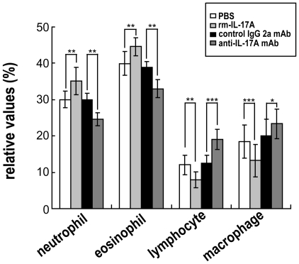
Figure 6 Percentages of granulomatous cells in S. japonicum infected mice administered rmIL-17A or anti-IL-17A neutralizing mAb. rmIL-17A, anti-mouse IL-17A mAb or controls were administered to mice, followed by challenge with 40 S. japonicum cercariae per mouse. Six weeks after the challenge, the mice were sacrificed, and the livers were processed for H&E staining. Percentages of neutrophils, eosinophils, lymphocytes and macrophages in the granulomas were calculated by microscopic analysis of the same granulomas analyzed for lesion size by randomly counting 200 cells (not including hepatocytes) in each granuloma. Ten sections for each mouse and five microscope fields for each section were counted. Values are given as mean ± SD of eight mice from two independent experiments. *P<0.05; **P<0.01; ***P<0.001, compared to control mice injected with PBS or isotype antibody.
Figure 6 Percentages of granulomatous cells in S. japonicum infected mice administered rmIL-17A or anti-IL-17A neutralizing mAb. rmIL-17A, anti-mouse IL-17A mAb or controls were administered to mice, followed by challenge with 40 S. japonicum cercariae per mouse. Six weeks after the challenge, the mice were sacrificed, and the livers were processed for H&E staining. Percentages of neutrophils, eosinophils, lymphocytes and macrophages in the granulomas were calculated by microscopic analysis of the same granulomas analyzed for lesion size by randomly counting 200 cells (not including hepatocytes) in each granuloma. Ten sections for each mouse and five microscope fields for each section were counted. Values are given as mean ± SD of eight mice from two independent experiments. *P<0.05; **P<0.01; ***P<0.001, compared to control mice injected with PBS or isotype antibody.CC BY · PubMed Central
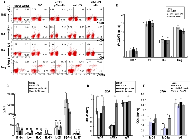
Figure 7 Immune responses in S. japonicum infected mice after administration of rmIL-17A or anti-IL-17A neutralizing mAb. rmIL-17A, anti-mouse IL-17A mAb or controls were administered to mice, followed by challenge with 40 S. japonicum per mouse. Six weeks after the challenge, the mice were sacrificed, and the splenocytes were prepared. A. Flow cytometric analysis of Th17, Th1, Th2 and Treg cells from one representative experiment. Single cell suspensions of splenocytes were stimulated with PMA/ionomycin in the presence of Golgistop for 6 h, followed by surface staining with anti-CD3-APC and anti-CD4-FITC and intracellularly staining with PE-conjugated antibodies against IL-17A, IFN-γ, IL-4 or isotype IgG2a control antibody for FACS analysis of Th17, Th1 or Th2 cells. Splenocytes were also stained with the Mouse Regulatory T Cell Staining Kit for detection of Treg cells. Cells were gated on the CD3+ population for analysis of Th17, Th1, Th2 cells analysis, or gated on the CD3+CD4+ population for analysis of Treg cells. B. The proportions of Th17, Th1, Th2 and Treg cells in CD4+ T cells. Results are expressed as mean ± SD of eight mice from two independent experiments. *P<0.05; **P<0.01; ***P<0.001, compared to control mice injected with PBS or isotype antibody. C. ELISA detection of cytokines in the supernatant produced by splenocytes from S. japonicum infected mice after administration of rmIL-17A or anti-IL-17A neutralizing mAb. Splenocytes (2×10⁵/well in 200 µl of complete media) were cultured in 96 well plates with PMA/ionomycin. Culture supernatants were collected after 72 h, and cytokines were detected by ELISA. Results are expressed as mean ± SD of eight mice from two independent experiments. *P<0.05; **P<0.01; ***P<0.001, compared to control mice injected with PBS or isotype antibody. SEA (D) and SWA (E) specific IgG, IgG1 and IgG2a antibodies in serum from S. japonicum infected mice after administration of rmIL-17A or anti-IL-17A neutralizing mAb were detected by ELISA. Results are expressed as mean ± SD of eight mice from two independent experiments. *P<0.05; **P<0.01; ***P<0.001, compared to control mice injected with PBS or isotype antibody.
Figure 7 Immune responses in S. japonicum infected mice after administration of rmIL-17A or anti-IL-17A neutralizing mAb. rmIL-17A, anti-mouse IL-17A mAb or controls were administered to mice, followed by challenge with 40 S. japonicum per mouse. Six weeks after the challenge, the mice were sacrificed, and the splenocytes were prepared. A. Flow cytometric analysis of Th17, Th1, Th2 and Treg cells from one representative experiment. Single cell suspensions of splenocytes were stimulated with PMA/ionomycin in the presence of Golgistop for 6 h, followed by surface staining with anti-CD3-APC and anti-CD4-FITC and intracellularly staining with PE-conjugated antibodies against IL-17A, IFN-γ, IL-4 or isotype IgG2a control antibody for FACS analysis of Th17, Th1 or Th2 cells. Splenocytes were also stained with the Mouse Regulatory T Cell Staining Kit for detection of Treg cells. Cells were gated on the CD3+ population for analysis of Th17, Th1, Th2 cells analysis, or gated on the CD3+CD4+ population for analysis of Treg cells. B. The proportions of Th17, Th1, Th2 and Treg cells in CD4+ T cells. Results are expressed as mean ± SD of eight mice from two independent experiments. *P<0.05; **P<0.01; ***P<0.001, compared to control mice injected with PBS or isotype antibody. C. ELISA detection of cytokines in the supernatant produced by splenocytes from S. japonicum infected mice after administration of rmIL-17A or anti-IL-17A neutralizing mAb. Splenocytes (2×10⁵/well in 200 µl of complete media) were cultured in 96 well plates with PMA/ionomycin. Culture supernatants were collected after 72 h, and cytokines were detected by ELISA. Results are expressed as mean ± SD of eight mice from two independent experiments. *P<0.05; **P<0.01; ***P<0.001, compared to control mice injected with PBS or isotype antibody. SEA (D) and SWA (E) specific IgG, IgG1 and IgG2a antibodies in serum from S. japonicum infected mice after administration of rmIL-17A or anti-IL-17A neutralizing mAb were detected by ELISA. Results are expressed as mean ± SD of eight mice from two independent experiments. *P<0.05; **P<0.01; ***P<0.001, compared to control mice injected with PBS or isotype antibody.CC BY · PubMed Central

Verification

Claims the paper makes

The claims and statistics of this paper haven’t been checked yet.

Run the check — free, no account needed. It reads the open-access text and checks the sources it cites.

A few free checks a day · papers already verified open instantly · results are public

Claims other papers make about this one

Each is checked against this paper’s own text: does it say what the citing paper says it does?

Self-check

Percentages, sums and reported differences re-computed from the paper’s own numbers.

The self-check needs the paper’s full text.

Citations

Cites

Show all 3 references

Research works printed but not linked: 57

  1. TR Mosmann, 1989, TH1 and TH2 cells: different patterns of lymphokine secretion lead to different functional properties., Annu Rev Immunol, 7, 145, 10.1146/annurev.iy.07.040189.001045 · DOI · Paper page
  2. LE Harrington, 2005, Interleukin 17-producing CD4+ effector T cells develop via a lineage distinct from the T helper type 1 and 2 lineages., Nat Immunol, 6, 1123, 10.1038/ni1254 · DOI · Paper page
  3. LE Harrington, 2006, Expanding the effector CD4 T-cell repertoire: the Th17 lineage., Curr Opin Immunol, 18, 349, 10.1016/j.coi.2006.03.017 · DOI
  4. YK Lee, 2009, Developmental plasticity of Th17 and Treg cells., Curr Opin Immunol, 21, 274, 10.1016/j.coi.2009.05.021 · DOI
  5. H Park, 2005, A distinct lineage of CD4 T cells regulates tissue inflammation by p roducing interleukin 17., Nat Immunol, 6, 1133, 10.1038/ni1261 · DOI · Paper page
  6. P Ye, 2001, Requirement of interleukin 17 receptor signaling for lung CXC chemokine and granulocyte colony-stimulating factor expression, neutrophil recruitment, and host defense., J Exp Med, 194, 519, 10.1084/jem.194.4.519 · DOI
  7. PJ Dubin, 2007, IL-23 mediates inflammatory responses to mucoid Pseudomonas aeruginosa lung infection in mice., Am J Physiol Lung Cell Mol Physiol, 292, L519, 10.1152/ajplung.00312.2006 · DOI
  8. JJ Yu, 2007, An essential role for IL-17 in preventing pathogen-initiated bone destruction: recruitment of neutrophils to inflamed bone requires IL-17 receptor-dependent signals., Blood, 109, 3794, 10.1182/blood-2005-09-010116 · DOI
  9. DR Chung, 2003, CD4+ T cells mediate abscess formation in intra-abdominal sepsis by an IL-17-dependent mechanism., J Immunol, 170, 1958, 10.4049/jimmunol.170.4.1958 · DOI
  10. M Umemura, 2007, IL-17-mediated regulation of innate and acquired immune response against pulmonary Mycobacterium bovis bacille Calmette-Guerin infection., J Immunol, 178, 3786, 10.4049/jimmunol.178.6.3786 · DOI
  11. PJ Gaddi, 2007, Cytokine regulation of immunopathology in toxoplasmosis., Immunol Cell Biol, 85, 155, 10.1038/sj.icb.7100038 · DOI
  12. LI Rutitzky, 2005, Severe CD4 T cell-mediated immunopathology in murine schistosomiasis is dependent on IL-12p40 and correlates with high levels of IL-17., J Immunol, 175, 3920, 10.4049/jimmunol.175.6.3920 · DOI
  13. LI Rutitzky, 2006, CD4 T cells producing pro-inflammatory interleukin-17 mediate high pathology in schistosomiasis., Mem Inst Oswaldo Cruz, 101, 327, 10.1590/S0074-02762006000900052 · DOI
  14. LI Rutitzky, 2009, T-bet protects against exacerbation of schistosome egg-induced immunopathology by regulating Th17-mediated inflammation., Eur J Immunol, 39, 2470, 10.1002/eji.200939325 · DOI
  15. PM Smith, 2009, Genetic control of severe egg-induced immunopathology and IL-17 production in murine schistosomiasis., J Immunol, 183, 3317, 10.4049/jimmunol.0901504 · DOI
  16. MG Shainheit, 2008, Dendritic cell IL-23 and IL-1 production in response to schistosome eggs induces Th17 cells in a mouse strain prone to severe immunopathology., J Immunol, 181, 8559, 10.4049/jimmunol.181.12.8559 · DOI
  17. LI Rutitzky, 2011, Exacerbated egg-induced immunopathology in murine Schistosoma mansoni infection is primarily mediated by IL-17 and restrained by IFN-gamma., Eur J Immunol., 41, 2677, 10.1002/eji.201041327 · DOI
  18. Y Belkaid, 2001, The role of interleukin (IL)-10 in the persistence of Leishmania major in the skin after healing and the therapeutic potential of anti-IL-10 receptor antibody for sterile cure., J Exp Med, 194, 1497, 10.1084/jem.194.10.1497 · DOI · Paper page
  19. Y Belkaid, 2002, CD4+CD25+ regulatory T cells control Leishmania major persistence and immunity., Nature, 420, 502, 10.1038/nature01152 · DOI
  20. M Hesse, 2004, The pathogenesis of schistosomiasis is controlled by cooperating IL-10-producing innate effector and regulatory T cells., J Immunol, 172, 3157, 10.4049/jimmunol.172.5.3157 · DOI
  21. S Mendez, 2004, Role for CD4(+) CD25(+) regulatory T cells in reactivation of persistent leishmaniasis and control of concomitant immunity., J Exp Med, 200, 201, 10.1084/jem.20040298 · DOI
  22. S Sakaguchi, 2000, Regulatory T cells: key controllers of immunologic self-tolerance., Cell, 101, 455, 10.1016/S0092-8674(00)80856-9 · DOI
  23. E Bettelli, 2006, Reciprocal developmental pathways for the generation of pathogenic effector TH17 and regulatory T cells., Nature, 441, 235, 10.1038/nature04753 · DOI · Paper page
  24. PR Mangan, 2006, Transforming growth factor-beta induces development of the T(H)17 lineage., Nature, 441, 231, 10.1038/nature04754 · DOI · Paper page
  25. M Veldhoen, 2006, TGF beta in the context of an inflammatory cytokine milieu supports de novo differentiation of IL-17-producing T cells., Immunity, 24, 179, 10.1016/j.immuni.2006.01.001 · DOI · Paper page
  26. S Wan, 2007, IL-6 produced by dendritic cells from lupus-prone mice inhibits CD4+CD25+ T cell regulatory functions., J Immunol, 178, 271, 10.4049/jimmunol.178.1.271 · DOI
  27. L Zhou, 2007, IL-6 programs TH-17 cell differentiation by promoting sequential engagement of the IL-21 and IL-23 pathways., Nat Immunol, 8, 967, 10.1038/ni1488 · DOI
  28. T Korn, 2007, IL-21 initiates an alternate pathway to induce proinflammatory Th17 cells., Nature, 448, 484, 10.1038/nature05970 · DOI
  29. CL Langrish, 2005, IL-23 drives a pathogenic T cell population that induces autoimmune inflammation., J Exp Med, 201, 233, 10.1084/jem.20041257 · DOI · Paper page
  30. L Zhou, 2008, TGF-β-induced Foxp3 inhibits TH17 cell differentiation by antagonizing RORγt function., Nature, 453, 236, 10.1038/nature06878 · DOI
  31. MA Gavin, 2007, Foxp3-dependent programme of regulatory T-cell differentiation., Nature, 445, 771, 10.1038/nature05543 · DOI
  32. II Ivanov, 2006, The orphan nuclear receptor ROR gammat directs the differentiation program of proinflammatory IL-17(+) T helper cells., Cell, 126, 1121, 10.1016/j.cell.2006.07.035 · DOI · Paper page
  33. ML Burke, 2009, Immunopathogenesis of human schistosomiasis., Parasite Immunol, 31, 163, 10.1111/j.1365-3024.2009.01098.x · DOI
  34. LI Rutitzky, 2008, IL-23 is required for the development of severe egg-induced immunopathology in schistosomiasis and for lesional expression of IL-17., J Immunol, 180, 2486, 10.4049/jimmunol.180.4.2486 · DOI
  35. RA Abdul-Ghani, 2010, Murine schistosomiasis as a model for human schistosomiasis mansoni: similarities and discrepancies., Parasitol Res, 107, 1, 10.1007/s00436-010-1855-5 · DOI
  36. DW Dunne, 2005, A worm's eye view of the immune system: consequences for evolution of human autoimmune disease., Nat Rev Immunol, 5, 420, 10.1038/nri1601 · DOI
  37. GN Gobert, 2007, Biology of the schistosome lung-stage schistosomulum., Parasitology, 134, 453, 10.1017/S0031182006001648 · DOI
  38. JM Grzych, 1991, Egg deposition is the major stimulus for the production of Th2 cytokines in murine schistosomiasis mansoni., J Immunol, 146, 1322, 10.4049/jimmunol.146.4.1322 · DOI
  39. W Zhang, 2011, Schistosoma mansoni antigen Sm-p80: Prophylactic efficacy of a vaccine formulated in human approved plasmid vector and adjuvant (VR 1020 and alum)., Acta Trop, 118, 142, 10.1016/j.actatropica.2011.01.010 · DOI
  40. YS Li, 1999, Antibody isotype responses, infection and re-infection for Schistosoma japonicum in a marshland area of China., Acta Trop, 73, 79, 10.1016/S0001-706X(99)00019-4 · DOI
  41. TA Wynn, 1995, An IL-12-based vaccination method for preventing fibrosis induced by schistosome infection., Nature, 376, 594, 10.1038/376594a0 · DOI
  42. LI Rutitzky, 2001, Th1-polarizing immunization with egg antigens correlates with severe exacerbation of immunopathology and death in schistosome infection., Proc Natl Acad Sci, 98, 13243, 10.1073/pnas.231258498 · DOI
  43. MJ Edwards, 2005, Reciprocal immunomodulation in a schistosome and hepatotropic virus coinfection model., J Immunol, 175, 6275, 10.4049/jimmunol.175.10.6275 · DOI
  44. AB de Almeida, 1998, Differences in the frequency of cytokine-producing cells in antigenemic and nonantigenemic individuals with bancroftian filariasis., Infect Immun, 66, 1377, 10.1128/IAI.66.4.1377-1383.1998 · DOI
  45. F Dieli, 2003, Characterization of lung gamma delta T cells following intranasal infection with Mycobacterium bovis bacillus Calmette-Guerin., J Immunol, 170, 463, 10.4049/jimmunol.170.1.463 · DOI
  46. J Ismaili, 2003, Plasmodium falciparum infection of the placenta affects newborn immune responses., Clin Exp Immunol, 133, 414, 10.1046/j.1365-2249.2003.02243.x · DOI
  47. A Ogawa, 2004, Neutralization of interleukin-17 aggravates dextran sulfate sodium-induced colitis in mice., Clin Immunol, 110, 55, 10.1016/j.clim.2003.09.013 · DOI
  48. YL Cheng, 2008, The effects of T cell deficiency on the development of worms and granuloma formation in mice infected with Schistosoma japonicum., Parasitol Res, 102, 1129, 10.1007/s00436-008-0880-0 · DOI
  49. T Baba, 1988, Electron microscopic cytochemical analysis of hepatic granuloma induced by Cryptococcus neoformans., Mycopathologia, 104, 37, 10.1007/BF00437922 · DOI
  50. Y Zhu, 2004, The protective effect of a Schistosoma japonicum Chinese strain 23 kDa plasmid DNA vaccine in pigs is enhanced with IL-12., Vaccine, 23, 78, 10.1016/j.vaccine.2004.04.031 · DOI
  51. SA Khader, 2007, IL-23 and IL-17 in the establishment of protective pulmonary CD4+ T cell responses after vaccination and during Mycobacterium tuberculosis challenge., Nat Immunol, 8, 369, 10.1038/ni1449 · DOI
  52. JK Kolls, 2004, Interleukin-17 family members and inflammation., Immunity, 21, 467, 10.1016/j.immuni.2004.08.018 · DOI
  53. MN Kelly, 2005, Interleukin-17/interleukin-17 receptor-mediated signaling is important for generation of an optimal polymorphonuclear response against Toxoplasma gondii infection., Infect Immun, 73, 617, 10.1128/IAI.73.1.617-621.2005 · DOI
  54. EJ Pearce, 2002, The immunobiology of schistosomiasis., Nat Rev Immunol, 2, 499, 10.1038/nri843 · DOI
  55. T Korn, 2009, IL-17 and Th17 Cells., Annu Rev Immunol, 27, 485, 10.1146/annurev.immunol.021908.132710 · DOI · Paper page
  56. E Bettelli, 2007, Th17: the third member of the effector T cell trilogy., Curr Opin Immunol, 19, 652, 10.1016/j.coi.2007.07.020 · DOI
  57. TM Kariuki, 2005, Resistance to re-infection after exposure to normal and attenuated schistosome parasites in the baboon model., Parasite Immunol, 27, 281, 10.1111/j.1365-3024.2005.00783.x · DOI

Cited by

Sorted by citations

Metadata

Authors
Wen, X., He, L., Chi, Y., Zhou, S., Hoellwarth, J., Zhang, C., Zhu, J., Wu, C., Dhesi, S., Wang, X., Liu, F., & Su, C.
Venue
PLoS Neglected Tropical Diseases
Published
2011-11-15
Volume
5
Issue
11
Pages
e1399
ISSN
1935-2735
Cited by
70
References
3
Language
en

About these checks

Registry, retraction, Cites and statistics results are computed, not judged, with one exception: a printed reference that no index lists is read once by a model before it counts as a citation error. Claims are extracted by a model and kept only when their quote appears verbatim in the text; each cited source is then checked, with a model’s help, for whether it says what the claim relies on. Those verdicts can be wrong — read the claim’s quote and the cited source before relying on one.

Statistics and arithmetic are recomputed, not judged.

Confidence bands are shown only for fields calibrated on our annotated test set. None is calibrated yet, so no band is shown.

What counts as a claim ›How we measure confidence ›How we check this ›

Are you an author, or do you see a problem with this page? Request a review