

The DOI matches its Crossref record; 68 of the 68 references it cites were found in a registry or an index.
NLRP3 Controls Trypanosoma cruzi Infection through a Caspase-1-Dependent IL-1R-Independent NO Production
Open accessAbstract
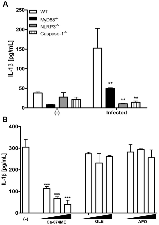
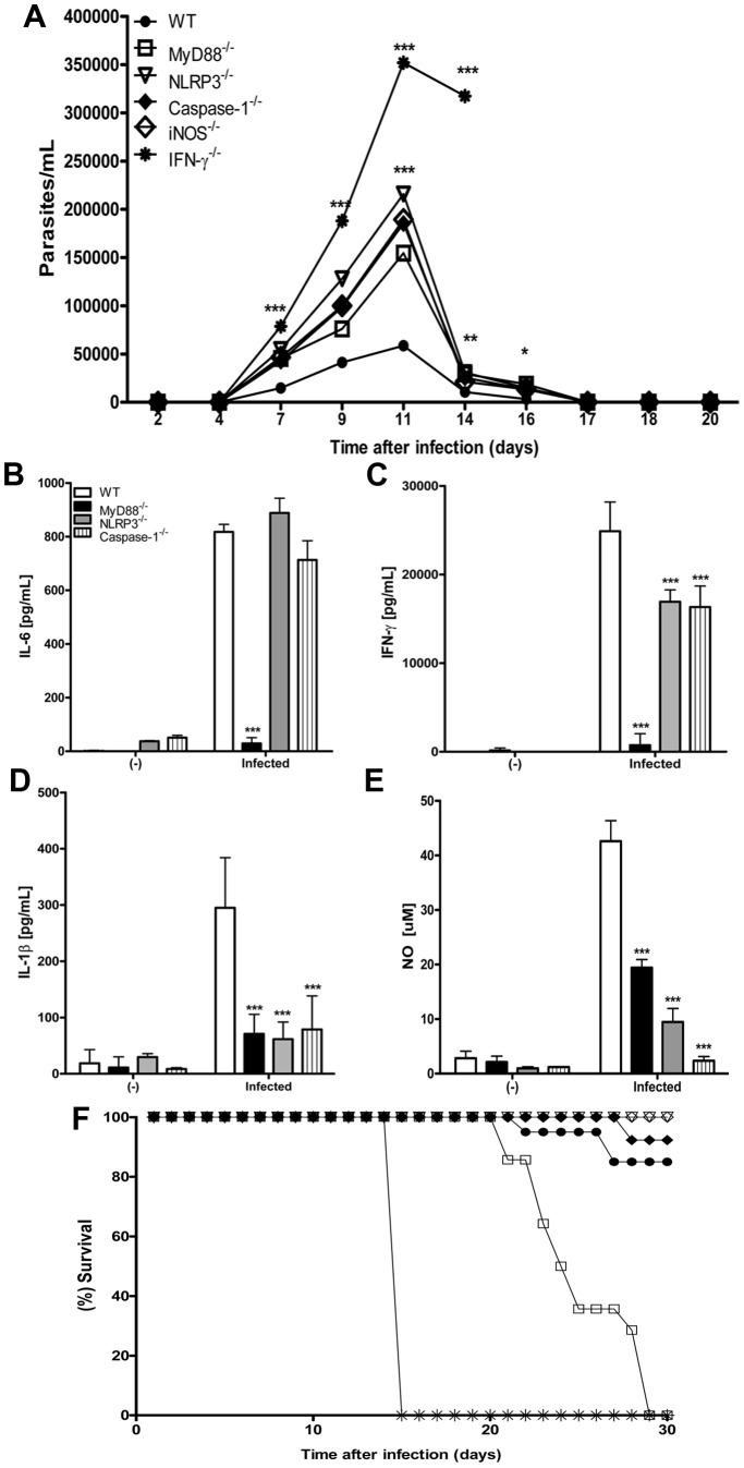
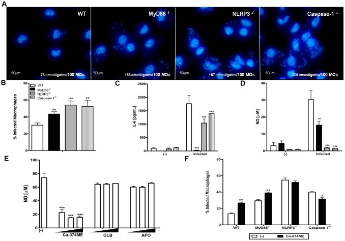
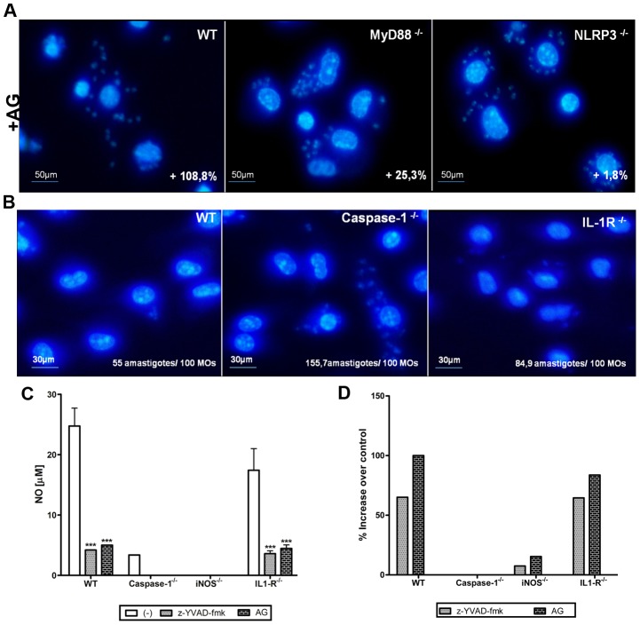
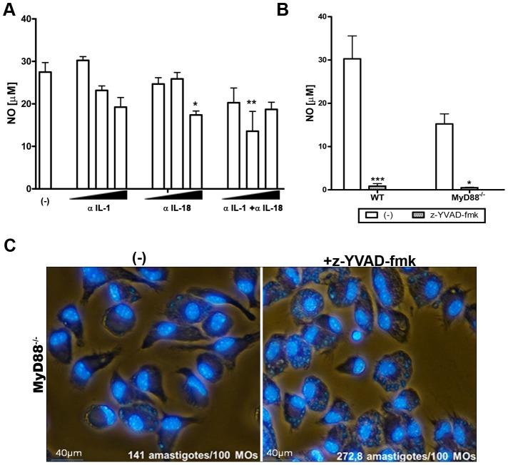
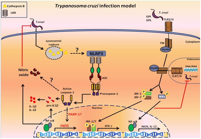
Trypanosoma cruzi (T. cruzi) is an intracellular protozoan parasite and the etiological agent of Chagas disease, a chronic infectious illness that affects millions of people worldwide. Although the role of TLR and Nod1 in the control of T. cruzi infection is well-established, the involvement of inflammasomes remains to be elucidated. Herein, we demonstrate for the first time that T. cruzi infection induces IL-1β production in an NLRP3- and caspase-1-dependent manner. Cathepsin B appears to be required for NLRP3 activation in response to infection with T. cruzi, as pharmacological inhibition of cathepsin B abrogates IL-1β secretion. NLRP3(-/-) and caspase1(-/-) mice exhibited high numbers of T. cruzi parasites, with a magnitude of peak parasitemia comparable to MyD88(-/-) and iNOS(-/-) mice (which are susceptible models for T. cruzi infection), indicating the involvement of NLRP3 inflammasome in the control of the acute phase of T. cruzi infection. Although the inflammatory cytokines IL-6 and IFN-γ were found in spleen cells from NLRP3(-/-) and caspase1(-/-) mice infected with T. cruzi, these mice exhibited severe defects in nitric oxide (NO) production and an impairment in macrophage-mediated parasite killing. Interestingly, neutralization of IL-1β and IL-18, and IL-1R genetic deficiency demonstrate that these cytokines have a minor effect on NO secretion and the capacity of macrophages to control T. cruzi infection. In contrast, inhibition of caspase-1 with z-YVAD-fmk abrogated NO production by WT and MyD88(-/-) macrophages and rendered them as susceptible to T. cruzi infection as NLRP3(-/-) and caspase-1(-/-) macrophages. Taken together, our results demonstrate a role for the NLRP3 inflammasome in the control of T. cruzi infection and identify NLRP3-mediated, caspase-1-dependent and IL-1R-independent NO production as a novel effector mechanism for these innate receptors.
Overview
The claims and statistics of this paper haven’t been checked yet.
Run the check — free, no account needed. It reads the open-access text and checks the sources it cites.
Integrity signals
Registry
Cites
Statistics
Figures
Verification
Claims the paper makes
The claims and statistics of this paper haven’t been checked yet.
Run the check — free, no account needed. It reads the open-access text and checks the sources it cites.
Claims other papers make about this one
Each is checked against this paper’s own text: does it say what the citing paper says it does?
The source backs part of the claim, not all of it — or backs it for a different population, condition or comparison.
In the citing paper
“Remarkably, the master regulator Caspase, which is a family of protease enzymes, is associated mainly with regulating the reduction of the load of intracellular parasites, induction of nitric oxide production, increasing the level of CD4 and CD8+ T-cells, secretion of IFNγ, and control of trypanosome infection by macrophages (Gonçalves et al., 2013).”
From Identification of Candidate Signature Genes and Key Regulators Associated With Trypanotolerance in the Sheko Breed (2019) · cites this paper as Gonçalves et al., 2013
Experimental — accuracy not yet measured
Self-check
Percentages, sums and reported differences re-computed from the paper’s own numbers.
The self-check needs the paper’s full text.
Citations
Cites
Schroder, K., & Tschopp, J. (2010) · Cell · doi:10.1016/j.cell.2010.01.040 · Cited by 5,475
Cryopyrin activates the inflammasome in response to toxins and ATP
Mariathasan, S., Weiss, D. S., Newton, K., McBride, J., O'Rourke, K., Roose-Girma, M., Lee, W. P., Weinrauch, Y., Monack, D. M., & Dixit, V. M. (2006) · Nature · doi:10.1038/nature04515 · Cited by 3,080
Hornung, V., Bauernfeind, F., Halle, A., Samstad, E. O., Kono, H., Rock, K. L., Fitzgerald, K. A., & Latz, E. (2008) · Nature Immunology · doi:10.1038/ni.1631 · Cited by 3,065
Crucial role for the Nalp3 inflammasome in the immunostimulatory properties of aluminium adjuvants
Eisenbarth, S. C., Colegio, O. R., O’Connor, W., Sutterwala, F. S., & Flavell, R. A. (2008) · Nature · doi:10.1038/nature06939 · Cited by 1,542
Caspase-1-induced pyroptosis is an innate immune effector mechanism against intracellular bacteria
Miao, E. A., Leaf, I. A., Treuting, P. M., Mao, D. P., Dors, M., Sarkar, A., Warren, S. E., Wewers, M. D., & Aderem, A. (2010) · Nature Immunology · doi:10.1038/ni.1960 · Cited by 1,336
The AIM2 inflammasome is essential for host defense against cytosolic bacteria and DNA viruses
Rathinam, V. A. K., Jiang, Z., Waggoner, S. N., Sharma, S., Cole, L. E., Waggoner, L., Vanaja, S. K., Monks, B. G., Ganesan, S., Latz, E., et al. (2010) · Nature Immunology · doi:10.1038/ni.1864 · Cited by 1,331
Franchi, L., Amer, A., Body-Malapel, M., Kanneganti, T.-D., Özören, N., Jagirdar, R., Inohara, N., Vandenabeele, P., Bertin, J., Coyle, A., et al. (2006) · Nature Immunology · doi:10.1038/ni1346 · Cited by 1,131
Allen, I. C., Scull, M. A., Moore, C. B., Holl, E. K., McElvania-TeKippe, E., Taxman, D. J., Guthrie, E. H., Pickles, R. J., & Ting, J. P.-Y. (2009) · Immunity · doi:10.1016/j.immuni.2009.02.005 · Cited by 1,113
Sensing and reacting to microbes through the inflammasomes
Franchi, L., Muñoz-Planillo, R., & Núñez, G. (2012) · Nature Immunology · doi:10.1038/ni.2231 · Cited by 1,046
Syk kinase signalling couples to the Nlrp3 inflammasome for anti-fungal host defence
Groß, O., Poeck, H., Bscheider, M., Dostert, C., Hannesschläger, N., Endres, S., Hartmann, G., Tardivel, A., Schweighoffer, E., Tybulewicz, V. L. J., et al. (2009) · Nature · doi:10.1038/nature07965 · Cited by 947
Cited by
Sorted by citations
Man, S. M., Karki, R., & Kanneganti, T. (2017) · Immunological Reviews · doi:10.1111/imr.12534 · Cited by 1,684
How is inflammation initiated? Individual influences of IL-1, IL-18 and HMGB1
Keyel, P. A. (2014) · Cytokine · doi:10.1016/j.cyto.2014.03.007 · Cited by 182
Cytosolic Recognition of Microbes and Pathogens: Inflammasomes in Action
Hayward, J. A., Mathur, A., Ngo, C., & Man, S. M. (2018) · Microbiology and Molecular Biology Reviews · doi:10.1128/mmbr.00015-18 · Cited by 168
Nasoohi, S., Ismael, S., & Ishrat, T. (2018) · Molecular Neurobiology · doi:10.1007/s12035-018-0917-z · Cited by 162
Transcriptome Remodeling in Trypanosoma cruzi and Human Cells during Intracellular Infection
Li, Y., Tomko, S. S., Okrah, K., Belew, A. T., Choi, J., Caradonna, K. L., Padmanabhan, P. K., Ndegwa, D. M., Temanni, M.-R., Bravo, H. C., et al. (2016) · PLoS Pathogens · doi:10.1371/journal.ppat.1005511 · Cited by 160
How Inflammasomes Inform Adaptive Immunity
Evavold, C. L., & Kagan, J. C. (2017) · Journal of Molecular Biology · doi:10.1016/j.jmb.2017.09.019 · Cited by 155
Inflammasomes in host response to protozoan parasites
Zamboni, D. S., & Lima-Junior, D. S. (2015) · Immunological Reviews · doi:10.1111/imr.12291 · Cited by 118
ROS and Trypanosoma cruzi: Fuel to infection, poison to the heart
Paiva, C. N., Medei, E. H., & Bozza, M. T. (2018) · PLoS Pathogens · doi:10.1371/journal.ppat.1006928 · Cited by 112
Wang, L., Chen, Y., Li, X., Zhang, Y., Gulbins, E., & Zhang, Y. (2016) · Oncotarget · doi:10.18632/oncotarget.12302 · Cited by 111
Chen, Y., Li, X., Boini, K. M., Pitzer, A. L., Gulbins, E., Zhang, Y., & Li, P. (2014) · Biochimica et Biophysica Acta (BBA) - Molecular Cell Research · doi:10.1016/j.bbamcr.2014.11.012 · Cited by 105
Shio, M. T., Christian, J. G., Jung, J. Y., Chang, K. P., & Olivier, M. (2015) · PLoS neglected tropical diseases · doi:10.1371/journal.pntd.0003868 · Cited by 109
Inflammasome: A Double-Edged Sword in Liver Diseases
Luan, J., & Ju, D. (2018) · Frontiers in Immunology · doi:10.3389/fimmu.2018.02201 · Cited by 96
Ribeiro, K. S., Vasconcelos, C. I., Soares, R. P., Mendes, M. T., Ellis, C. C., Aguilera-Flores, M., de Almeida, I. C., Schenkman, S., Iwai, L. K., & Torrecilhas, A. C. (2018) · Journal of Extracellular Vesicles · doi:10.1080/20013078.2018.1463779 · Cited by 88
Self-citationBabamale, A. O., & Chen, S.-T. (2021) · International Journal of Molecular Sciences · doi:10.3390/ijms222111398 · Cited by 78
Wang, Z., Hu, W., Lu, C., Ma, Z., Jiang, S., Gu, C., Acuña-Castroviejo, D., & Yang, Y. (2018) · Arteriosclerosis Thrombosis and Vascular Biology · doi:10.1161/atvbaha.118.311916 · Cited by 72
Chevillard, C., Nunes, J. P. S., Frade, A. F., Almeida, R. R., Pandey, R. P., Nascimento, M. S., Kalil, J. E., & Cunha‐Neto, E. (2018) · Frontiers in Immunology · doi:10.3389/fimmu.2018.02791 · Cited by 71
Immune responses against protozoan parasites: a focus on the emerging role of Nod-like receptors
Gurung, P., & Kanneganti, T. (2016) · Cellular and Molecular Life Sciences · doi:10.1007/s00018-016-2212-3 · Cited by 68
Global Metabolomic Profiling of Acute Myocarditis Caused by Trypanosoma cruzi Infection
Gironès, N., Carbajosa, S., Guerrero, N. A., Poveda, C., Chillón-Marinas, C., & Fresno, M. (2014) · PLoS neglected tropical diseases · doi:10.1371/journal.pntd.0003337 · Cited by 66
NLR proteins and parasitic disease
Clay, G. M., Sutterwala, F. S., & Wilson, M. E. (2014) · Immunologic Research · doi:10.1007/s12026-014-8544-x · Cited by 67
Reassessing the Evolutionary Importance of Inflammasomes
Maltez, V. I., & Miao, E. A. (2016) · The Journal of Immunology · doi:10.4049/jimmunol.1502060 · Cited by 62
Innate immunomodulation to trypanosomatid parasite infections
Dos-Santos, A. L. A., Carvalho‐Kelly, L. F., Dick, C. F., & Meyer‐Fernandes, J. R. (2016) · Experimental Parasitology · doi:10.1016/j.exppara.2016.05.005 · Cited by 61
Honda, T. S. B., Ku, J., & Anders, H. (2023) · Frontiers in Immunology · doi:10.3389/fimmu.2023.1214289 · Cited by 63
Chaves, M. M., Sinflorio, D. A., Thorstenberg, M. L., Martins, M. D. A., Moreira-Souza, A. C. A., Rangel, T. P., Silva, C. L. M., Bellio, M., Canetti, C. D. A., & Coutinho‐Silva, R. (2019) · PLoS Pathogens · doi:10.1371/journal.ppat.1007887 · Cited by 60
Dey, N., Sinha, M., Gupta, S., Gonzalez, M. N., Fang, R., Endsley, J. J., Luxon, B. A., & Garg, N. J. (2014) · PLoS ONE · doi:10.1371/journal.pone.0111539 · Cited by 56
Sanmarco, L. M., Eberhardt, N., Ponce, N. E., Cano, R. C., Bonacci, G., & Aoki, M. P. (2018) · Frontiers in Immunology · doi:10.3389/fimmu.2017.01921 · Cited by 55
NLRP3 Inflammasome Participates in Host Response to Neospora caninum Infection
Wang, X., Gong, P., Zhang, X., Li, S., Lu, X.-Y., Zhao, C., Yu, Q., Wei, Z., Yang, Y., Liu, Q., et al. (2018) · Frontiers in Immunology · doi:10.3389/fimmu.2018.01791 · Cited by 51
Cai, S., Ge, M., Mennone, A., Hoque, R., Ouyang, X., & Boyer, J. L. (2019) · Cellular and Molecular Gastroenterology and Hepatology · doi:10.1016/j.jcmgh.2019.12.008 · Cited by 51
Davanso, M. R., Crisma, A. R., Braga, T. T., Masi, L. N., do Amaral, C. L., Leal, V. N. C., de Lima, D. S., Patente, T. A., Barbuto, J. A. M., Corrêa‐Giannella, M. L., et al. (2020) · Clinical Science · doi:10.1042/cs20201348 · Cited by 50
Signaling pathways that regulate Trypanosoma cruzi infection and immune response
Cerbán, F. M., Stempin, C. C., Volpini, X., Silva, E. A. C., Gea, S., & Motrán, C. C. (2020) · Biochimica et Biophysica Acta (BBA) - Molecular Basis of Disease · doi:10.1016/j.bbadis.2020.165707 · Cited by 53
Inflammasome Activation in Response to Intracellular Protozoan Parasites
de Carvalho, R. A., & Zamboni, D. S. (2020) · Trends in Parasitology · doi:10.1016/j.pt.2020.02.006 · Cited by 50
Gupta, S., & Garg, N. J. (2015) · PLoS Pathogens · doi:10.1371/journal.ppat.1004828 · Cited by 48
Activation of NLRP3 inflammasomes in mouse hepatic stellate cells during Schistosoma J. infection
Meng, N., Xia, M., Lu, Y., Wang, M., Boini, K. M., Li, P., & Tang, W.-X. (2016) · Oncotarget · doi:10.18632/oncotarget.10044 · Cited by 46
The role of NLRP3 inflammasome in type 2 inflammation related diseases
Xu, S., Wang, D., Tan, L., & Lu, J. (2024) · Autoimmunity · doi:10.1080/08916934.2024.2310269 · Cited by 43
Buzzo, C. D. L., Medina, T. D. S., Branco, L. M., Lage, S. L., Ferreira, L. C. D. S., Amarante-Mendes, G. P., Hottiger, M. O., Carvalho, D. D. D., & Bortoluci, K. R. (2017) · Scientific Reports · doi:10.1038/srep41686 · Cited by 44
Self-citationChagas Disease: Still Many Unsolved Issues
Alvarez, J., Fonseca, R., da Silva, H. B., Marinho, C. R. F., Bortoluci, K. R., Sardinha, L. R., Epiphânio, S., & Lima, M. R. D. (2014) · Mediators of Inflammation · doi:10.1155/2014/912965 · Cited by 41
Self-citationMárquez, J. D. R., Ana, Y., Baigorrí, R. E., Stempin, C. C., & Cerbán, F. M. (2018) · Frontiers in Immunology · doi:10.3389/fimmu.2018.00313 · Cited by 42
Programmed Cell Death in the Evolutionary Race against Bacterial Virulence FactorsPreprint
Lacey, C. A., & Miao, E. A. (2019) · Cold Spring Harbor Perspectives in Biology · doi:10.1101/cshperspect.a036459 · Cited by 43
Mekonnen, Y. A., Gültas, M., Effa, K., Hanotte, O., & Schmitt, A. O. (2019) · Frontiers in Genetics · doi:10.3389/fgene.2019.01095 · Cited by 37
“Remarkably, the master regulator Caspase, which is a family of protease enzymes, is associated mainly with regulating the reduction of the load of intracellular parasites, induction of nitric oxide production, increasing the level of CD4 and CD8+ T-cells, secretion of IFNγ, and control of trypanosome infection by macr…”
BackgroundHost‐parasite dynamics in Chagas disease from systemic to hyper‐local scales
Pérez‐Mazliah, D., Ward, A. I., & Lewis, M. D. (2020) · Parasite Immunology · doi:10.1111/pim.12786 · Cited by 37
Gu, N. Y., Kim, J., Han, I., Im, S., Seo, M., Chung, Y., & Ryu, J. (2016) · The Prostate · doi:10.1002/pros.23178 · Cited by 36
Queiroga, T. B. D., Pereira, N. D. S., da Silva, D. D., Andrade, C. D. M., Júnior, R. F. D. A., Brito, C. R. D. N., Galvão, L. M. D. C., da Câmara, A. C. J., Nascimento, M. S. L., & Guedes, P. M. D. M. (2021) · Frontiers in Cellular and Infection Microbiology · doi:10.3389/fcimb.2021.696719 · Cited by 35
Paroli, A. F., Gonzalez, P. V., Díaz-Luján, C. M., Onofrio, L. I., Arocena, A. R., Cano, R. C., Silva, E. A. C., & Gea, S. (2018) · Frontiers in Immunology · doi:10.3389/fimmu.2018.00913 · Cited by 33
Pereira, N. D. S., Queiroga, T. B. D., Nunes, D. F., Andrade, C. D. M., Nascimento, M. S. L., Matta, M. A. D. V., da Câmara, A. C. J., Galvão, L. M. D. C., Guedes, P. M. D. M., & Chiari, É. (2018) · PLoS neglected tropical diseases · doi:10.1371/journal.pntd.0006589 · Cited by 33
NLRP3 Inflammasome Activation in THP-1 Target Cells Triggered by Pathogenic Naegleria fowleri
Kim, J.-H., Sohn, H., Yoo, J.-K., Kang, H., Seong, G.-S., Chwae, Y., Kim, K. H., Park, S., & Shin, H. (2016) · Infection and Immunity · doi:10.1128/iai.00275-16 · Cited by 32
Host inflammatory responses to intracellular invaders: Review study
Chandra, R. (2019) · Life Sciences · doi:10.1016/j.lfs.2019.117084 · Cited by 33
NLRP3 played a role in Trichinella spiralis-triggered Th2 and regulatory T cells response
Jin, X., Bai, X., Yang, Y., Ding, J., Shi, H., Fu, B., Boireau, P., Liu, M., & Liu, X. (2020) · Veterinary Research · doi:10.1186/s13567-020-00829-2 · Cited by 25
Stahl, P., Ruppert, V., Schwarz, R. Τ., & Meyer, T. (2014) · PLoS ONE · doi:10.1371/journal.pone.0110512 · Cited by 24
Xanthine oxidoreductase activation is implicated in the onset of metabolic arthritis
Aibibula, Z., Ailixiding, M., Iwata, M., Piao, J., Hara, Y., Okawa, A., & Asou, Y. (2016) · Biochemical and Biophysical Research Communications · doi:10.1016/j.bbrc.2016.02.039 · Cited by 23
Ebi3 Prevents Trypanosoma cruzi-Induced Myocarditis by Dampening IFN-γ-Driven Inflammation
Medina, T. D. S., de Oliveira, G. G., Silva, M. C., David, B. A., Silva, G. K., da Fonseca, D. M., Sesti‐Costa, R., Frade, A. F., Baron, M., Ianni, B. M., et al. (2017) · Frontiers in Immunology · doi:10.3389/fimmu.2017.01213 · Cited by 22
Swain, B., & Miryala, K. R. (2025) · Frontiers in Immunology · doi:10.3389/fimmu.2025.1665071 · Cited by 23
Matteucci, K. C., Pereira, G. J. S., Weinlich, R., & Bortoluci, K. R. (2019) · Journal of Leukocyte Biology · doi:10.1002/jlb.hi1118-461r · Cited by 20
Self-citationAge and Chronodisruption in Mouse Heart: Effect of the NLRP3 Inflammasome and Melatonin Therapy
Fernández‐Ortiz, M., Sayed, R. K. A., Montoya, Y. R., Rol, M. A., Fernández‐Martínez, J., Ramírez‐Casas, Y., Florido, J., Rusanova, I., Escames, G., & Acuña-Castroviejo, D. (2022) · International Journal of Molecular Sciences · doi:10.3390/ijms23126846 · Cited by 21
Vasconcelos, J. F., Meira, C. S., Silva, D. N., Nonaka, C. K. V., Daltro, P. S., Macambira, S. G., Domizi, P., Borges, V. M., Ribeiro‐dos‐Santos, R., Souza, B. S. D. F., et al. (2017) · Scientific Reports · doi:10.1038/s41598-017-06275-z · Cited by 19
Molecular regulation of NLRP3 inflammasome activation during parasitic infection
Alonaizan, R. (2024) · Bioscience Reports · doi:10.1042/bsr20231918 · Cited by 18
Gasdermins in Innate Host Defense Against Entamoeba histolytica and Other Protozoan Parasites
Wang, S., Moreau, F., & Chadee, K. (2022) · Frontiers in Immunology · doi:10.3389/fimmu.2022.900553 · Cited by 15
Pacheco, A. D. O. L., Vicentini, G., Matteucci, K. C., Rosa‐Ribeiro, R., Weinlich, R., & Bortoluci, K. R. (2019) · Journal of Leukocyte Biology · doi:10.1002/jlb.4ab1118-416rr · Cited by 16
Self-citationWozniak, J. M., Silva, T. A., Thomas, D. M., Siqueira-Neto, J. L., McKerrow, J. H., Gonzalez, D. J., & Calvet, C. M. (2020) · PLoS neglected tropical diseases · doi:10.1371/journal.pntd.0007980 · Cited by 18
Vieira, R. D. S., Nascimento, M. S., Noronha, I. H., Vasconcelos, J. R., Benvenuti, L. A., Barber, G. N., Câmara, N. O. S., Kalil, J. E., Cunha‐Neto, E., & Almeida, R. R. (2022) · Frontiers in Immunology · doi:10.3389/fimmu.2021.775346 · Cited by 15
Rodríguez, T., Pacheco‐Fernández, T., Vázquez-Mendoza, A., Nieto-Yáñez, Ó., Juárez‐Avelar, I., Reyes, J. L., Terrazas, L. I., & Rodríguez‐Sosa, M. (2020) · Cells · doi:10.3390/cells9010108 · Cited by 15
Vorraro, F., Cabrera, W. H. K., Ribeiro, O. G., Jensen, J. R., Franco, M. D., Ibañez, O. M., & Starobinas, N. (2014) · Mediators of Inflammation · doi:10.1155/2014/952857 · Cited by 14
Avian leukosis virus subgroup J triggers caspase-1-mediated inflammatory response in chick livers
Liu, X., Shan, W.-J., Jia, L., Yang, X., Zhang, J., Wu, Y., Xu, F., & Li, J. (2016) · Virus Research · doi:10.1016/j.virusres.2016.01.011 · Cited by 14
Gil‐Jaramillo, N., Rocha, A. P., Raiol, T., Motta, F. N., Favali, C. B. F., Brígido, M. M., Bastos, I. M. D., & Santana, J. M. (2021) · Frontiers in Immunology · doi:10.3389/fimmu.2021.638020 · Cited by 13
Salvador, R., Aridgides, D. S., & PereiraPerrin, M. (2014) · Infection and Immunity · doi:10.1128/iai.02098-14 · Cited by 12
Deviant Behavior: Tick-Borne Pathogens and Inflammasome Signaling
Shaw, D. K., McClure, E. E., Wang, X., & Pedra, J. H. F. (2016) · Veterinary Sciences · doi:10.3390/vetsci3040027 · Cited by 12
Role of inflammasomes in innate host defense against Entamoeba histolytica
Begum, S., Gorman, H., Chadha, A., & Chadee, K. (2020) · Journal of Leukocyte Biology · doi:10.1002/jlb.3mr0420-465r · Cited by 11
Oliveira, A. E. R., Pereira, M. C. A., Belew, A. T., Ferreira, L. R. P., Pereira, L. M. N., Neves, E. G. A., Nunes, M. D. C. P., Burleigh, B. A., Dutra, W. O., El-Sayed, N. M., et al. (2020) · PLoS Pathogens · doi:10.1371/journal.ppat.1008781 · Cited by 11
IFN-induced cell-autonomous immune mechanisms in the control of intracellular protozoa
Skariah, S., Sultan, A. A., & Mordue, D. G. (2022) · Parasitology Research · doi:10.1007/s00436-022-07514-7 · Cited by 10
Nájera, C. A., Batista, M. F., Meneghelli, I., & Bahia, D. (2021) · Journal of Cell Science · doi:10.1242/jcs.255687 · Cited by 10
Chen, Y., Boini, K. M., Pitzer, A. L., Zhang, Y., & Li, P. (2014) · The FASEB Journal · doi:10.1096/fasebj.28.1_supplement.851.5 · Cited by 10
Fu, K. Y., Zamudio, R., Henderson-Frost, J., Almuedo-Riera, A., Steinberg, H. E., Clipman, S. J., Duran, G., Marcus, R. R., Crawford, T., Alyesh, D. M., et al. (2017) · Revista da Sociedade Brasileira de Medicina Tropical · doi:10.1590/0037-8682-0015-2017 · Cited by 10
Chen, C.-S., Zhang, Y.-G., Wang, H., & Fan, H.-N. (2023) · World Journal of Gastroenterology · doi:10.3748/wjg.v29.i14.2153 · Cited by 9
Protective host response to Trypanosoma cruzi and its limitations
Truyens, C., & Carlier, Y. (2017) · Elsevier eBooks · doi:10.1016/b978-0-12-801029-7.00026-5 · Cited by 9
Angiostrongylus cantonensis activates inflammasomes in meningoencephalitic BALB/c mice
Lam, H. Y. P., Chen, T. T.-W., Chen, C.-C., Yang, T.-H., Cheng, P.-C., & Peng, S.-Y. (2020) · Parasitology International · doi:10.1016/j.parint.2020.102119 · Cited by 9
de Souza, W. (2019) · IntechOpen eBooks · doi:10.5772/intechopen.80373 · Cited by 8
Amaral, M. P., Cardoso, F. D., de Farias, I. S., de Souza, R. Q., Matteucci, K. C., Torrecilhas, A. C., & Bortoluci, K. R. (2023) · Frontiers in Immunology · doi:10.3389/fimmu.2023.1282856 · Cited by 7
Self-citationYin, F., Liu, J., Gao, S., Liu, A., Zhao, S., Li, S., Wang, J., Li, Y., Luo, J., Guan, G., et al. (2018) · Parasitology Research · doi:10.1007/s00436-018-6026-0 · Cited by 7
Albornoz, S. P. P., Fraga‐Silva, T. F. D. C., de Carvalho, R. A., Rodrigues, T. S., Gembre, A. F., de Oliveira, R. S., de Souza, F. M., Corrêa, G. F., Ramalho, L. N. Z., Carlos, D., et al. (2022) · The Journal of Pathology · doi:10.1002/path.6041 · Cited by 6
Vellasco, L., Svensjö, E., Bulant, C. A., Blanco, P. J., Nogueira, F. C. S., Domont, G. B., de Almeida, N. P., Nascimento, C. R., dos Santos, D. S., Carvalho-Pinto, C. E., et al. (2022) · Pathogens · doi:10.3390/pathogens11020187 · Cited by 6
Jin, X., Yang, Y., Ding, J., Liu, X., Shi, H., Luo, X., Jia, W., Cai, X., Vallée, I., Boireau, P., et al. (2020) · Veterinary Parasitology · doi:10.1016/j.vetpar.2020.109159 · Cited by 5
São Paulo School of Advanced Sciences on Vaccines: an overview
Sorgi, S., Bonezi, V., Dominguez, M. R., Gimenez, A. M., Dobrescu, I., Boscardin, S. B., Nakaya, H. I., Bargieri, D. Y., Soares, I. S., & Silveira, E. L. V. (2020) · The Journal of venomous animals and toxins including tropical diseases · doi:10.1590/1678-9199-jvatitd-2019-0061 · Cited by 4
Rosero, D. Y. S., Salgado, R. M., Fevereiro, M., do Nascimento, R. S., Fonseca, R., Câmara, N. O. S., Epiphânio, S., Marinho, C. R. F., Barreto‐Chaves, M. L. M., Lima, M. R. D., et al. (2018) · PLoS neglected tropical diseases · doi:10.1371/journal.pntd.0006617 · Cited by 3
IL-1β is not critical to chronic heart dysfunction in mice with Chagas disease
Oliveira, C. V. S., Moreno-Loaiza, O., Vanzan, D. F., Ramos, I. P., dos Santos, H. A. M., Bozza, M. T., Paiva, C. N., & Medei, E. H. (2022) · Frontiers in Immunology · doi:10.3389/fimmu.2022.1010257 · Cited by 3
Chagas disease: host responses, parasite evasion and vaccine advances
Coll, H. A. S. (2025) · Transactions of the Royal Society of Tropical Medicine and Hygiene · doi:10.1093/trstmh/traf109 · Cited by 2
Novel Insights into the Pathophysiology of Chagas' Cardiomyopathy
Stahl, P., & Meyer, T. (2017) · InTech eBooks · doi:10.5772/65945 · Cited by 2
Lovo‐Martins, M. I., Martins‐Pinge, M. C., & Pinge‐Filho, P. (2019) · IntechOpen eBooks · doi:10.5772/intechopen.84792 · Cited by 2
Roles of ADP-Ribosylation during Infection Establishment by Trypanosomatidae Parasites
Dowling, J. L., & Doig, C. L. (2023) · Pathogens · doi:10.3390/pathogens12050708 · Cited by 1
Liu, X., Zhang, B., Zhang, Z., Wang, X., Zhang, T., Huang, H., Shi, C., Yang, W., Jiang, Y., Cao, X., et al. (2024) · Veterinary Parasitology · doi:10.1016/j.vetpar.2024.110386 · Cited by 1
de Melo, F. M., Maricato, J. T., Freire, B. M., de Souza, R. Q., Feng, A., dos Santos, P. C. F., Bortoluci, K. R., Moraes‐Vieira, P. M., Scumpia, P. O., Smale, S. T., et al. (2025) · The Journal of Immunology · doi:10.1093/jimmun/vkaf285 · Cited by 1
Self-citationChen, Y., Shimoda, N., Nihei, C.-I., Sawa, R., Takahashi, Y., Nishigori, M., Nakamura, S., Liu, J., Koshiba, T., Ushio-Watanabe, N., et al. (2025) · Frontiers in Immunology · doi:10.3389/fimmu.2025.1408992 · Cited by 1
Chowdhury, D. R., Pramanik, P. K., & Chakraborti, T. (2019) · Springer eBooks · doi:10.1007/978-981-13-8763-0_12 · Cited by 1
NLRP3 sets the table for a parasitic meal
Kumar, S., & Lukens, J. R. (2019) · Journal of Leukocyte Biology · doi:10.1002/jlb.4ce0319-088r · Cited by 1
Riul, T. B., Sales‐Campos, H., Lima-Junior, D. S., Azzolini, A. E. C. S., Cardoso, C. R. D. B., Stowell, S. R., Cummings, R. D., Lucisano-Valim, Y. M., & Dias‐Baruffi, M. (2020) · bioRxiv (Cold Spring Harbor Laboratory) · doi:10.1101/2020.09.11.294199 · Cited by 1
Carina Cristina Oliveira de, L. I. M. A. (2014) · Digital Library of Theses and Dissertations (Universidade de São Paulo) · doi:10.11606/t.42.2014.tde-11072014-093448
Ersching, J. (2015) · Digital Library of Theses and Dissertations (Universidade de São Paulo) · doi:10.11606/t.42.2015.tde-06102015-135509
Genes de cisteíno-proteases de Trypanosoma spp. de mamíferos: polimorfismo e relações filogenéticas.
Vargas, P. A. (2014) · Digital Library of Theses and Dissertations (Universidade de São Paulo) · doi:10.11606/t.42.2014.tde-22092014-175527
Salgado, R. M. (2014) · Digital Library of Theses and Dissertations (Universidade de São Paulo) · doi:10.11606/d.42.2014.tde-11072014-135353
- Self-citation
Papel Bim na resposta imune ao Trypanosoma cruzi durante a infecção experimental
Hernández-Torres, M. (2016) · Digital Library of Theses and Dissertations (Universidade de São Paulo) · doi:10.11606/d.42.2016.tde-18042016-150200
Corrêa, R. (2016) · RIUnB Institutional Repository (University of Brasília) · doi:10.26512/2016.02.d.21081
Metadata
- Authors
- Gonçalves, V. M., Matteucci, K. C., Buzzo, C. L., Miollo, B. H., Ferrante, D., Torrecilhas, A. C., Rodrigues, M. M., Alvarez, J. M., & Bortoluci, K. R.
- Affiliations
- Universidade Federal de São Paulo (BR)
- Instituto de Ciências Ambientais, Químicas e Farmacêuticas (BR)
- Corresponding author
- Bortoluci, Karina R.
- Venue
- PLoS Neglected Tropical Diseases
- Publisher
- Public Library of Science (PLoS)
- Published
- 2013-10-03
- Volume
- 7
- Issue
- 10
- Pages
- e2469
- ISSN
- 1935-2727 (ISSN-L)
- 1935-2735
- PMC
- PMC3789781
- OpenAlex
- W2140577495
- Cited by
- 123
- References
- 68
- Citations by year
- 2026: 4 · 2025: 7 · 2024: 4 · 2023: 4 · 2022: 7 · 2021: 5 · 2020: 15 · 2019: 14 · 2018: 17 · 2017: 12 · 2016: 14 · 2015: 6 · 2014: 14
- FWCI
- 5.08
- Type
- journal article
- Language
- en
- Open access
- gold
- doi.org
- Locations
- PLoS neglected tropical diseases (publishedVersion, cc-by)
- Europe PMC (PubMed Central) (publishedVersion, cc-by)
- PubMed Central (publishedVersion, cc-by)
- PubMed (publishedVersion)
- LA Referencia (Red Federada de Repositorios Institucionales de Publicaciones Científicas) (publishedVersion, other-oa)
- Americanae (AECID Library) (submittedVersion, other-oa)
- DOAJ (DOAJ: Directory of Open Access Journals) (submittedVersion, cc-by-sa)
- Figshare (submittedVersion, cc-by)
- Funders
- Fundação de Amparo à Pesquisa do Estado de São Paulo (08/50958-8)
- Conselho Nacional de Desenvolvimento Científico e Tecnológico
- Topics
- Life Sciences › Biochemistry, Genetics and Molecular Biology › Molecular Biology › Inflammasome and immune disorders
- Health Sciences › Medicine › Epidemiology › Trypanosoma species research and implications
- Life Sciences › Immunology and Microbiology › Immunology › Immune Response and Inflammation
- Keywords
- Trypanosoma cruzi, Chagas disease, NLRP3 inflammasome, caspase-1, nitric oxide production, IL-1β, cathepsin B, innate immunity, MyD88, IL-18, parasitemia control
- MeSH terms
- NLR Family, Pyrin Domain-Containing 3 Protein, Animals, Carrier Proteins, Disease Models, Animal, Female, Gene Expression Regulation, Nitric Oxide, Trypanosoma cruzi, Chagas Disease, Mice, Knockout, Caspase 1, Mice, Interleukin-1beta, Host-Pathogen Interactions
About these checks
Registry, retraction, Cites and statistics results are computed, not judged, with one exception: a printed reference that no index lists is read once by a model before it counts as a citation error. Claims are extracted by a model and kept only when their quote appears verbatim in the text; each cited source is then checked, with a model’s help, for whether it says what the claim relies on. Those verdicts can be wrong — read the claim’s quote and the cited source before relying on one.
Statistics and arithmetic are recomputed, not judged.
Confidence bands are shown only for fields calibrated on our annotated test set. None is calibrated yet, so no band is shown.
What counts as a claim ›How we measure confidence ›How we check this ›
Are you an author, or do you see a problem with this page? Request a review
