

The DOI matches its Crossref record; 36 of the 37 references it cites were found in a registry or an index.
Melatonin Promotes Oligodendroglial Maturation of Injured White Matter in Neonatal Rats
Open accessAbstract
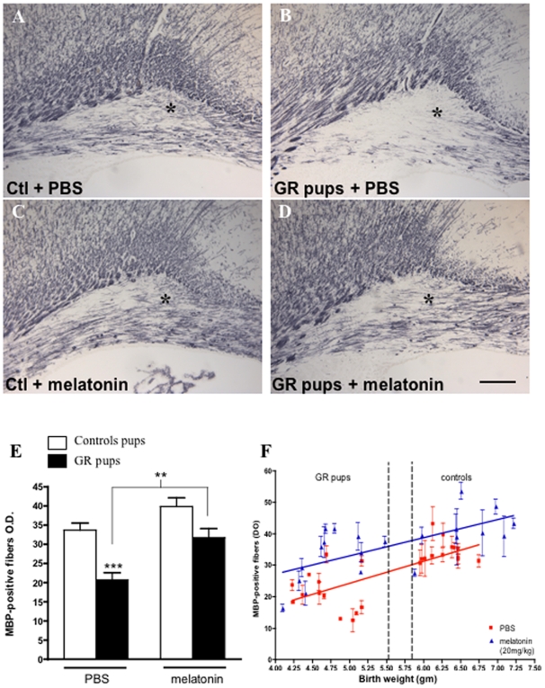
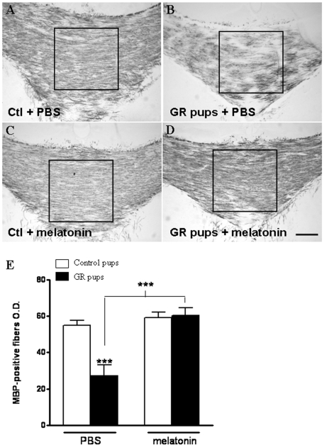
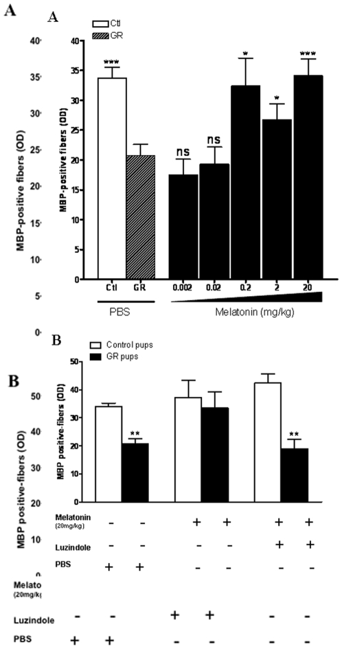
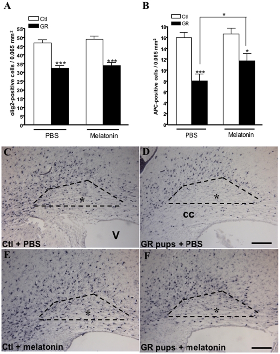
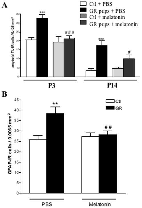
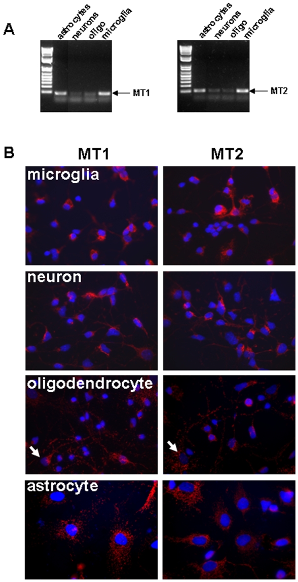
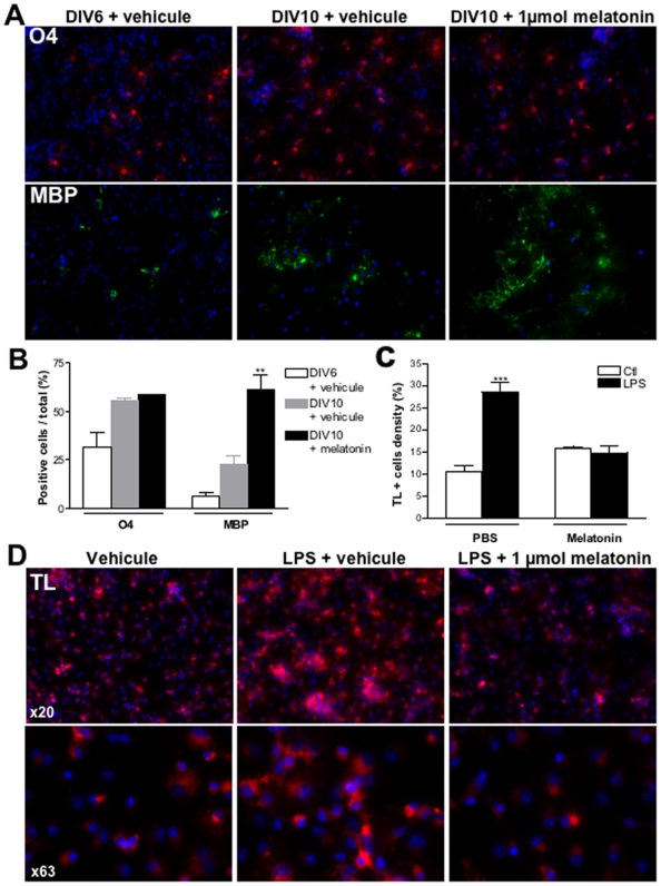
OBJECTIVE: To investigate the effects of melatonin treatment in a rat model of white matter damage (WMD) in the developing brain. Additionally, we aim to delineate the cellular mechanisms of melatonin effect on the oligodendroglial cell lineage. METHODS: A unilateral ligation of the uterine artery in pregnant rat at the embryonic day 17 induces fetal hypoxia and subsequent growth restriction (GR) in neonatal pups. GR and control pups received a daily intra-peritoneal injection of melatonin from birth to post-natal day (P) 3. RESULTS: Melatonin administration was associated with a dramatic decrease in microglial activation and astroglial reaction compared to untreated GR pups. At P14, melatonin prevented white matter myelination defects with an increased number of mature oligodendrocytes (APC-immunoreactive) in treated GR pups. Conversely, melatonin was not found to be associated with an increased density of total oligodendrocytes (Olig2-immunoreactive), suggesting that melatonin is able to promote oligodendrocyte maturation but not proliferation. These effects appear to be melatonin-receptor dependent and were reproduced in vitro. INTERPRETATION: These data suggest that melatonin has a strong protective effect on developing damaged white matter through decreased microglial activation and oligodendroglial maturation leading to a normalization of the myelination process. Consequently, melatonin should be a considered as an effective neuroprotective candidate not only in perinatal brain damage but also in inflammatory and demyelinating diseases observed in adults.
Overview
The claims and statistics of this paper haven’t been checked yet.
Run the check — free, no account needed. It reads the open-access text and checks the sources it cites.
Integrity signals
Registry
Cites
Statistics
Figures
Verification
Claims the paper makes
The claims and statistics of this paper haven’t been checked yet.
Run the check — free, no account needed. It reads the open-access text and checks the sources it cites.
Claims other papers make about this one
Each is checked against this paper’s own text: does it say what the citing paper says it does?
In the citing paper
“promotes oligodendroglial maturation in the newborn rat with abnormal white matter related to fetal hypoxia.105”
From Autism risk factors: genes, environment, and gene-environment interactions (2012) · cites this paper as [105]
Self-check
Percentages, sums and reported differences re-computed from the paper’s own numbers.
The self-check needs the paper’s full text.
Citations
Cites
Single-Step Method of RNA Isolation by Acid Guanidinium Thiocyanate–Phenol–Chloroform Extraction
Chomczynski, P., & Sacchi, N. (1987) · Analytical Biochemistry · doi:10.1006/abio.1987.9999 · Cited by 65,515
Preparation of separate astroglial and oligodendroglial cell cultures from rat cerebral tissue.
McCarthy, K. D., & de Vellis, J. (1980) · The Journal of Cell Biology · doi:10.1083/jcb.85.3.890 · Cited by 3,845
Tan, D., Manchester, L. C., Terron, M. P., Flores, L. J., & Reiter, R. J. (2007) · Journal of Pineal Research · doi:10.1111/j.1600-079x.2006.00407.x · Cited by 1,601
Pathogenesis of cerebral white matter injury of prematurity
Khwaja, O., & Volpe, J. J. (2008) · Archives of Disease in Childhood Fetal & Neonatal · doi:10.1136/adc.2006.108837 · Cited by 673
Readhead, C. W., Popko, B., Takahashi, N., Shine, H. D., Saavedra, R. A., Sidman, R. L., & Hood, L. (1987) · Cell · doi:10.1016/0092-8674(87)90248-0 · Cited by 397
Mayo, J. C., Sainz, R. M., Tan, D., Hardeland, R., León, J., Rodrı́guez, C., & Reiter, R. J. (2005) · Journal of Neuroimmunology · doi:10.1016/j.jneuroim.2005.05.002 · Cited by 332
Baud, O., Greene, A. E., Li, J., Wang, H., Volpe, J. J., & Rosenberg, P. A. (2004) · The Journal of Neuroscience · doi:10.1523/jneurosci.3989-03.2004 · Cited by 292
Self-citationMatuszak, Z., Reszka, K. J., & Chignell, C. F. (1997) · Free Radical Biology and Medicine · doi:10.1016/s0891-5849(96)00614-4 · Cited by 288
Brain macrophages synthesize interleukin‐1 and interleukin‐1 mRNAs in vitro
Hetier, E., Ayala‐Sanmartín, J., Denèfle, P., Bousseau, A., Rouget, P., Mallat, M., & Prochiantz, A. (1988) · Journal of Neuroscience Research · doi:10.1002/jnr.490210230 · Cited by 287
Cerebral palsy and restricted growth status at birth: population‐based case–control study
Jacobsson, B., Ahlin, K., Francis, A., Hagberg, G., Hagberg, H., & Gardosi, J. O. (2008) · BJOG An International Journal of Obstetrics & Gynaecology · doi:10.1111/j.1471-0528.2008.01827.x · Cited by 234
Cited by
Sorted by citations
The role of inflammation in perinatal brain injury
Hagberg, H., Mallard, C. E., Ferriero, D. M., Vannucci, S. J., Levison, S. W., Vexler, Z. S., & Gressèns, P. (2015) · Nature Reviews Neurology · doi:10.1038/nrneurol.2015.13 · Cited by 852
Autism risk factors: genes, environment, and gene-environment interactions
Chaste, P., & Leboyer, M. (2012) · Dialogues in Clinical Neuroscience · doi:10.31887/dcns.2012.14.3/pchaste · Cited by 807
Tarocco, A., Caroccia, N., Morciano, G., Wieckowski, M. R., Ancora, G., Garani, G., & Pinton, P. (2019) · Cell Death and Disease · doi:10.1038/s41419-019-1556-7 · Cited by 307
Alzheimer’s disease: pathological mechanisms and the beneficial role of melatonin
Rosales‐Corral, S., Acuña-Castroviejo, D., Coto‐Montes, A. M., Boga, J. A., Manchester, L. C., Fuentes‐Broto, L., Korkmaz, A., Ma, S., Tan, D. X., & Reiter, R. J. (2011) · Journal of Pineal Research · doi:10.1111/j.1600-079x.2011.00937.x · Cited by 299
Shah, S. A., Khan, M., Jo, M. H., Jo, M. G., Amin, F. U., & Kim, M. O. (2016) · CNS Neuroscience & Therapeutics · doi:10.1111/cns.12588 · Cited by 243
Li, Y., Zhang, J., Wan, J., Liu, A., & Sun, J. (2020) · Biomedicine & Pharmacotherapy · doi:10.1016/j.biopha.2020.110887 · Cited by 214
Miller, S., Yawno, T., Alers, N. O., Castillo-Meléndez, M., Supramaniam, V. G., VanZyl, N., Sabaretnam, T., Loose, J. M., Drummond, G. R., Walker, D. W., et al. (2014) · Journal of Pineal Research · doi:10.1111/jpi.12121 · Cited by 169
Which Neuroprotective Agents are Ready for Bench to Bedside Translation in the Newborn Infant?
Robertson, N. J., Tan, S., Groenendaal, F., van Bel, F., Juul, S. E., Bennet, L., Derrick, M., Back, S. A., Chavez‐Valdez, R., Northington, F. J., et al. (2012) · The Journal of Pediatrics · doi:10.1016/j.jpeds.2011.12.052 · Cited by 159
Impaired oligodendrocyte maturation in preterm infants: Potential therapeutic targets
van Tilborg, E., Heijnen, C. J., Benders, M. J. N. L., van Bel, F., Fleiss, B., Gressèns, P., & Nijboer, C. H. A. (2015) · Progress in Neurobiology · doi:10.1016/j.pneurobio.2015.11.002 · Cited by 159
Rodríguez-Giraldo, M., González-Reyes, R. E., Ramírez-Guerrero, S., Bonilla-Trilleras, C. E., Guardo-Maya, S., & Nava-Mesa, M. O. (2022) · International Journal of Molecular Sciences · doi:10.3390/ijms232113630 · Cited by 122
Oligodendrocyte Pathophysiology and Treatment Strategies in Cerebral Ischemia
Mifsud, G., Zammit, C., Muscat, R., Giovanni, G. D., & Valentino, M. (2014) · CNS Neuroscience & Therapeutics · doi:10.1111/cns.12263 · Cited by 123
Melatonin Promotes Myelination by Decreasing White Matter Inflammation After Neonatal Stroke
Villapol, S., Fau, S., Renolleau, S., Biran, V., Charriaut‐Marlangue, C., & Baud, O. (2010) · Pediatric Research · doi:10.1203/pdr.0b013e3181fcb40b · Cited by 103
Self-citationNeuroprotective Effect of Melatonin: A Novel Therapy against Perinatal Hypoxia-Ischemia
Alconada, D. A., Alvarez, A. A., Arteaga, O., Martínez-Ibargüen, A., & Hilario, E. (2013) · International Journal of Molecular Sciences · doi:10.3390/ijms14059379 · Cited by 103
Role of mitochondria in apoptotic and necroptotic cell death in the developing brain
Thornton, C., & Hagberg, H. (2015) · Clinica Chimica Acta · doi:10.1016/j.cca.2015.01.026 · Cited by 102
Pharmacokinetics of melatonin in preterm infants
Merchant, N. M., Azzopardi, D., Hawwa, A. F., McElnay, J. C., Middleton, B., Arendt, J., Arichi, T., Gressèns, P., & Edwards, A. D. (2013) · British Journal of Clinical Pharmacology · doi:10.1111/bcp.12092 · Cited by 95
Wang, F.-W., Wang, Z., Zhang, Y.-M., Du, Z., Zhang, X., Liu, Q., Guo, Y., Li, X., & Hao, A. (2013) · Journal of Cellular Biochemistry · doi:10.1002/jcb.24582 · Cited by 92
Is melatonin ready to be used in preterm infants as a neuroprotectant?
Biran, V., Duy, A. P., Decobert, F., Bednarek, N., Alberti, C., & Baud, O. (2014) · Developmental Medicine & Child Neurology · doi:10.1111/dmcn.12415 · Cited by 78
Self-citationCreatine for women in pregnancy for neuroprotection of the fetus
Dickinson, H., Bain, E., Wilkinson, D. J. C., Middleton, P., Crowther, C. A., & Walker, D. W. (2014) · Cochrane Database of Systematic Reviews · doi:10.1002/14651858.cd010846.pub2 · Cited by 77
A new neurological focus in neonatal intensive care
Bonifacio, S. L., Glass, H. C., Peloquin, S., & Ferriero, D. M. (2011) · Nature Reviews Neurology · doi:10.1038/nrneurol.2011.119 · Cited by 77
Huang, C., Chiang, R. P., Chen, C.-L., & Tsai, Y. (2014) · SLEEP · doi:10.5665/sleep.4002 · Cited by 75
Experimental treatments for hypoxic ischaemic encephalopathy
Kelen, D., & Robertson, N. J. (2010) · Early Human Development · doi:10.1016/j.earlhumdev.2010.05.011 · Cited by 72
Yawno, T., Castillo-Meléndez, M., Jenkin, G., Wallace, E. M., Walker, D. W., & Miller, S. (2012) · Developmental Neuroscience · doi:10.1159/000346323 · Cited by 69
Zhou, Q., Lin, L., Li, H., Wang, H., Jiang, S., Huang, P., Lin, Q., Chen, X., & Deng, Y. (2021) · Molecular Neurobiology · doi:10.1007/s12035-021-02568-7 · Cited by 67
Mitochondria, Bioenergetics and Excitotoxicity: New Therapeutic Targets in Perinatal Brain Injury
Leaw, B., Nair, S., Lim, R., Thornton, C., Mallard, C. E., & Hagberg, H. (2017) · Frontiers in Cellular Neuroscience · doi:10.3389/fncel.2017.00199 · Cited by 64
Denihan, N. M., Kirwan, J., Walsh, B. H., Dunn, W. B., Broadhurst, D., Boylan, G. B., & Murray, D. M. (2017) · Journal of Cerebral Blood Flow & Metabolism · doi:10.1177/0271678x17726502 · Cited by 62
Wang, Z., Liu, D., Wang, J., Liu, S., Gao, M. J., Ling, E., & Hao, A. (2011) · Journal of Pineal Research · doi:10.1111/j.1600-079x.2011.00952.x · Cited by 60
Basilious, A., Yager, J. Y., & Fehlings, M. G. (2014) · Developmental Medicine & Child Neurology · doi:10.1111/dmcn.12599 · Cited by 60
The Beneficial Effects of Melatonin Administration Following Hypoxia-Ischemia in Preterm Fetal Sheep
Yawno, T., Mahen, M., Li, J., Fahey, M. C., Jenkin, G., & Miller, S. (2017) · Frontiers in Cellular Neuroscience · doi:10.3389/fncel.2017.00296 · Cited by 59
Patrício, P., Mateus-Pinheiro, A., Irmler, M., Alves, N. D., Machado-Santos, A. R., Morais, M., Correia, J. S., Korostynski, M., Piechota, M., Stoffel, R., et al. (2015) · Neuropsychopharmacology · doi:10.1038/npp.2014.176 · Cited by 57
Hormonal Regulation of Oligodendrogenesis II: Implications for Myelin Repair
Breton, J., Long, K. L. P., Barraza, M., Perloff, O. S., & Kaufer, D. (2021) · Biomolecules · doi:10.3390/biom11020290 · Cited by 57
Melatonin for women in pregnancy for neuroprotection of the fetus
Wilkinson, D. J. C., Shepherd, E. S., & Wallace, E. M. (2016) · Cochrane Database of Systematic Reviews · doi:10.1002/14651858.cd010527.pub2 · Cited by 55
Hormonal Regulation of Oligodendrogenesis I: Effects across the Lifespan
Long, K. L. P., Breton, J., Barraza, M., Perloff, O. S., & Kaufer, D. (2021) · Biomolecules · doi:10.3390/biom11020283 · Cited by 56
Wu, U., Mai, F., Sheu, J., Chen, L., Liu, Y., Huang, H., & Chang, H. (2010) · Journal of Pineal Research · doi:10.1111/j.1600-079x.2010.00825.x · Cited by 53
AboTaleb, H. A., & Alghamdi, B. S. A. (2019) · Journal of Molecular Neuroscience · doi:10.1007/s12031-019-01425-6 · Cited by 54
Azedi, F., Mehrpour, M., Talebi, S., Zendedel, A., Kazemnejad, S., Mousavizadeh, K., Beyer, C., Zarnani, A., & Joghataei, M. T. (2019) · Brain Research · doi:10.1016/j.brainres.2019.146401 · Cited by 53
Melatonin Increases Oligodendrocyte Differentiation in Cultured Neural Stem Cells
Ghareghani, M., Sadeghi, H., Zibara, K., Danaei, N., Azari, H., & Ghanbari, A. (2016) · Cellular and Molecular Neurobiology · doi:10.1007/s10571-016-0450-4 · Cited by 51
New Antioxidant Drugs for Neonatal Brain Injury
Tataranno, M. L., Perrone, S., Longini, M., & Buonocore, G. (2015) · Oxidative Medicine and Cellular Longevity · doi:10.1155/2015/108251 · Cited by 48
Ghareghani, M., Scavo, L., Jand, Y., Farhadi, N., Sadeghi, H., Ghanbari, A., Mondello, S., Arnoult, D., Gharaghani, S., & Zibara, K. (2019) · Frontiers in Pharmacology · doi:10.3389/fphar.2019.00147 · Cited by 48
Tolcos, M., Petratos, S., Hirst, J. J., Wong, F. Y., Spencer, S. J., Azhan, A., Emery, B., & Walker, D. W. (2017) · Progress in Neurobiology · doi:10.1016/j.pneurobio.2017.03.009 · Cited by 46
Microglia in developing white matter and perinatal brain injury
McNamara, N. B., & Miron, V. E. (2019) · Neuroscience Letters · doi:10.1016/j.neulet.2019.134539 · Cited by 46
Jiang, S., Wang, H., Zhou, Q., Li, Q., Liu, N., Li, Z., Chen, C., & Deng, Y. (2021) · Journal of Inflammation Research · doi:10.2147/jir.s337499 · Cited by 45
Bossuyt, J., Herrewegen, Y. V. D., Nestor, L., Buckinx, A., Bundel, D. D., & Smolders, I. J. (2023) · Glia · doi:10.1002/glia.24390 · Cited by 44
Melatonin and the newborn brain
Colella, M., Biran, V., & Baud, O. (2016) · Early Human Development · doi:10.1016/j.earlhumdev.2016.09.001 · Cited by 37
Self-citationMelatonin Administration from 2000 to 2020 to Human Newborns with Hypoxic-Ischemic Encephalopathy
D’Angelo, G., Cannavò, L., Reiter, R. J., & Gitto, E. (2020) · American Journal of Perinatology · doi:10.1055/s-0040-1719151 · Cited by 36
Melatonin ameliorates brain injury induced by systemic lipopolysaccharide in neonatal rats
Wong, C., Jow, G., Kaizaki, A., Fan, L.-W., & Tien, L. (2014) · Neuroscience · doi:10.1016/j.neuroscience.2014.02.032 · Cited by 36
Berkiks, I., Benmhammed, H., Mesfioui, A., Ouichou, A., hasnaoui, A. E., Mouden, S., Touil, T., Bahbiti, Y., Nakache, R., & Hessni, A. E. (2017) · International Journal of Neuroscience · doi:10.1080/00207454.2017.1398156 · Cited by 35
Ball, G., Boardman, J. P., Arichi, T., Merchant, N. M., Rueckert, D., Edwards, A. D., & Counsell, S. J. (2013) · PLoS ONE · doi:10.1371/journal.pone.0067706 · Cited by 34
Hypoxia and myelination deficits in the developing brain
Singh, D. K., Ling, E., & Kaur, C. (2018) · International Journal of Developmental Neuroscience · doi:10.1016/j.ijdevneu.2018.06.012 · Cited by 34
Ciprofloxacin prevents myelination delay in neonatal rats subjected to E. coli sepsis
Loron, G., Olivier, P., See, H., Saché, N. L., Angulo, L., Biran, V., Brunelle, N., Besson-Lescure, B., Kitzis, M.-D., Pansiot, J., et al. (2010) · Annals of Neurology · doi:10.1002/ana.22190 · Cited by 30
Self-citationChiang, R. P., Huang, C., & Tsai, Y. (2012) · Journal of Pineal Research · doi:10.1111/jpi.12029 · Cited by 28
Ruff, C. A., Faulkner, S. D., Rumajogee, P., Beldick, S. R., Foltz, W. D., Corrigan, J., Basilious, A., Jiang, S., Thiyagalingam, S., Yager, J. Y., et al. (2017) · PLoS ONE · doi:10.1371/journal.pone.0184653 · Cited by 26
Malhotra, A., Rocha, A. K. A. D. A., Yawno, T., Sutherland, A. E., Allison, B. J., Nitsos, I., Pham, Y., Jenkin, G., Castillo-Meléndez, M., & Miller, S. (2024) · Pediatric Research · doi:10.1038/s41390-024-03027-4 · Cited by 25
Jiang, L., Shen, F., Degos, V., Schonemann, M. D., Pleasure, S. J., Mellon, S. H., Young, W. L., & Su, H. (2011) · Translational Stroke Research · doi:10.1007/s12975-011-0078-0 · Cited by 25
Wang, Z., Liu, D., Zhan, J., Xie, K., Wang, X., Xian, X., Gu, J., Chen, W., & Hao, A. (2013) · Pharmacological Research · doi:10.1016/j.phrs.2013.07.008 · Cited by 24
Ribeiro, R. T., Zanatta, Â., Amaral, A. U., Leipnitz, G., de Oliveira, F. H., Seminotti, B., & Wajner, M. (2018) · Neurotoxicity Research · doi:10.1007/s12640-018-9874-6 · Cited by 24
Therapeutic potential of melatonin in glioblastoma: Current knowledge and future prospects
Madhu, N. R., Sarkar, B., Biswas, P., Roychoudhury, S., Behera, B. K., & Acharya, C. K. (2024) · Elsevier eBooks · doi:10.1016/b978-0-323-95114-2.00002-9 · Cited by 23
Moura, A. P., Parmeggiani, B., Grings, M., Alvorcem, L. D. M., Boldrini, R. M., Bumbel, A. P., Motta, M. M., Seminotti, B., Wajner, M., & Leipnitz, G. (2015) · Molecular Neurobiology · doi:10.1007/s12035-015-9493-7 · Cited by 23
Klosen, P. (2024) · Journal of Pineal Research · doi:10.1111/jpi.12955 · Cited by 22
Ghareghani, M., Zibara, K., Sadeghi, H., & Farhadi, N. (2018) · Cellular and Molecular Neurobiology · doi:10.1007/s10571-018-0580-y · Cited by 22
Pang, R., Meehan, C., Maple, G., Norris, G., Campbell, E., Tucker, K., Mintoft, A., Torrealdea, F. J., Bainbridge, A. P., Hristova, M., et al. (2024) · Journal of Pineal Research · doi:10.1111/jpi.12962 · Cited by 20
Costa, E. M., Rocha, S., Rocha‐Pereira, P., Castro, E. M. B., Miranda, V. M. P., Faria, M., Loureiro, A., Quintanilha, A. T., Belo, L., & Santos‐Silva, A. (2008) · Blood Purification · doi:10.1159/000126922 · Cited by 20
Melatonin as a Therapy for Preterm Brain Injury: What Is the Evidence?
Häusler, S., Robertson, N. J., Golhen, K., van den Anker, J. N., Tucker, K., & Felder, T. K. (2023) · Antioxidants · doi:10.3390/antiox12081630 · Cited by 20
Gasperoni, F., Turini, P., & Agostinelli, E. (2019) · Amino Acids · doi:10.1007/s00726-019-02718-1 · Cited by 19
Jin, S., Liu, Y., Deng, S., Liao, L., Lin, T., Ning, Q., & Luo, X. (2015) · International Journal of Molecular Medicine · doi:10.3892/ijmm.2015.2136 · Cited by 18
Huang, C., Chen, S., Chang, C.-F., Lin, S., Lue, J., & Tsai, Y. (2020) · Neurochemistry International · doi:10.1016/j.neuint.2020.104827 · Cited by 18
Melatonin Exerts an Anti‐Panoptoic Role in Spinal Cord Ischemia‐Reperfusion Injured Rats
Xie, L., Wu, H., Shi, W., Zhang, J., Huang, X., & Yu, T. (2023) · Advanced Biology · doi:10.1002/adbi.202300424 · Cited by 17
Bench to Cribside: the Path for Developing a Neuroprotectant
Ramanantsoa, N., Fleiss, B., Bouslama, M., Matrot, B., Schwendimann, L., Cohen‐Salmon, C., Gressèns, P., & Gallego, J. (2012) · Translational Stroke Research · doi:10.1007/s12975-012-0233-2 · Cited by 17
Maternal Melatonin Deficiency Leads to Endocrine Pathologies in Children in Early Ontogenesis
Иванов, Д. О., Evsyukova, I. I., Mironova, E. S., Polyakova, V. O., Kvetnoy, I. M., & Nasyrov, R. A. (2021) · International Journal of Molecular Sciences · doi:10.3390/ijms22042058 · Cited by 18
Partial protective effects of melatonin on developing brain in a rat model of chorioamnionitis
Favrais, G., Saliba, É., Savary, L., Bodard, S., Gulhan, Z., Gressèns, P., & Chalon, S. (2021) · Scientific Reports · doi:10.1038/s41598-021-01746-w · Cited by 17
Embracing oligodendrocyte diversity in the context of perinatal injury
Cunningham, L. A., Newville, J., & Jantzie, L. L. (2017) · Neural Regeneration Research · doi:10.4103/1673-5374.217320 · Cited by 20
Fetal Neuroprotective Strategies: Therapeutic Agents and Their Underlying Synaptic Pathways
Elsayed, N. A., Boyer, T. M., & Burd, I. D. (2021) · Frontiers in Synaptic Neuroscience · doi:10.3389/fnsyn.2021.680899 · Cited by 16
Melatonin signalling in Schwann cells during neuroregeneration
Klymenko, A., & Lutz, D. (2022) · Frontiers in Cell and Developmental Biology · doi:10.3389/fcell.2022.999322 · Cited by 16
Diurnal Melatonin Patterns in Children: Ready to Apply in Clinical Practice?
Praninskienė, R., Dumalakienė, I., Kemežys, R., Mauricas, M., & Jučaitė, A. (2012) · Pediatric Neurology · doi:10.1016/j.pediatrneurol.2011.11.018 · Cited by 16
Ribeiro, R. T., Seminotti, B., Zanatta, Â., de Oliveira, F. H., Amaral, A. U., Leipnitz, G., & Wajner, M. (2021) · Neuroscience · doi:10.1016/j.neuroscience.2021.07.024 · Cited by 15
Beyond Hypothermia: Alternative Therapies for Hypoxic Ischemic Encephalopathy
Hobson, A., Baines, J., & Weiss, M. D. (2013) · The Open Pharmacology Journal · doi:10.2174/1874143620130805001 · Cited by 15
MELATONIN AND ITS ROLE IN NEURODEVELOPMENT DURING THE PERINATAL PERIOD: A REVIEW
Hassell, K. J., Reiter, R. J., & Robertson, N. J. (2013) · Fetal and Maternal Medicine Review · doi:10.1017/s0965539513000089 · Cited by 15
Ramarao, S. M., Pang, Y., Carter, K., & Bhatt, A. (2021) · Developmental Neuroscience · doi:10.1159/000519874 · Cited by 14
Melatonin secretion in children with epilepsy
Praninskienė, R., Dumalakienė, I., Kemežys, R., Mauricas, M., & Jučaitė, A. (2012) · Epilepsy & Behavior · doi:10.1016/j.yebeh.2012.08.012 · Cited by 14
Al-Griw, M. A., Salter, M., & Wood, I. (2022) · Acta Neurobiologiae Experimentalis · doi:10.55782/ane-2022-005 · Cited by 13
Abiramalatha, T., Ramaswamy, V. V., Ponnala, A. K., Kallem, V. R., Murkunde, Y. V., Punnoose, A. M., Aravindhan, V., Pullattayil, A. K., & Amboiram, P. (2022) · Expert Opinion on Investigational Drugs · doi:10.1080/13543784.2022.2040479 · Cited by 12
Short photoperiods attenuate central responses to an inflammogen
Fonken, L. K., Bedrosian, T. A., Michaels, H. D., Weil, Z. M., & Nelson, R. J. (2012) · Brain Behavior and Immunity · doi:10.1016/j.bbi.2012.01.017 · Cited by 12
Jallouli, S., Ghroubi, S., Damak, M., Sakka, S., Elleuch, M. H., Mhiri, C., Yahia, A., Driss, T., de Marco, G., & Hammouda, O. (2024) · Behavioural Brain Research · doi:10.1016/j.bbr.2024.115191 · Cited by 12
Cabrera-Muñoz, E. A., Ramírez-Rodríguez, G. B., Díaz-Yañez, L., Reyes‐Galindo, V., Juan, D. M.-S., & Vega‐Rivera, N. M. (2023) · International Journal of Molecular Sciences · doi:10.3390/ijms241713376 · Cited by 10
Иванов, Д. О., Mironova, E. S., Polyakova, V. O., Evsyukova, I. I., Osetrov, M., Kvetnoy, I. M., & Nasyrov, R. A. (2021) · PLoS ONE · doi:10.1371/journal.pone.0256197 · Cited by 10
RetractedRibeiro, R. T., Carvalho, A. V. S., Palavro, R., Durán‐Carabali, L. E., Zemniaçak, Â. B., Amaral, A. U., Netto, C. A., & Wajner, M. (2022) · Neurotoxicity Research · doi:10.1007/s12640-022-00625-0 · Cited by 9
Therapeutic opportunities for targeting cellular senescence in progressive multiple sclerosis
Sutter, P. A., McKenna, M. G., Imitola, J., Pijewski, R. S., & Crocker, S. J. (2022) · Current Opinion in Pharmacology · doi:10.1016/j.coph.2022.102184 · Cited by 8
Melatonin: a safe nutraceutical and clinical agent
Anadón, A., Arés, I., Martínez‐Larrañaga, M., & Martínez, M. (2021) · Elsevier eBooks · doi:10.1016/b978-0-12-821038-3.00034-3 · Cited by 8
Choi, M. S., Chung, Y. Y., Kim, D. J., Kim, S. T., & JUN, Y. H. (2020) · In Vivo · doi:10.21873/invivo.11819 · Cited by 8
Suofu, Y., Jauhari, A., Nirmala, E. S., Mullins, W. A., Wang, X., Li, F., Carlisle, D. L., & Friedlander, R. M. (2022) · Neuroscience Letters · doi:10.1016/j.neulet.2022.137043 · Cited by 8
Joshi, M., Singh, S., Patel, S. P., Shah, D. P., & Krishnakumar, A. (2018) · Computational Toxicology · doi:10.1016/j.comtox.2018.08.004 · Cited by 6
Is there a role for melatonin in the intensive care unit?
Richards, G. A., Bentley, A., Gopalan, D. P., Brannigan, L., & Paruk, F. (2021) · Southern African Journal of Critical Care · doi:10.7196/sajcc.2021.v37i2.490 · Cited by 6
Kühne, B. A., Vázquez‐Aristizabal, P., Fuentes-Amell, M., Plà, L., Loreiro, C., Gómez-Catalán, J., Gratacós, E., Illa, M., & Barenys, M. (2022) · Biomedicines · doi:10.3390/biomedicines10051205 · Cited by 5
Melatonin ameliorates astrogliosis and microgliosis in a cuprizone demyelinating mouse model
Alidadi, M., Omidi, N., Abdi, M., Mohammadi, M., Shabani, M., & Kashani, I. R. (2025) · Biochemistry and Biophysics Reports · doi:10.1016/j.bbrep.2025.101929 · Cited by 4
Neonatal hypoxic ischaemic encephalopathy: current and future treatment options
Merchant, N. M., Azzopardi, D., & Edwards, A. D. (2015) · Expert Opinion on Orphan Drugs · doi:10.1517/21678707.2015.1021776 · Cited by 4
Ma, R., Ma, Y., Zhang, X., Geng, Y., Chen, L., Zhang, X., & Wang, X. (2021) · PubMed Central · doi:10.7499/j.issn.1008-8830.2011132 · Cited by 4
Al-Griw, M. A., Salter, M. G., & Wood, I. (2021) · Acta Neurobiologiae Experimentalis · doi:10.21307/ane-2021-022 · Cited by 4
The Role of Melatonin in Morphofunctional Development of the Brain in Early Ontogeny
Arutjunyan, A. V., Evsyukova, I. I., & Polyakova, V. O. (2019) · Neurochemical Journal · doi:10.1134/s1819712419030036 · Cited by 4
The Role of Melatonin in Prenatal Ontogenesis
Evsyukova, I. I. (2021) · Journal of Evolutionary Biochemistry and Physiology · doi:10.1134/s0022093021010038 · Cited by 3
REDOX Balance in Oligodendrocytes Is Important for Zebrafish Visual System Regeneration
Pérez‐Montes, C., Jiménez-Cubides, J. P., Velasco, A., Arévalo, R., Santos‐Ledo, A., & Macia, M. G. (2023) · Antioxidants · doi:10.3390/antiox12122026 · Cited by 3
A study on the effect of JNJ-10397049 on proliferation and differentiation of neural precursor cells
Karami, N., Azari, H., Rahimi, M., Aligholi, H., & Kalantari, T. (2022) · Anatomy & Cell Biology · doi:10.5115/acb.21.202 · Cited by 2
Metadata
- Authors
- Olivier, P., Fontaine, R. H., Loron, G., Van Steenwinckel, J., Biran, V., Massonneau, V., Kaindl, A., Dalous, J., Charriaut-Marlangue, C., Aigrot, M.-S., Pansiot, J., Verney, C., Gressens, P., & Baud, O.
- Affiliations
- Inserm (FR)
- Centre National de la Recherche Scientifique (FR)
- Corresponding author
- Baud, Olivier
- ORCID
- Venue
- PLoS ONE
- Publisher
- Public Library of Science (PLoS)
- Published
- 2009-09-22
- Volume
- 4
- Issue
- 9
- Pages
- e7128
- ISSN
- 1932-6203 (ISSN-L)
- PMC
- PMC2742165
- OpenAlex
- W1966357480
- Cited by
- 119
- References
- 37
- Citations by year
- 2025: 3 · 2024: 7 · 2023: 5 · 2022: 11 · 2021: 17 · 2020: 6 · 2019: 8 · 2018: 4 · 2017: 7 · 2016: 6 · 2015: 7 · 2014: 9 · 2013: 9 · 2012: 9
- FWCI
- 5.64
- Type
- journal article
- Language
- en
- Update notices
- correction (2009-10-15)
- Open access
- gold
- doi.org
- Locations
- PLoS ONE (publishedVersion, cc-by)
- PubMed Central (publishedVersion, cc-by)
- PubMed (publishedVersion)
- Research Portal (King's College London) (publishedVersion, other-oa)
- RePEc: Research Papers in Economics (submittedVersion)
- DOAJ (DOAJ: Directory of Open Access Journals) (submittedVersion, cc-by-sa)
- Figshare (submittedVersion, cc-by)
- Funders
- Ikaria
- Institut National de la Santé et de la Recherche Médicale
- Centre National de la Recherche Scientifique (UMR 7101)
- Topics
- Health Sciences › Medicine › Pediatrics, Perinatology and Child Health › Neonatal and fetal brain pathology
- Life Sciences › Neuroscience › Developmental Neuroscience › Neurogenesis and neuroplasticity mechanisms
- Life Sciences › Neuroscience › Endocrine and Autonomic Systems › Circadian rhythm and melatonin
- Keywords
- melatonin, oligodendrocyte maturation, white matter injury, fetal hypoxia, growth restriction, uterine artery ligation, myelination, microglial activation, astroglial reaction, APC, Olig2, neuroprotection, perinatal brain injury, melatonin receptors
- MeSH terms
- Animals, Animals, Newborn, Brain, Cells, Cultured, Immunohistochemistry, Melatonin, Microscopy, Fluorescence, Nerve Fibers, Myelinated, Oligodendroglia, RNA, Messenger, Time Factors, Rats, Sprague-Dawley, Gene Expression Regulation, Developmental, Cell Lineage, Rats
About these checks
Registry, retraction, Cites and statistics results are computed, not judged, with one exception: a printed reference that no index lists is read once by a model before it counts as a citation error. Claims are extracted by a model and kept only when their quote appears verbatim in the text; each cited source is then checked, with a model’s help, for whether it says what the claim relies on. Those verdicts can be wrong — read the claim’s quote and the cited source before relying on one.
Statistics and arithmetic are recomputed, not judged.
Confidence bands are shown only for fields calibrated on our annotated test set. None is calibrated yet, so no band is shown.
What counts as a claim ›How we measure confidence ›How we check this ›
Are you an author, or do you see a problem with this page? Request a review
