

The DOI matches its Crossref record; 29 of the 29 references it cites were found in a registry or an index.
Muscle-Specific Adaptations, Impaired Oxidative Capacity and Maintenance of Contractile Function Characterize Diet-Induced Obese Mouse Skeletal Muscle
Open accessAbstract
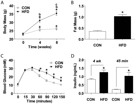
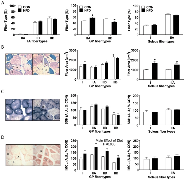
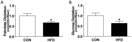
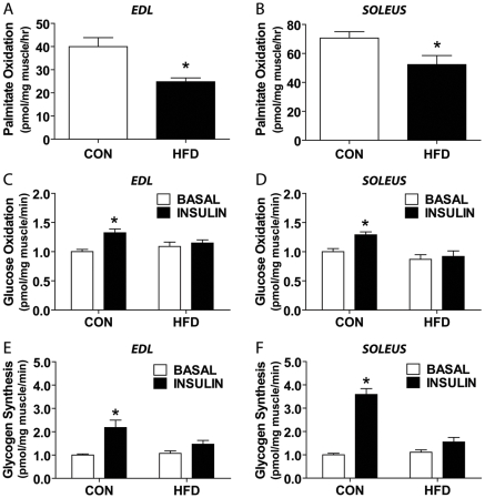
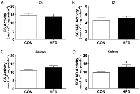
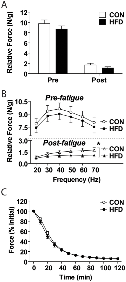
BACKGROUND: The effects of diet-induced obesity on skeletal muscle function are largely unknown, particularly as it relates to changes in oxidative metabolism and morphology. PRINCIPAL FINDINGS: Compared to control fed mice, mice fed a high fat diet (HFD; 60% kcal: fat) for 8 weeks displayed increased body mass and insulin resistance without overt fasting hyperglycemia (i.e. pre-diabetic). Histological analysis revealed a greater oxidative potential in the HFD gastrocnemius/plantaris (increased IIA, reduced IIB fiber-type percentages) and soleus (increased I, IIA cross-sectional areas) muscles, but no change in fiber type percentages in tibialis anterior muscles compared to controls. Intramyocellular lipid levels were significantly increased relative to control in HFD gastrocnemius/plantaris, but were similar to control values in the HFD soleus. Using a novel, single muscle fiber approach, impairments in complete palmitate and glucose oxidation (72.8+/-6.6% and 61.8+/-9.1% of control, respectively; p<0.05) with HFD were detected. These reductions were consistent with measures made using intact extensor digitorum longus and soleus muscles. Compared to controls, no difference in succinate dehydrogenase or citrate synthase enzyme activities were observed between groups in any muscle studied, however, short-chain fatty acyl CoA dehydrogenase (SCHAD) activity was elevated in the HFD soleus, but not tibialis anterior muscles. Despite these morphological and metabolic alterations, no significant difference in peak tetanic force or low-frequency fatigue rates were observed between groups. CONCLUSIONS: These findings indicate that HFD induces early adaptive responses that occur in a muscle-specific pattern, but are insufficient to prevent impairments in oxidative metabolism with continued high-fat feeding. Moreover, the morphological and metabolic changes which occur with 8 weeks of HFD do not significantly impact muscle contractile properties.
Overview
The claims and statistics of this paper haven’t been checked yet.
Run the check — free, no account needed. It reads the open-access text and checks the sources it cites.
Integrity signals
Registry
Cites
Statistics
Figures
Verification
Claims the paper makes
The claims and statistics of this paper haven’t been checked yet.
Run the check — free, no account needed. It reads the open-access text and checks the sources it cites.
Claims other papers make about this one
Each is checked against this paper’s own text: does it say what the citing paper says it does?
In the citing paper
“muscle β-oxidation increases in response to a high-fat diet (HFD) [83–86], although excessive lipid content or duration of such diets can exceed this capacity, leading to ectopic lipid accumulation and impaired lipid metabolism [87–91].”
From Targeting Intramyocellular Lipids to Improve Aging Muscle Function (2025) · cites this paper as [90]
Self-check
Percentages, sums and reported differences re-computed from the paper’s own numbers.
The self-check needs the paper’s full text.
Citations
Cites
Koves, T. R., Ussher, J. R., Noland, R. C., Slentz, D., Mosedale, M., Ilkayeva, O., Bain, J., Stevens, R., Dyck, J. R. B., Newgard, C. B., et al. (2008) · Cell Metabolism · doi:10.1016/j.cmet.2007.10.013 · Cited by 1,990
Bonnard, C., Durand, A., Peyrol, S., Chanseaume, E., Chauvin, M.-A., Morio, B., Vidal, H., & Rieusset, J. (2008) · Journal of Clinical Investigation · doi:10.1172/jci32601 · Cited by 819
Sparks, L. M., Xie, H., Koza, R. A., Mynatt, R., Hulver, M. W., Bray, G. A., & Smith, S. R. (2005) · Diabetes · doi:10.2337/diabetes.54.7.1926 · Cited by 629 · cited 1×
“Furthermore, an increase in oxidative phosphorylation protein complexes have been measured in the gastrocnemius muscles following 4 weeks of high fat feeding [6], while a decreased expression of oxidative phosphorylation genes and cytochrome C protein has been demonstrated in the quadriceps muscles in response to 3 we…”
BackgroundInfluentialBurkholder, T. J., Fingado, B., Baron, S., & Lieber, R. L. (1994) · Journal of Morphology · doi:10.1002/jmor.1052210207 · Cited by 527
Excess Lipid Availability Increases Mitochondrial Fatty Acid Oxidative Capacity in Muscle
Turner, N., Bruce, C. R., Beale, S. M., Hoehn, K. L., So, T., Rolph, M. S., & Cooney, G. J. (2007) · Diabetes · doi:10.2337/db07-0093 · Cited by 520
Koopman, R., Schaart, G., & Hesselink, M. K. C. (2001) · Histochemistry and Cell Biology · doi:10.1007/s004180100297 · Cited by 471
Type 2 Diabetes, Muscle Strength, and Impaired Physical Function
Sayer, A. A., Dennison, E. M., Syddall, H. E., Gilbody, H. J., Phillips, D. I. W., & Cooper, C. (2005) · Diabetes Care · doi:10.2337/diacare.28.10.2541 · Cited by 366
Hämäläinen, N., & Pette, D. (1993) · Journal of Histochemistry & Cytochemistry · doi:10.1177/41.5.8468455 · Cited by 350
Muscle Fiber Composition and Capillary Density in Women and Men With NIDDM
Mårin, P., Andersson, B., Krotkiewski, M., & Björntorp, P. A. (1994) · Diabetes Care · doi:10.2337/diacare.17.5.382 · Cited by 342
Lee, J. S., Pinnamaneni, S. K., Eo, S. J., Cho, I. H., Pyo, J. H., Kim, C. K., Sinclair, A. J., Febbraio, M. A., & Watt, M. J. (2005) · Journal of Applied Physiology · doi:10.1152/japplphysiol.01438.2005 · Cited by 325
Cited by
Sorted by citations
Items 101–129 of 129
Ex-vivo Nuclear Magnetic Resonance Characterization of Pig Muscles
Zancanaro, C., Fontanella, M. M., Nicolato, E., Mosconi, E., Pellicciar, C., & Malatesta, M. (2011) · International Journal of Meat Science · doi:10.3923/ijmeat.2011.93.107 · Cited by 2
Tallis, J. A., James, R. S., Eyre, E. L. J., Shelley, S. P., Hill, C., Renshaw, D. W., & Hurst, J. (2024) · Experimental Physiology · doi:10.1113/ep091832 · Cited by 1
Botezelli, J. D., Overby, P., Lindo, L., Wang, S., Haïda, O., Lim, G. E., Templeman, N. M., Pauli, J. R., & Johnson, J. D. (2020) · bioRxiv (Cold Spring Harbor Laboratory) · doi:10.1101/2020.03.31.018432 · Cited by 2
Skeletal Muscle Function, Morphology, and Biochemistry in Ts65Dn Mice: A Model of Down Syndrome
Cowley, P. M. (2011) · Syracuse University Libraries (Syracuse University) · Cited by 1
Jeon, Y., Ha, K., Seo, D. Y., Yeo, J. Y., Choi, S. J., & Han, J. (2017) · IJASS(International Journal of Applied Sports Sciences) · doi:10.24985/ijass.2017.29.1.42 · Cited by 1
Investigating the potential relationship between skeletal muscle atrophy and obesity
Elmore, C. (2012) · doi:10.17077/etd.vkfadurp · Cited by 1
Dunford, E. C. (2016) · YorkSpace (York University) · Cited by 1
Diet Induced Obesity Alters Muscle Spindle Afferent Function in Adult Mice
Elahi, L. S. (2017) · doi:10.31979/etd.5hc9-8tjn · Cited by 1
van de Weijer, T. (2013) · Research Publications (Maastricht University) · doi:10.26481/dis.20130705tw
The Effects of a High Fat Diet on Mitochondrial Biogenesis and Inflammation in the Brain
Vervaecke, L. S., Davis, J. M., Carson, J., Murphy, A., & Thompson, R. (2014) · Medicine & Science in Sports & Exercise · doi:10.1249/01.mss.0000495380.12776.95
Leung, M. (2011) · The Atrium (University of Guelph)
Schumm, S. (2011) · OhioLink ETD Center (Ohio Library and Information Network)
Gamu, D. (2012) · UWSpace (University of Waterloo)
Frias, F. D. T. (2016) · Digital Library of Theses and Dissertations (Universidade de São Paulo) · doi:10.11606/d.42.2016.tde-12092016-085246
Hill, C. (2018) · Coventry University Open Collections (Coventry university)
The impact on high-fat diet on skeletal muscle stem cell recruitment in CD36 deficient mice
Verpoorten, S. (2019) · Repository@Hull (Worktribe) (University of Hull)
Spagnol, A. R. (2022) · Digital Library of Theses and Dissertations (Universidade de São Paulo) · doi:10.11606/t.42.2022.tde-16112022-160054
Skeletal Muscle Mitochondrial Morphology Negatively Affected by Loss of XinPreprint
Martin, G., Al‐Sajee, D., Gingrich, M. A., Chattha, R., Akcan, M., Monaco, C. M. F., Hughes, M. C., Perry, C. G. R., Rebalka, I. A., Tarnopolsky, M. A., et al. (2023) · bioRxiv (Cold Spring Harbor Laboratory) · doi:10.1101/2023.09.14.557614
Self-citationSkeletal muscle mitochondrial morphology negatively affected in mice lacking Xin
Martin, G., Al‐Sajee, D., Gingrich, M. A., Chattha, R., Akcan, M., Monaco, C. M. F., Hughes, M. C., Perry, C. G. R., Rebalka, I. A., Tarnopolsky, M. A., et al. (2024) · Biochemistry and Cell Biology · doi:10.1139/bcb-2024-0034
Self-citationLawler, J. M., Kamal, K. Y., Botchlett, R. E., Woo, S., Li, H., Hord, J. M., Fluckey, J. D., & Wu, C. (2025) · Connective Tissue Research · doi:10.1080/03008207.2025.2471853 · Cited by 1
Kallabis, S., Abraham, L., Müller, S., Dzialas, V., Türk, C., Wiederstein, J. L., Bock, T., Nolte, H., Nogara, L., Blaauw, B., et al. (2020) · Figshare · doi:10.6084/m9.figshare.12021909
Kallabis, S., Abraham, L., Müller, S., Dzialas, V., Türk, C., Wiederstein, J. L., Bock, T., Nolte, H., Nogara, L., Blaauw, B., et al. (2020) · Figshare · doi:10.6084/m9.figshare.12021900
Kallabis, S., Abraham, L., Müller, S., Dzialas, V., Türk, C., Wiederstein, J. L., Bock, T., Nolte, H., Nogara, L., Blaauw, B., et al. (2020) · Figshare · doi:10.6084/m9.figshare.12021915
Kallabis, S., Abraham, L., Müller, S., Dzialas, V., Türk, C., Wiederstein, J. L., Bock, T., Nolte, H., Nogara, L., Blaauw, B., et al. (2020) · Figshare · doi:10.6084/m9.figshare.12021921
Kallabis, S., Abraham, L., Müller, S., Dzialas, V., Türk, C., Wiederstein, J. L., Bock, T., Nolte, H., Nogara, L., Blaauw, B., et al. (2020) · Figshare · doi:10.6084/m9.figshare.12021897
Effect of (−)-Epicatechin on Mitochondrial Homeostasis in Skeletal Muscle of Female Obese Rats
Herrera-Cogco, E. D. L. C., Meza, S. H., Martínez‐Meza, Y., Duran, J. P., Ceballos, G. M., Méndez-Bolaina, E., & Nájera, N. (2026) · Molecules · doi:10.3390/molecules31061050
Impact of diet induced obesity on mouse skeletal muscle health: metabolism, growth and regeneration.
Trajcevski, K. E. (2014) · MacSphere (McMaster University) · doi:10.71548/22585
Metadata
- Authors
- Shortreed, K. E., Krause, M. P., Huang, J. H., Dhanani, D., Moradi, J., Ceddia, R. B., & Hawke, T. J.
- Affiliations
- York University (CA)
- Corresponding author
- Hawke, Thomas J.
- ORCID
- Venue
- PLoS ONE
- Publisher
- Public Library of Science (PLoS)
- Published
- 2009-10-06
- Volume
- 4
- Issue
- 10
- Pages
- e7293
- ISSN
- 1932-6203 (ISSN-L)
- PMC
- PMC2752162
- OpenAlex
- W2116618150
- Cited by
- 131
- References
- 29
- Citations by year
- 2026: 1 · 2025: 5 · 2024: 5 · 2023: 6 · 2022: 7 · 2021: 4 · 2020: 15 · 2019: 7 · 2018: 8 · 2017: 11 · 2016: 6 · 2015: 12 · 2014: 13 · 2013: 11 · 2012: 4
- FWCI
- 2.66
- Type
- journal article
- Language
- en
- Open access
- gold
- doi.org
- Locations
- PLoS ONE (publishedVersion, cc-by)
- PubMed Central (publishedVersion, cc-by)
- PubMed (publishedVersion)
- DOAJ (DOAJ: Directory of Open Access Journals) (submittedVersion, cc-by-sa)
- Funders
- Ontario Innovation Trust
- Topics
- Life Sciences › Biochemistry, Genetics and Molecular Biology › Molecular Biology › Muscle Physiology and Disorders
- Health Sciences › Medicine › Physiology (medical) › Adipose Tissue and Metabolism
- Health Sciences › Medicine › Physiology (medical) › Cardiovascular and exercise physiology
- Keywords
- diet-induced obesity, high-fat diet, skeletal muscle, oxidative metabolism, muscle fiber type, intramyocellular lipid, palmitate oxidation, glucose oxidation, succinate dehydrogenase, citrate synthase, muscle contractility, insulin resistance, prediabetes, gastrocnemius, soleus muscle, tibialis anterior, extensor digitorum longus
- MeSH terms
- Animals, Animal Feed, Blood Glucose, Dietary Fats, Glucose, Lipids, Male, Mice, Inbred C57BL, Muscle Contraction, Obesity, Oxygen, Time Factors, Muscle, Skeletal, Mice, Animal Nutrition Sciences
About these checks
Registry, retraction, Cites and statistics results are computed, not judged, with one exception: a printed reference that no index lists is read once by a model before it counts as a citation error. Claims are extracted by a model and kept only when their quote appears verbatim in the text; each cited source is then checked, with a model’s help, for whether it says what the claim relies on. Those verdicts can be wrong — read the claim’s quote and the cited source before relying on one.
Statistics and arithmetic are recomputed, not judged.
Confidence bands are shown only for fields calibrated on our annotated test set. None is calibrated yet, so no band is shown.
What counts as a claim ›How we measure confidence ›How we check this ›
Are you an author, or do you see a problem with this page? Request a review
