

The DOI matches its Crossref record; 58 of the 60 references it cites were found in a registry or an index.
Cholesterol Crystals Activate the NLRP3 Inflammasome in Human Macrophages: A Novel Link between Cholesterol Metabolism and Inflammation
Open accessAbstract
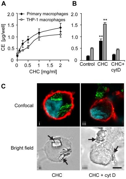
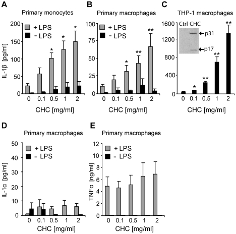
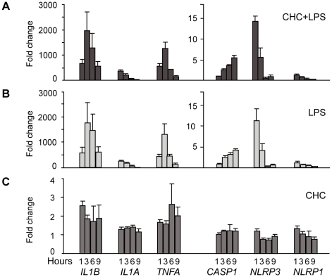
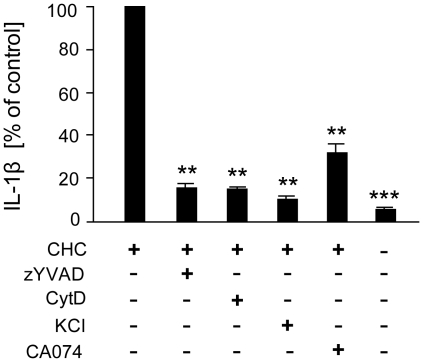
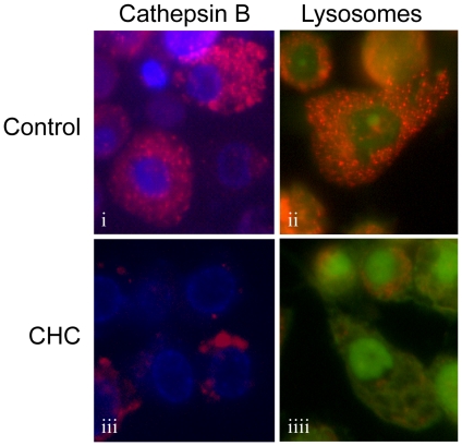
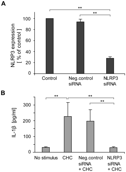
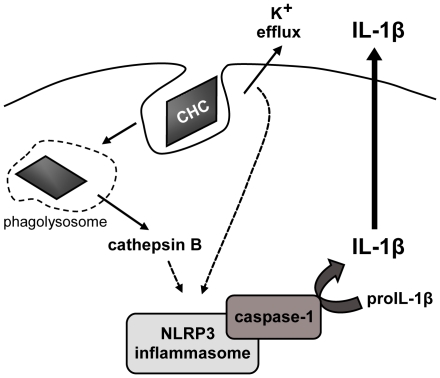
BACKGROUND: Chronic inflammation of the arterial wall is a key element in the pathogenesis of atherosclerosis, yet the factors that trigger and sustain the inflammation remain elusive. Inflammasomes are cytoplasmic caspase-1-activating protein complexes that promote maturation and secretion of the proinflammatory cytokines interleukin(IL)-1beta and IL-18. The most intensively studied inflammasome, NLRP3 inflammasome, is activated by diverse substances, including crystalline and particulate materials. As cholesterol crystals are abundant in atherosclerotic lesions, and IL-1beta has been linked to atherogenesis, we explored the possibility that cholesterol crystals promote inflammation by activating the inflammasome pathway. PRINCIPAL FINDINGS: Here we show that human macrophages avidly phagocytose cholesterol crystals and store the ingested cholesterol as cholesteryl esters. Importantly, cholesterol crystals induced dose-dependent secretion of mature IL-1beta from human monocytes and macrophages. The cholesterol crystal-induced secretion of IL-1beta was caspase-1-dependent, suggesting the involvement of an inflammasome-mediated pathway. Silencing of the NLRP3 receptor, the crucial component in NLRP3 inflammasome, completely abolished crystal-induced IL-1beta secretion, thus identifying NLRP3 inflammasome as the cholesterol crystal-responsive element in macrophages. The crystals were shown to induce leakage of the lysosomal protease cathepsin B into the cytoplasm and inhibition of this enzyme reduced cholesterol crystal-induced IL-1beta secretion, suggesting that NLRP3 inflammasome activation occurred via lysosomal destabilization. CONCLUSIONS: The cholesterol crystal-induced inflammasome activation in macrophages may represent an important link between cholesterol metabolism and inflammation in atherosclerotic lesions.
Overview
The claims and statistics of this paper haven’t been checked yet.
Run the check — free, no account needed. It reads the open-access text and checks the sources it cites.
Integrity signals
Registry
Cites
Statistics
Figures
Verification
Claims the paper makes
The claims and statistics of this paper haven’t been checked yet.
Run the check — free, no account needed. It reads the open-access text and checks the sources it cites.
Claims other papers make about this one
Each is checked against this paper’s own text: does it say what the citing paper says it does?
In the citing paper
“Primed macrophages secrete large amounts of IL‐1β in response to cholesterol crystals in an NLRP3 inflammasome‐dependent manner in mouse and human cells.34, 35”
From Trimethylamine‐N‐Oxide Induces Vascular Inflammation by Activating the NLRP3 Inflammasome Through the SIRT3‐SOD2‐mtROS Signaling Pathway (2017) · cites this paper as [34]
Self-check
Percentages, sums and reported differences re-computed from the paper’s own numbers.
The self-check needs the paper’s full text.
Citations
Cites
Analysis of Relative Gene Expression Data Using Real-Time Quantitative PCR and the 2−ΔΔCT Method
Livak, K. J., & Schmittgen, T. D. (2001) · Methods · doi:10.1006/meth.2001.1262 · Cited by 180,860
Gout-associated uric acid crystals activate the NALP3 inflammasome
Martinon, F., Pétrilli, V., Mayor, A., Tardivel, A., & Tschopp, J. (2006) · Nature · doi:10.1038/nature04516 · Cited by 5,326
NLRP3 inflammasomes are required for atherogenesis and activated by cholesterol crystals
Duewell, P., Kono, H., Rayner, K. J., Sirois, C. M., Vladimer, G., Bauernfeind, F. G., Abela, G. S., Franchi, L., Nuñez, G., Schnurr, M., et al. (2010) · Nature · doi:10.1038/nature08938 · Cited by 4,026 · cited 1×
“While this study was being finalized, Duewell and colleagues published a similar study reporting that cholesterol crystals activate the NLRP3 inflammasome in mouse macrophages through a lysosomal damage- and cathepsin-mediated mechanism”
BackgroundBauernfeind, F. G., Horvath, G. L., Stutz, A., Alnemri, E. S., MacDonald, K. L., Speert, D. P., Fernandes‐Alnemri, T., Wu, J., Monks, B. G., Fitzgerald, K. A., et al. (2009) · The Journal of Immunology · doi:10.4049/jimmunol.0901363 · Cited by 3,087
Cryopyrin activates the inflammasome in response to toxins and ATP
Mariathasan, S., Weiss, D. S., Newton, K., McBride, J., O'Rourke, K., Roose-Girma, M., Lee, W. P., Weinrauch, Y., Monack, D. M., & Dixit, V. M. (2006) · Nature · doi:10.1038/nature04515 · Cited by 3,080
Hornung, V., Bauernfeind, F., Halle, A., Samstad, E. O., Kono, H., Rock, K. L., Fitzgerald, K. A., & Latz, E. (2008) · Nature Immunology · doi:10.1038/ni.1631 · Cited by 3,065
Innate Immune Activation Through Nalp3 Inflammasome Sensing of Asbestos and Silica
Dostert, C., Pétrilli, V., Van Bruggen, R., Steele, C., Mossman, B. T., & Tschopp, J. (2008) · Science · doi:10.1126/science.1156995 · Cited by 2,658
The Inflammasomes: Guardians of the Body
Martinon, F., Mayor, A., & Tschopp, J. (2009) · Annual Review of Immunology · doi:10.1146/annurev.immunol.021908.132715 · Cited by 2,436
NLRP3 inflammasome activation: the convergence of multiple signalling pathways on ROS production?
Tschopp, J., & Schroder, K. (2010) · Nature reviews. Immunology · doi:10.1038/nri2725 · Cited by 1,859
Cytokines in Atherosclerosis: Pathogenic and Regulatory Pathways
Tedgui, A., & Mallat, Z. (2006) · Physiological Reviews · doi:10.1152/physrev.00024.2005 · Cited by 1,751
Cited by
Sorted by citations
Items 301–400 of 998
Varsano, N., Beghi, F., Elad, N., Pereiro, E., Dadosh, T., Pinkas, I., Perez-Berna, A. J., Jin, X., Kruth, H. S., Leiserowitz, L., et al. (2018) · Proceedings of the National Academy of Sciences · doi:10.1073/pnas.1803119115 · Cited by 67
Role of pyroptosis in atherosclerosis and its therapeutic implications
He, B., Nie, Q., Wang, F., Han, Y., Yang, B., Sun, M., Fan, X., Ye, Z., Liu, P., & Wen, J. (2021) · Journal of Cellular Physiology · doi:10.1002/jcp.30366 · Cited by 66
Do patients benefit from omega-3 fatty acids?
Sherratt, S. C. R., Mason, R. P., Libby, P., Steg, P. G., & Bhatt, D. L. (2023) · Cardiovascular Research · doi:10.1093/cvr/cvad188 · Cited by 75
Closing the loop on inflammation and atherothrombosis: why perform the CIRT and CANTOS trials?
Ridker, P. M. (2013) · Europe PMC (PubMed Central) · Cited by 67
Shaw, C., Li, Y., & Tomljenovic, L. (2013) · Journal of Inorganic Biochemistry · doi:10.1016/j.jinorgbio.2013.07.022 · Cited by 66
Inflammation modulation and cardiovascular disease prevention
Awan, Z. A., & Genest, J. J. (2014) · European Journal of Preventive Cardiology · doi:10.1177/2047487314529350 · Cited by 65
Chistiakov, D. A., Orekhov, A. N., & Bobryshev, Y. V. (2016) · Immunobiology · doi:10.1016/j.imbio.2016.05.010 · Cited by 66
Increased levels of legumain in plasma and plaques from patients with carotid atherosclerosis
Lunde, N. N., Holm, S., Dahl, T. B., Elyouncha, I., Sporsheim, B., Gregersen, I., Abbas, A., Skjelland, M., Espevik, T., Solberg, R., et al. (2016) · Atherosclerosis · doi:10.1016/j.atherosclerosis.2016.11.026 · Cited by 65
Orlowski, G. M., Sharma, S., Colbert, J. D., Bogyo, M. S., Robertson, S. A., Kataoka, H., Chan, F. K.-M., & Rock, K. L. (2017) · Journal of Leukocyte Biology · doi:10.1189/jlb.3hi0316-152r · Cited by 65
Expression of NLRP3 in subcutaneous adipose tissue is associated with coronary atherosclerosis
Bando, S., Fukuda, D., Soeki, T., Nishimoto, S., Uematsu, E., Matsuura, T., Ise, T., Tobiume, T., Yamaguchi, K., Yagi, S., et al. (2015) · Atherosclerosis · doi:10.1016/j.atherosclerosis.2015.07.043 · Cited by 64
Nurmi, K., Virkanen, J., Rajamäki, K., Niemi, K., Kovanen, P. T., & Eklund, K. K. (2013) · PLoS ONE · doi:10.1371/journal.pone.0078537 · Cited by 63
Self-citationIL-1 signaling in atherosclerosis: sibling rivalry
Sheedy, F. J., & Moore, K. J. (2013) · Nature Immunology · doi:10.1038/ni.2711 · Cited by 64
Weinstock, N. I., Shin, D., Dhimal, N., Hong, X., Irons, E. E., Silvestri, N. J., Reed, C. B., Nguyen, D. A., Sampson, O., Cheng, Y., et al. (2020) · Neuron · doi:10.1016/j.neuron.2020.03.031 · Cited by 62
Moonlighting chromatin: when DNA escapes nuclear control
Singh, J., Boettcher, M., Dölling, M., Heuer, A., Hohberger, B., Leppkes, M., Naschberger, E., Schapher, M., Schauer, C., Schoen, J., et al. (2023) · Cell Death and Differentiation · doi:10.1038/s41418-023-01124-1 · Cited by 66
Dai, J., Tian, J., Hou, J., Xing, L., Liu, S., Ma, L., Yu, H., Ren, X., Dong, N., & Yu, B. (2016) · Atherosclerosis · doi:10.1016/j.atherosclerosis.2016.02.010 · Cited by 63
Khambhati, J., Engels, M., Allard‐Ratick, M., Sandesara, P. B., Quyyumi, A. A., & Sperling, L. S. (2018) · Atherosclerosis · doi:10.1016/j.atherosclerosis.2018.07.007 · Cited by 65
NLRP3 Inflammasome and IL-33: Novel Players in Sterile Liver Inflammation
Neumann, K., Schiller, B., & Tiegs, G. (2018) · International Journal of Molecular Sciences · doi:10.3390/ijms19092732 · Cited by 64
Henkel, J., Coleman, C. D., Schraplau, A., Jöhrens, K., Weber, D., Castro, J. P., Hugo, M., Schulz, T. J., Krämer, S., Schürmann, A., et al. (2017) · Molecular Medicine · doi:10.2119/molmed.2016.00203 · Cited by 62
Thompson, P. L., & Nidorf, S. M. (2018) · Journal of Thoracic Disease · doi:10.21037/jtd.2018.01.119 · Cited by 61
Sosa, M. A. G., Gasperi, R. D., Pryor, D., Garcia, G. S. P., Perez, G. M., Abutarboush, R., Kawoos, U., Hogg, S., Ache, B., Janssen, W. G. M., et al. (2021) · Acta Neuropathologica Communications · doi:10.1186/s40478-021-01269-5 · Cited by 64
Tuning macrophages for atherosclerosis treatment
Fang, F., Xiao, C., Li, C., Liu, X., & Li, S. (2022) · Regenerative Biomaterials · doi:10.1093/rb/rbac103 · Cited by 65
NLRP3 inflammasomes that induce antitumor immunity
Zhivaki, D., & Kagan, J. C. (2021) · Trends in Immunology · doi:10.1016/j.it.2021.05.001 · Cited by 61
O'Rourke, S. A., Neto, N. G. B., Devilly, E., Shanley, L. C., Fitzgerald, H. K., Monaghan, M. G., & Dunne, A. (2022) · Atherosclerosis · doi:10.1016/j.atherosclerosis.2022.05.015 · Cited by 62
Savic, S., Dickie, L. J., Wittmann, M., & McDermott, M. (2012) · Best Practice & Research Clinical Rheumatology · doi:10.1016/j.berh.2012.07.009 · Cited by 62
Potassium in Hypertension and Cardiovascular Disease
Castro, H., & Raij, L. R. (2013) · Seminars in Nephrology · doi:10.1016/j.semnephrol.2013.04.008 · Cited by 59
Varsano, N., Dadosh, T., Kapishnikov, S., Pereiro, E., Shimoni, E., Jin, X., Kruth, H. S., Leiserowitz, L., & Addadi, L. (2016) · Journal of the American Chemical Society · doi:10.1021/jacs.6b07584 · Cited by 56
Tanshinone IIA attenuates atherosclerosis via inhibiting NLRP3 inflammasome activation
Wen, J., Chang, Y., Huo, S., Li, W., Huang, H., Gao, Y., Lin, H., Zhang, J., Zhang, Y., Zuo, Y., et al. (2020) · Aging · doi:10.18632/aging.202202 · Cited by 61
Bahri, F., Mansoori, M., Vafaei, S., Fooladi, S., Mir, Y., Mehrabani, M., Hozhabri, Y., Nematollahi, M. H., & Iravani, S. (2024) · Materials Advances · doi:10.1039/d3ma00856h · Cited by 63
Han, X., Wu, Y., Meng, M., Sun, Q., Gao, S., & Sun, H. (2018) · International Journal of Molecular Medicine · doi:10.3892/ijmm.2018.3710 · Cited by 59
Inflammation in atherosclerotic cardiovascular disease
Shah, P. K., & Lecis, D. (2019) · F1000Research · doi:10.12688/f1000research.18901.1 · Cited by 58
NOD-like Receptors in the Eye: Uncovering Its Role in Diabetic Retinopathy
Lim, R. R., Wieser, M. E., Ganga, R., Barathi, V. A., Lakshminarayanan, R., Mohan, R. R., Hainsworth, D. P., & Chaurasia, S. S. (2020) · International Journal of Molecular Sciences · doi:10.3390/ijms21030899 · Cited by 59
Johansson, M. E., Zhang, X., Edfeldt, K., Lundberg, A. M., Levin, M. C., Borén, J., Li, W., Yuan, X., Folkersen, L., Eriksson, P. O., et al. (2014) · European Journal of Immunology · doi:10.1002/eji.201444755 · Cited by 58
Li, S., Du, L., Zhang, L., Hu, Y., Xia, W., Wu, J., Zhu, J., Chen, L., Zhu, F., Li, C., et al. (2013) · Journal of Biological Chemistry · doi:10.1074/jbc.m113.494286 · Cited by 60
Varsano, N., Fargion, I., Wolf, S. G., Leiserowitz, L., & Addadi, L. (2015) · Journal of the American Chemical Society · doi:10.1021/ja511642t · Cited by 58
Lauterbach, M., Saavedra, V., Mangan, M., Penno, A., Thiele, C., Latz, E., & Kuerschner, L. (2020) · Autophagy · doi:10.1080/15548627.2020.1804677 · Cited by 57
Inflammasomes in the Pathophysiology of Aortic Disease
Wortmann, M., Peters, A. S., Erhart, P., Körfer, D., Böckler, D., & Dihlmann, S. (2021) · Cells · doi:10.3390/cells10092433 · Cited by 56
Foam Cell Formation In Vivo Converts Macrophages to a Pro-Fibrotic Phenotype
Thomas, A. C., Eijgelaar, W. J., Daemen, M. J. A. P., & Newby, A. C. (2015) · PLoS ONE · doi:10.1371/journal.pone.0128163 · Cited by 58
Bleda, S., Haro, J. D., Varela, C., Esparza, L., Ferruelo, A., & Acín, F. (2014) · International Journal of Cardiology · doi:10.1016/j.ijcard.2013.12.201 · Cited by 55
Afonso, M. S., Lavrador, M. S. F., Koike, M. K., Cintra, D. E., Ferreira, F. D., Nunes, V. S., Castilho, G., Gioielli, L. A., Bombo, R. P. A., Catanozi, S., et al. (2016) · The Journal of Nutritional Biochemistry · doi:10.1016/j.jnutbio.2016.01.005 · Cited by 56
The crystal-induced activation of NLRP3 inflammasomes in atherosclerosis
Karasawa, T., & Takahashi, M. (2017) · Inflammation and Regeneration · doi:10.1186/s41232-017-0050-9 · Cited by 59
Jiang, Y., Du, H., Liu, X., Fu, X., Li, X., & Cao, Q. (2019) · Journal of drug targeting · doi:10.1080/1061186x.2019.1616296 · Cited by 56
Lipid signature of advanced human carotid atherosclerosis assessed by mass spectrometry imaging
Moerman, A. M., Visscher, M., Slijkhuis, N., van Gaalen, K., Heijs, B., Klein, T., Burgers, P. C., de Rijke, Y. B., van Beusekom, H. M. M., Luider, T. M., et al. (2020) · Journal of Lipid Research · doi:10.1194/jlr.ra120000974 · Cited by 56
Opoku, E., Traughber, C. A., Zhang, D. K. Y., Iacano, A. J., Khan, M. R., Han, J., Smith, J. D., & Gulshan, K. (2021) · Frontiers in Cell and Developmental Biology · doi:10.3389/fcell.2021.715211 · Cited by 55
Munno, M., Mallia, A., Greco, A., Modafferi, G., Banfi, C., & Eligini, S. (2024) · Antioxidants · doi:10.3390/antiox13050583 · Cited by 63
Caspases and inflammasomes in metabolic inflammation
Skeldon, A. M., Faraj, M., & Saleh, M. (2014) · Immunology and Cell Biology · doi:10.1038/icb.2014.5 · Cited by 55
Cholesterol and nonalcoholic fatty liver disease: Renewed focus on an old villain
Kerr, T. A., Davidson, N. O., Groszmann, R. J., Iwakiri, Y., & Taddei, T. H. (2012) · Hepatology · doi:10.1002/hep.26088 · Cited by 55
Two-faced Janus: the dual role of macrophages in atherosclerotic calcification
Waring, O. J., Skenteris, N. T., Biessen, E. A. L., & Donners, M. M. P. C. (2021) · Cardiovascular Research · doi:10.1093/cvr/cvab301 · Cited by 56
The role of Cl− and K+ efflux in NLRP3 inflammasome and innate immune response activation
Koumangoye, R. B. (2022) · American Journal of Physiology-Cell Physiology · doi:10.1152/ajpcell.00421.2021 · Cited by 55
Implications of High-Density Cholesterol Metabolism for Oocyte Biology and Female Fertility
Arias, A., Quíroz, A., Santander, N. G., Morselli, E., & Busso, D. (2022) · Frontiers in Cell and Developmental Biology · doi:10.3389/fcell.2022.941539 · Cited by 54
Wolman disease associated with hemophagocytic lymphohistiocytosis: attempts for an explanation
Taurisano, R., Maiorana, A., Benedetti, F. D., Dionisi‐Vici, C., Boldrini, R., & Deodato, F. (2014) · European Journal of Pediatrics · doi:10.1007/s00431-014-2338-y · Cited by 54
Liu, K., Pi, F., Zhang, H., Ji, J., Xia, S., Cui, F., Sun, J., & Sun, X. (2017) · Journal of Agricultural and Food Chemistry · doi:10.1021/acs.jafc.7b01692 · Cited by 54
Nedd8 Regulates Inflammasome-Dependent Caspase-1 Activation
Segovia, J. A., Tsai, S.-Y., Chang, T.-H., Shil, N. K., Weintraub, S. T., Short, J. D., & Bose, S. (2014) · Molecular and Cellular Biology · doi:10.1128/mcb.00775-14 · Cited by 54
The spectrum of lysosomal stress and damage responses: from mechanosensing to inflammation
Scott, O., Saran, E., & Freeman, S. A. (2025) · EMBO Reports · doi:10.1038/s44319-025-00405-9 · Cited by 52
Matsuura, E., Lopez, L. R., Shoenfeld, Y., & Ames, P. R. J. (2012) · Autoimmunity Reviews · doi:10.1016/j.autrev.2012.04.003 · Cited by 53
Andrés, M., Bernal, J. A., Sivera, F., Quilis, N., Carmona, L., Vela, P., & Pascual, E. (2017) · Annals of the Rheumatic Diseases · doi:10.1136/annrheumdis-2016-210357 · Cited by 55
Awan, Z. A., Denis, M., Roubtsova, A., Essalmani, R., Marcinkiewicz, J., Awan, A., Gram, H., Seidah, N. G., & Genest, J. J. (2015) · Angiology · doi:10.1177/0003319715583205 · Cited by 51
Mechanisms of NLRP3 inflammasome activation and the development of peptide inhibitors
Tao, Y., Tao, W., Chen, X.-Y., Jiang, C., Di, B., & Xu, L. (2023) · Cytokine & Growth Factor Reviews · doi:10.1016/j.cytogfr.2023.09.007 · Cited by 55
Microautophagy regulated by STK38 and GABARAPs is essential to repair lysosomes and prevent aging
Ogura, M., Kaminishi, T., Shima, T., Torigata, M., Bekku, N., Tabata, K., Minami, S., Nishino, K., Nezu, A., Hamasaki, M., et al. (2023) · EMBO Reports · doi:10.15252/embr.202357300 · Cited by 50
Inflammasome activation in response to dead cells and their metabolites
Kono, H., Kimura, Y., & Latz, E. (2014) · Current Opinion in Immunology · doi:10.1016/j.coi.2014.09.001 · Cited by 52
Reframing the link between metabolism and NLRP3 inflammasome: therapeutic opportunities
Ortega, M. A., León-Oliva, D. D., Montero, C. G., Fraile‐Μartinez, O., Boaru, D. L., de Castro, A. V., Sáez, M. A., López-González, L., Buján, J., Álvarez-Mon, M. Á., et al. (2023) · Frontiers in Immunology · doi:10.3389/fimmu.2023.1232629 · Cited by 58
Lee, S.-I., Kang, S., Jung, H.-J., Chun, Y., Kwon, Y.-D., & Kim, E.-C. (2014) · Clinical Oral Investigations · doi:10.1007/s00784-014-1361-8 · Cited by 51
A clear and present danger: inflammasomes DAMPing down disorders of pregnancy
Khan, R. N., & Hay, D. P. (2014) · Human Reproduction Update · doi:10.1093/humupd/dmu059 · Cited by 49
Acute Atherosis of the Uterine Spiral Arteries: Clinicopathologic Implications
Kim, J.-Y., & Kim, Y. M. (2015) · Journal of Pathology and Translational Medicine · doi:10.4132/jptm.2015.10.23 · Cited by 50
Spotlight on NLRP3 Inflammasome: Role in Pathogenesis and Therapies of Atherosclerosis
Jiang, C., Xie, S., Yang, G., & Wang, N. (2021) · Journal of Inflammation Research · doi:10.2147/jir.s344730 · Cited by 49
Molecular Features of CA-074 pH-Dependent Inhibition of Cathepsin B
Yoon, M. C., Christy, M. P., Phan, V. V., Gerwick, W. H., Hook, G., O’Donoghue, A. J., & HOOK, V. Y. H. (2022) · Biochemistry · doi:10.1021/acs.biochem.1c00684 · Cited by 49
Pilely, K., Rosbjerg, A., Genster, N., Gál, P., Pál, G., Halvorsen, B. L., Holm, S., Aukrust, P., Bakke, S. S., Sporsheim, B., et al. (2016) · The Journal of Immunology · doi:10.4049/jimmunol.1502595 · Cited by 49
Hoyt, L. R., Ather, J. L., Randall, M. J., DePuccio, D. P., Landry, C. C., Wewers, M. D., Gavrilin, M. A., & Poynter, M. E. (2016) · The Journal of Immunology · doi:10.4049/jimmunol.1600406 · Cited by 49
Cyclodextrin Reduces Cholesterol Crystal–Induced Inflammation by Modulating Complement Activation
Bakke, S. S., Aune, M. H., Niyonzima, N., Pilely, K., Ryan, L., Skjelland, M., Garred, P., Aukrust, P., Halvorsen, B. L., Latz, E., et al. (2017) · The Journal of Immunology · doi:10.4049/jimmunol.1700302 · Cited by 49
Verma, S., Bhatt, K., Lovey, A., Ribeiro‐Rodrigues, R., Durbin, J. E., Jones‐López, E. C., Palaci, M., Vinhas, S. A., Alland, D., Dietze, R., et al. (2019) · PLoS Pathogens · doi:10.1371/journal.ppat.1007613 · Cited by 49
NLRP3 Inflammasome: A Potential Alternative Therapy Target for Atherosclerosis
Liu, Y., Li, C., Yin, H., Zhang, X., & Li, Y. (2020) · Evidence-based Complementary and Alternative Medicine · doi:10.1155/2020/1561342 · Cited by 49
Modified Lipoprotein-Derived Lipid Particles Accumulate in Human Stenotic Aortic Valves
Lehti, S., Käkelä, R., Hörkkö, S., Kummu, O., Suihko, S., Kupari, M. M., Werkkala, K., Kovanen, P. T., & Öörni, K. (2013) · PLoS ONE · doi:10.1371/journal.pone.0065810 · Cited by 48
Self-citationCoveney, S. A., Murphy, S., Belton, O., Cassidy, T., Crowe, M., Dolan, E., de Gaetano, M., Harbison, J. A., Horgan, G. W., Marnane, M., et al. (2021) · International Journal of Stroke · doi:10.1177/1747493021995595 · Cited by 46
The contribution of sterile inflammation to the fatty liver disease and the potential therapies
Shaker, M. E. (2022) · Biomedicine & Pharmacotherapy · doi:10.1016/j.biopha.2022.112789 · Cited by 51
Mitochondrial bioenergetics and redox dysfunctions in hypercholesterolemia and atherosclerosis
Oliveira, H. C. F., & Vercesi, A. E. (2019) · Molecular Aspects of Medicine · doi:10.1016/j.mam.2019.100840 · Cited by 49
Thacker, S. G., Rousset, X., Esmail, S., Zarzour, A., Jin, X., Collins, H. L., Sampson, M. L., Stonik, J. A., Demosky, S. J., Malide, D. A., et al. (2015) · Journal of Lipid Research · doi:10.1194/jlr.m048629 · Cited by 46
Nidorf, S. M., Eikelboom, J. W., & Thompson, P. L. (2013) · Journal of Cardiovascular Pharmacology and Therapeutics · doi:10.1177/1074248413499972 · Cited by 46
Bleda, S., de Haro, J., Varela, C., Ferruelo, A., & Acin, F. (2016) · International Journal of Cardiology · doi:10.1016/j.ijcard.2016.06.193 · Cited by 47
Liu, E. A., Schultz, M. L., Mochida, C., Chung, C., Paulson, H. L., & Lieberman, A. P. (2020) · JCI Insight · doi:10.1172/jci.insight.136676 · Cited by 51
Jenkins, A. J., Grant, M. B., & Busik, J. V. (2022) · Diabetologia · doi:10.1007/s00125-022-05655-z · Cited by 46
Cholesterol crystal-induced endothelial cell activation is complement-dependent and mediated by TNF
Nymo, S. H., Niyonzima, N., Espevik, T., & Mollnes, T. E. (2014) · Immunobiology · doi:10.1016/j.imbio.2014.06.006 · Cited by 46
From intraplaque haemorrhages to plaque vulnerability
Michel, J., Delbosc, S., Ho‐Tin‐Noé, B., Lesèche, G., Nicoletti, A., Meilhac, O., & Martı́n-Ventura, J. L. (2012) · Journal of Cardiovascular Medicine · doi:10.2459/jcm.0b013e328357face · Cited by 46
Cholesterol crystals activate Syk and PI3 kinase in human macrophages and dendritic cells
Corr, E. M., Cunningham, C. C., & Dunne, A. (2016) · Atherosclerosis · doi:10.1016/j.atherosclerosis.2016.06.035 · Cited by 46
Current perspectives on the interleukin‐1 family as targets for inflammatory disease
Hernandez-Santana, Y., Giannoudaki, E., León, G., Lucitt, M. B., & Walsh, P. T. (2019) · European Journal of Immunology · doi:10.1002/eji.201848056 · Cited by 47
Parker, H., Ellison, S. M., Holley, R. J., O’Leary, C., Liao, A., Asadi, J., Glover, E., Ghosh, A., Jones, S. A., Wilkinson, F. L., et al. (2020) · EMBO Molecular Medicine · doi:10.15252/emmm.201911185 · Cited by 45
Boini, K. M., Xia, M., Koka, S., Gehr, T. W. B., & Li, P. (2016) · Oncotarget · doi:10.18632/oncotarget.8023 · Cited by 45
Liu, D., Richardson, G. D., Benli, F. M., Park, C., de Souza, J. V. B., Bronowska, A. K., & Spyridopoulos, I. (2020) · Clinical Science · doi:10.1042/cs20191213 · Cited by 47
Emerging Roles of Inflammasomes in Cardiovascular Diseases
Liao, Y., Liu, K., & Zhu, L. (2022) · Frontiers in Immunology · doi:10.3389/fimmu.2022.834289 · Cited by 53
Mehta, S., Bongcaron, V., Nguyen, T. K., Jirwanka, Y., Maluenda, A. C., Walsh, A. P. G., Palasubramaniam, J., Hulett, M. D., Srivastava, R., Bobik, A., et al. (2022) · Small · doi:10.1002/smll.202200967 · Cited by 46
NLRP3 inflammasome activation, metabolic danger signals, and protein binding partners
Leu, S., Tsang, Y.-L., Ho, L., Yang, C.-C., Shao, A., Chang, C.-Y., Lin, H.-K., Tsai, P.-J., Sung, J., & Tsai, Y.-S. (2023) · Journal of Endocrinology · doi:10.1530/joe-22-0184 · Cited by 46
IL-1 and atherosclerosis: a murine twist to an evolving human story
Rader, D. J. (2011) · Journal of Clinical Investigation · doi:10.1172/jci61163 · Cited by 45
Are there Negative CNS Impacts of Aluminum Adjuvants Used in Vaccines and Immunotherapy?
Shaw, C. A., Li, D., & Tomljenovic, L. (2014) · Immunotherapy · doi:10.2217/imt.14.81 · Cited by 46
Caspase-1 mediates hyperlipidemia-weakened progenitor cell vessel repair
Huang, C.-X. (2015) · Frontiers in bioscience · doi:10.2741/4383 · Cited by 44
Nurmi, K., Kareinen, I., Virkanen, J., Rajamäki, K., Kouri, V., Vaali, K., Levonen, A., Fyhrquist, N., Matikainen, S., Kovanen, P. T., et al. (2016) · Journal of Innate Immunity · doi:10.1159/000448894 · Cited by 44
Self-citationSubclinical Atherosclerosis in Primary Sjögren's Syndrome: Does Inflammation Matter?
Bartoloni, E., Alunno, A., Cafaro, G., Valentini, V., Bistoni, O., Bonifacio, A. F., & Gerli, R. (2019) · Frontiers in Immunology · doi:10.3389/fimmu.2019.00817 · Cited by 44
Puylaert, P., Praet, M. V., Vaes, F., Neutel, C. H. G., Roth, L., Guns, P., Meyer, G. R. Y. D., & Martinet, W. (2022) · Biomedicines · doi:10.3390/biomedicines10051171 · Cited by 45
Välimäki, E., Miettinen, J. J., Lietzén, N., Matikainen, S., & Nyman, T. A. (2013) · Molecular & Cellular Proteomics · doi:10.1074/mcp.m112.024661 · Cited by 43
Self-citationCompound K Attenuates the Development of Atherosclerosis in ApoE−/− Mice via LXRα Activation
Zhou, L., Zheng, Y., Li, Z., Bao, L., Dou, Y., Tang, Y., Zhang, J., Zhou, J., Liu, Y., Jia, Y., et al. (2016) · International Journal of Molecular Sciences · doi:10.3390/ijms17071054 · Cited by 43
Zhu, J., Wu, S., Hu, S., Li, H., Li, M., Xu, G., & Wang, H. (2019) · Molecular Medicine Reports · doi:10.3892/mmr.2019.10382 · Cited by 45
MicroRNAs as the actors in the atherosclerosis scenario
Hajibabaie, F., Kouhpayeh, S., Mirian, M., Rahimmanesh, I., Boshtam, M., Sadeghian, L., Gheibi, A., Khanahmad, H., & Shariati, L. (2019) · Journal of physiology and biochemistry · doi:10.1007/s13105-019-00710-7 · Cited by 42
Infections, immunity and atherosclerosis: Pathogenic mechanisms and unsolved questions
Pedicino, D., Giglio, A. F., Galiffa, V. A., Cialdella, P., Trotta, F., Graziani, F., & Liuzzo, G. (2012) · International Journal of Cardiology · doi:10.1016/j.ijcard.2012.05.098 · Cited by 44
Metadata
- Authors
- Rajamäki, K., Lappalainen, J., Öörni, K., Välimäki, E., Matikainen, S., Kovanen, P. T., & Eklund, K. K.
- Affiliations
- Wihuri Research Institute (FI)
- Finnish Institute of Occupational Health (FI)
- Corresponding author
- Rajamäki, Kristiina
- ORCID
- Venue
- PLoS ONE
- Publisher
- Public Library of Science (PLoS)
- Published
- 2010-07-23
- Volume
- 5
- Issue
- 7
- Pages
- e11765
- ISSN
- 1932-6203 (ISSN-L)
- PMC
- PMC2909263
- OpenAlex
- W2028545985
- Semantic Scholar
- fa6cd20e147dd066a3358c77aa3fc9b4dc9ff451
- Cited by
- 1,039
- References
- 60
- Citations by year
- 2026: 65 · 2025: 51 · 2024: 45 · 2023: 77 · 2022: 80 · 2021: 74 · 2020: 72 · 2019: 57 · 2018: 66 · 2017: 77 · 2016: 52 · 2015: 76 · 2014: 75 · 2013: 70 · 2012: 59
- FWCI
- 21.86
- Type
- journal article
- Language
- en
- Open access
- gold
- doi.org
- Locations
- PLoS ONE (publishedVersion, cc-by)
- PubMed Central (publishedVersion, cc-by)
- PubMed (publishedVersion)
- DOAJ (DOAJ: Directory of Open Access Journals) (submittedVersion)
- Funders
- Jenny ja Antti Wihurin Rahasto
- Topics
- Life Sciences › Biochemistry, Genetics and Molecular Biology › Molecular Biology › Inflammasome and immune disorders
- Life Sciences › Immunology and Microbiology › Immunology › Atherosclerosis and Cardiovascular Diseases
- Life Sciences › Biochemistry, Genetics and Molecular Biology › Biochemistry › Cholesterol and Lipid Metabolism
- Keywords
- cholesterol crystals, NLRP3 inflammasome, macrophages, atherosclerosis, IL-1β secretion, caspase-1, lysosomal destabilization, cathepsin B, cholesterol esters, cholesterol metabolism, inflammation, monocytes
- MeSH terms
- NLR Family, Pyrin Domain-Containing 3 Protein, Carrier Proteins, Cathepsin B, Cells, Cultured, Cholesterol, Chromatography, Thin Layer, Cytoplasm, Humans, Inflammation, Lipopolysaccharides, Lysosomes, Macrophages, Monocytes, Phagocytosis, Potassium, Blotting, Western, Cytochalasin D, Microscopy, Confocal, Reverse Transcriptase Polymerase Chain Reaction, Caspase 1, RNA, Small Interfering, Interleukin-1beta, Caspase Inhibitors
About these checks
Registry, retraction, Cites and statistics results are computed, not judged, with one exception: a printed reference that no index lists is read once by a model before it counts as a citation error. Claims are extracted by a model and kept only when their quote appears verbatim in the text; each cited source is then checked, with a model’s help, for whether it says what the claim relies on. Those verdicts can be wrong — read the claim’s quote and the cited source before relying on one.
Statistics and arithmetic are recomputed, not judged.
Confidence bands are shown only for fields calibrated on our annotated test set. None is calibrated yet, so no band is shown.
What counts as a claim ›How we measure confidence ›How we check this ›
Are you an author, or do you see a problem with this page? Request a review
