Cited by 40

The DOI matches its Crossref record.

Infection-Associated Nuclear Degeneration in the Rice Blast Fungus Magnaporthe oryzae Requires Non-Selective Macro-Autophagy

He, Min, Kershaw, Michael J., Soanes, Darren M., Xia, Yuxian, Talbot, Nicholas J.·2012·PLoS ONE

Overview

The claims and statistics of this paper haven’t been checked yet.

Run the check — free, no account needed. It reads the open-access text and checks the sources it cites.

A few free checks a day · papers already verified open instantly · results are public

Integrity signals

Registry

Crossref · 10.1371/journal.pone.0033270

Cites

Statistics

Figures

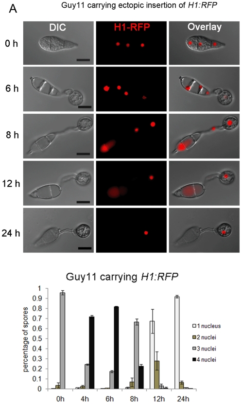
Figure 1 Nuclear degeneration occurs during appressorium development in M. oryzae. (A). Upper panel: time course live cell imaging experiment showing nuclear division and nuclear degeneration during appressorium development in M. oryzae. Guy11 conidia expressing H1:RFP were examined by epifluorescence microscopy at indicated time points during appressorium development. Lower panel: bar charts showing the percentage of spore germlings in Guy11 containing between 0 and 4 nuclei (mean ± SD, n>100, triple replications) during a timecourse of appressorium development. Scale bar = 10 µm.
Figure 1 Nuclear degeneration occurs during appressorium development in M. oryzae. (A). Upper panel: time course live cell imaging experiment showing nuclear division and nuclear degeneration during appressorium development in M. oryzae. Guy11 conidia expressing H1:RFP were examined by epifluorescence microscopy at indicated time points during appressorium development. Lower panel: bar charts showing the percentage of spore germlings in Guy11 containing between 0 and 4 nuclei (mean ± SD, n>100, triple replications) during a timecourse of appressorium development. Scale bar = 10 µm.CC BY · PubMed Central
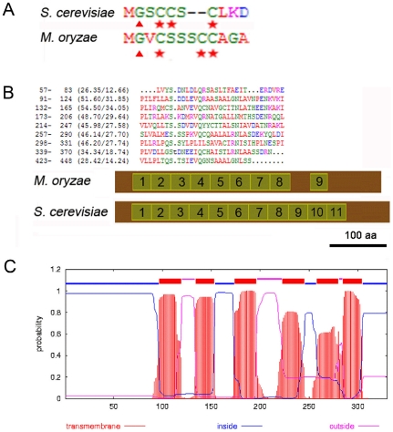
Figure 2 Bioinformatic identification of components of the S. cerevisiae PMN pathway in M. oryzae. (A) Alignment of acylation modification amino acids in the N-terminus of Vac8p from M. oryzae and S. cerevisiae. Triangles indicate putative myristoylation modification site (Gly) and stars indicate palmitoylation modification sites (Cys) that have been shown to be important for localisation and function of Vac8p in S. cerevisiae. Similar acylation modification sites are present in MoVac8p. (B) ARM repeat organization in MoVac8p is distinct from that of S. cerevisiae Vac8p. Upper panel: alignment of 9 ARM repeats from M. oryzae Vac8p. A dot indicates a space. Lower panel: Comparison of distribution of the ARM repeats in Vac8p between M. oryzae and S. cerevisiae. Scale bar indicate 100 amino acids. (C) MoTsc13p shares the same topology of six tramsmembrane domains as S. cerevisiae Tsc13p.
Figure 2 Bioinformatic identification of components of the S. cerevisiae PMN pathway in M. oryzae. (A) Alignment of acylation modification amino acids in the N-terminus of Vac8p from M. oryzae and S. cerevisiae. Triangles indicate putative myristoylation modification site (Gly) and stars indicate palmitoylation modification sites (Cys) that have been shown to be important for localisation and function of Vac8p in S. cerevisiae. Similar acylation modification sites are present in MoVac8p. (B) ARM repeat organization in MoVac8p is distinct from that of S. cerevisiae Vac8p. Upper panel: alignment of 9 ARM repeats from M. oryzae Vac8p. A dot indicates a space. Lower panel: Comparison of distribution of the ARM repeats in Vac8p between M. oryzae and S. cerevisiae. Scale bar indicate 100 amino acids. (C) MoTsc13p shares the same topology of six tramsmembrane domains as S. cerevisiae Tsc13p.CC BY · PubMed Central
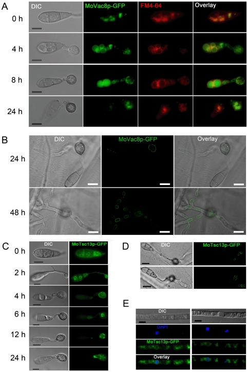
Figure 3 Sub-cellular localisation of MoVac8p and MoTsc13p in Magnaporthe oryzae. (A) MoVac8p-GFP is localised at the vacuolar membrane in both conidia and appressoria. FM4–64 was used to stain vacuoles and endosomes of Guy11, expressing MoVAC8:GFP. Conidia were collected and resuspended in 50 µl of CM with 7.5 µM FM4–64. Appressorium development was observed on coverslips at indicated time points. (B) MoVac8p-GFP is localised on the vacuolar membrane in penetration pegs and invasive hyphae. Penetration of onion epidermis was examined at indicated time points. (C) Tsc13p-GFP is associated with of perinuclear and peripheral ER in both conidium and appressorium. (D) Tsc13p-GFP is associated with perinuclear and peripheral ER in penetration pegs and invasive hyphae. Images were taken from onion epidermis infected with Guy11 expressing MoTSC13:GFP at 24 hpi. (E) MoTsc13p-GFP is localised at the perinuclear and peripheral ER in hyphae grown in CM. DAPI was used to stain nuclei of Guy11 expressing MoTSC13:GFP. Scale bar = 10 µm.
Figure 3 Sub-cellular localisation of MoVac8p and MoTsc13p in Magnaporthe oryzae. (A) MoVac8p-GFP is localised at the vacuolar membrane in both conidia and appressoria. FM4–64 was used to stain vacuoles and endosomes of Guy11, expressing MoVAC8:GFP. Conidia were collected and resuspended in 50 µl of CM with 7.5 µM FM4–64. Appressorium development was observed on coverslips at indicated time points. (B) MoVac8p-GFP is localised on the vacuolar membrane in penetration pegs and invasive hyphae. Penetration of onion epidermis was examined at indicated time points. (C) Tsc13p-GFP is associated with of perinuclear and peripheral ER in both conidium and appressorium. (D) Tsc13p-GFP is associated with perinuclear and peripheral ER in penetration pegs and invasive hyphae. Images were taken from onion epidermis infected with Guy11 expressing MoTSC13:GFP at 24 hpi. (E) MoTsc13p-GFP is localised at the perinuclear and peripheral ER in hyphae grown in CM. DAPI was used to stain nuclei of Guy11 expressing MoTSC13:GFP. Scale bar = 10 µm.CC BY · PubMed Central
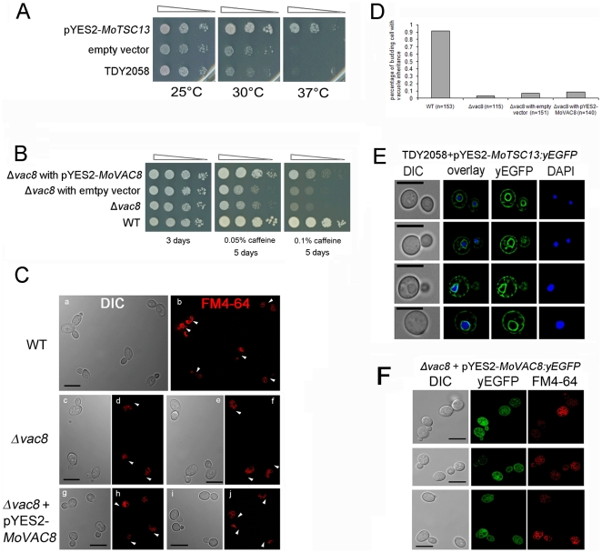
Figure 4 MoTSC13 is functionally equivalent to S. cerevisiae TSC13, while MoVAC8 only partially substitutes for VAC8. (A) MoTSC13 complemented the temperature-sensitive lethality of the yeast strain TDY2058 (double mutant tsc13-1 Δelo). Yeast tsc13-1 Δelo mutants were transformed either with empty vector or a plasmid expressing MoTSC13 cDNA under control of the galactose-inducible GAL1 promoter. Cells were grown in YPDA overnight, normalized to and subjected to 10-fold serial dilutions, spotted onto SD+Gal plates and incubated at 26°C, 30°C or 37°C for 3 days prior to photographing. The experiments were carried out in triplicate, examining two independent yeast transformants. (B) MoVAC8 complemented the caffeine sensitivity of yeast strain BY4741 vac8Δ::KANMX4. Yeast vac8Δ::KANMX4 mutants were transformed with empty vector or a plasmid expressing MoVAC8 cDNA under the control of the GAL1 promoter. Cells were grown in YPDA overnight, normalized and subjected to 10-fold serial dilutions, spotted onto SD+Gal plates containing either 0.05% or 0.1% caffeine, and incubated at 26°C for the indicated period of time. The experiments were carried out in triplicate, examining two independent yeast transformants. (C) MoVAC8 does not complement vacuole morphology and vacuole inheritance defects of yeast mutant vac8Δ::KANMX4. FM4–64 was used to stain cells of S. cerevisiae, vac8Δ::KANMX4 and vac8Δ::KANMX4 strain, expressing MoVAC8 by pulse-chase labelling. Arrows indicates presence (b) or absence (d, f, h, j) of segregating vacuoles. (D) MoVAC8 failed to complement vacuole inheritance defects of yeast mutant vac8Δ::KANMX4 during budding. Pulse-chase labelled S. cerevisiae cells by FM4–64 were counted for the proportion of daughter cells carrying vacuoles inherited from the mother cell during budding. (N indicates the total number of cells counted). (E) MoTSC13p localises to the perinuclear and peripheral ER membrane in S. cerevisiae. yEGFP tagged MoTSC13p was expressed in yeast strain TDY2058 under control of the GAL1 promoter in plasmid pYES2. Cells were stained with DAPI to visualise nuclei. (F) MoVAC8p failed to accumulate at the vacuolar membrane in S. cerevisiae. yEGFP tagged MoVAC8p was expressed in yeast strain vac8Δ::KANMX4 under control of GAL1p promoter in plasmid pYES2.
Figure 4 MoTSC13 is functionally equivalent to S. cerevisiae TSC13, while MoVAC8 only partially substitutes for VAC8. (A) MoTSC13 complemented the temperature-sensitive lethality of the yeast strain TDY2058 (double mutant tsc13-1 Δelo). Yeast tsc13-1 Δelo mutants were transformed either with empty vector or a plasmid expressing MoTSC13 cDNA under control of the galactose-inducible GAL1 promoter. Cells were grown in YPDA overnight, normalized to and subjected to 10-fold serial dilutions, spotted onto SD+Gal plates and incubated at 26°C, 30°C or 37°C for 3 days prior to photographing. The experiments were carried out in triplicate, examining two independent yeast transformants. (B) MoVAC8 complemented the caffeine sensitivity of yeast strain BY4741 vac8Δ::KANMX4. Yeast vac8Δ::KANMX4 mutants were transformed with empty vector or a plasmid expressing MoVAC8 cDNA under the control of the GAL1 promoter. Cells were grown in YPDA overnight, normalized and subjected to 10-fold serial dilutions, spotted onto SD+Gal plates containing either 0.05% or 0.1% caffeine, and incubated at 26°C for the indicated period of time. The experiments were carried out in triplicate, examining two independent yeast transformants. (C) MoVAC8 does not complement vacuole morphology and vacuole inheritance defects of yeast mutant vac8Δ::KANMX4. FM4–64 was used to stain cells of S. cerevisiae, vac8Δ::KANMX4 and vac8Δ::KANMX4 strain, expressing MoVAC8 by pulse-chase labelling. Arrows indicates presence (b) or absence (d, f, h, j) of segregating vacuoles. (D) MoVAC8 failed to complement vacuole inheritance defects of yeast mutant vac8Δ::KANMX4 during budding. Pulse-chase labelled S. cerevisiae cells by FM4–64 were counted for the proportion of daughter cells carrying vacuoles inherited from the mother cell during budding. (N indicates the total number of cells counted). (E) MoTSC13p localises to the perinuclear and peripheral ER membrane in S. cerevisiae. yEGFP tagged MoTSC13p was expressed in yeast strain TDY2058 under control of the GAL1 promoter in plasmid pYES2. Cells were stained with DAPI to visualise nuclei. (F) MoVAC8p failed to accumulate at the vacuolar membrane in S. cerevisiae. yEGFP tagged MoVAC8p was expressed in yeast strain vac8Δ::KANMX4 under control of GAL1p promoter in plasmid pYES2.CC BY · PubMed Central
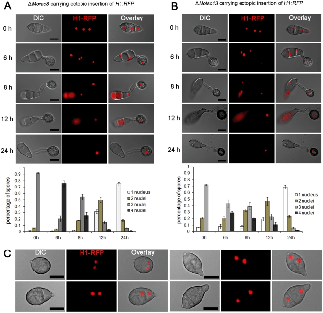
Figure 5 MoVAC8 and MoTSC13 are not required for nuclear degeneration in M. oryzae. (A) Nuclear degeneration during appressorium development occurs independently of MoVAC8. Upper panel: time course live cell images showing nuclear division and nuclear degeneration during appressorium development in M. oryzae ΔMovac8 deletion mutants. ΔMovac8 expressing H1:RFP were examined by epifluorescence microscopy at indicated time points during appressorium development. Lower panel: time series of bar charts showing the percentage of spore germlings of ΔMovac8 containing between 0 and 4 nuclei (mean ± SD, n>100, triple replications). (B) Nuclear degeneration during appressorium development occurs independently of MoTSC13. Upper panel: time course live cell images showing nuclear division and nuclear degeneration during appressorium development in M. oryzae MoTSC13 deletion mutants. ΔMotsc13 expressing H1:RFP were examined by epifluorescence microscopy at indicated time points during appressorium development. Lower panel: time series of bar charts showing the percentage of spores in ΔMotsc13 containing between 0 and 4 nuclei (mean ± SD, n>100, triple replications). (C) Micrographs showing abnormal conidia of ΔMotsc13 containing two nuclei. Scale bar = 10 µm.
Figure 5 MoVAC8 and MoTSC13 are not required for nuclear degeneration in M. oryzae. (A) Nuclear degeneration during appressorium development occurs independently of MoVAC8. Upper panel: time course live cell images showing nuclear division and nuclear degeneration during appressorium development in M. oryzae ΔMovac8 deletion mutants. ΔMovac8 expressing H1:RFP were examined by epifluorescence microscopy at indicated time points during appressorium development. Lower panel: time series of bar charts showing the percentage of spore germlings of ΔMovac8 containing between 0 and 4 nuclei (mean ± SD, n>100, triple replications). (B) Nuclear degeneration during appressorium development occurs independently of MoTSC13. Upper panel: time course live cell images showing nuclear division and nuclear degeneration during appressorium development in M. oryzae MoTSC13 deletion mutants. ΔMotsc13 expressing H1:RFP were examined by epifluorescence microscopy at indicated time points during appressorium development. Lower panel: time series of bar charts showing the percentage of spores in ΔMotsc13 containing between 0 and 4 nuclei (mean ± SD, n>100, triple replications). (C) Micrographs showing abnormal conidia of ΔMotsc13 containing two nuclei. Scale bar = 10 µm.CC BY · PubMed Central
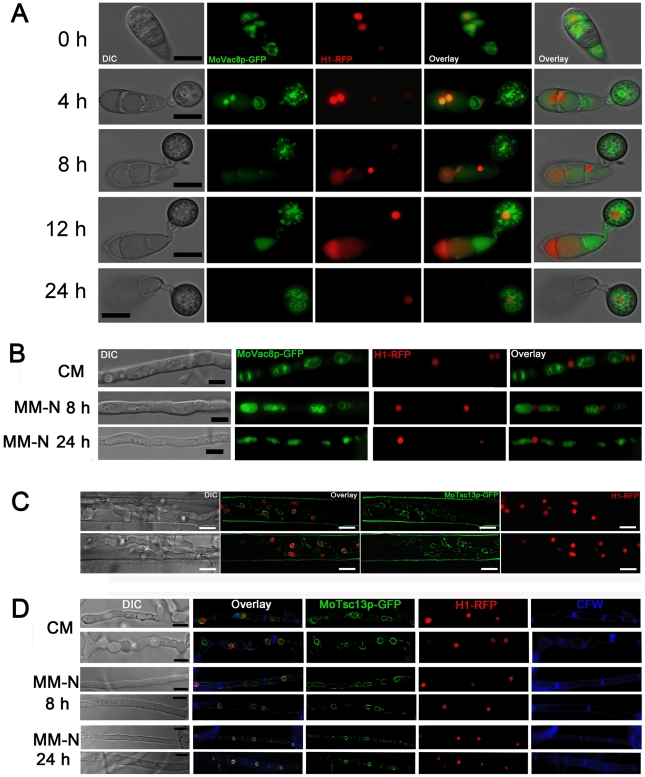
Figure 6 Nuclear-vacuolar (NV) junctions are not present in M. oryzae during appressorium development or starvation stress. (A) Time course live cell imaging of appressorium development in M. oryzae expressing both MoVAC:GFP and H1:RFP. (B) Micrographs of hyphae of M. oryzae expressing both MoVAC:GFP and H1:RFP. M. oryzae was grown in nutrient-rich medium (CM) or nitrogen stress minimal medium (MM-N) for the indicated period of times, and hyphae collected for epifluorescence microscopy. Hyphae cultured in CM for 40 h were washed with distilled water, before being transferred into MM-N. (C) MoTsc13p is associated with perinuclear and peripheral ER membrane in invasive hyphae. M. oryzae expressing both MoTSC13:GFP and H1:RFP was inoculated onto rice sheath epidermis for 36 h before epifluorescence microscopy. (D) Distribution of MoTsc13p-GFP is not affected by environmental nutritional status. Hyphae of M. oryzae expressing both MoTSC13:GFP and H1:RFP were grown in CM or MM-N for epifluorescence microscopy. Pictures shown are representatives of at least 30 different hyphae cell compartments analyzed for each time point. Scale bar = 10 µm.
Figure 6 Nuclear-vacuolar (NV) junctions are not present in M. oryzae during appressorium development or starvation stress. (A) Time course live cell imaging of appressorium development in M. oryzae expressing both MoVAC:GFP and H1:RFP. (B) Micrographs of hyphae of M. oryzae expressing both MoVAC:GFP and H1:RFP. M. oryzae was grown in nutrient-rich medium (CM) or nitrogen stress minimal medium (MM-N) for the indicated period of times, and hyphae collected for epifluorescence microscopy. Hyphae cultured in CM for 40 h were washed with distilled water, before being transferred into MM-N. (C) MoTsc13p is associated with perinuclear and peripheral ER membrane in invasive hyphae. M. oryzae expressing both MoTSC13:GFP and H1:RFP was inoculated onto rice sheath epidermis for 36 h before epifluorescence microscopy. (D) Distribution of MoTsc13p-GFP is not affected by environmental nutritional status. Hyphae of M. oryzae expressing both MoTSC13:GFP and H1:RFP were grown in CM or MM-N for epifluorescence microscopy. Pictures shown are representatives of at least 30 different hyphae cell compartments analyzed for each time point. Scale bar = 10 µm.CC BY · PubMed Central
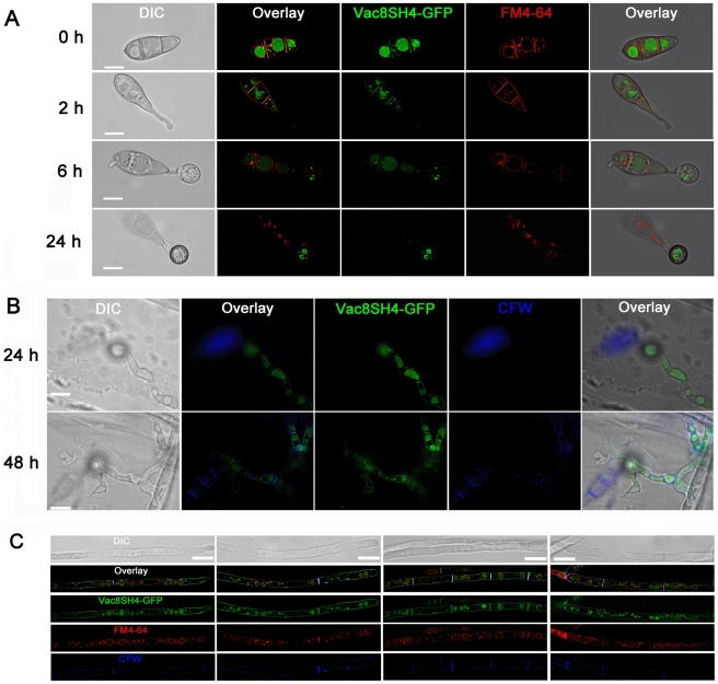
Figure 7 The N-terminus of MoVAC8p contains a SH4 domain. (A) SH4:GFP fusion proteins are anchored at the vacuole, septa and the plasma membrane in both conidia and appressoria. The first 21 amino acid residues of MoVAC8p were fused into the N-terminus of GFP to produce SH4:GFP and subcellular localisation of the GFP fusion analysed by epifluorescence microscopy. Guy11 conidia expressing SH4:GFP fusion proteins were stained with FM4–64 and allowed to germinate and undergo appressorium development. SH4:GFP fusions colocalise with the FM4–64-stained vacuole, septa and plasma membrane. (B) SH4:GFP fusion proteins associate with the vacuole, septa and the plasma membrane in penetration pegs and invasive hyphae. Penetration of onion epidermis was examined at indicated time points and Calcofluor White (CFW) used to stain the cell wall. SH4:GFP fusion proteins colocalise with the vacuole (as seen in the bright field image) and CFW-stained cell wall. (C) SH4:GFP fusion proteins were targeted to the vacuole, septa and the plasma membrane in vegetative hyphae. Vegetative hyphae expressing SH4:GFP fusion proteins were grown in CM for 24 h, and 200 µl of the cultures incubated with 7.5 µM FM4–64 at 26°C for 1 h, followed by CFW staining. In vegetative hyphae, SH4:GFP fusion proteins also colocalise with FM4–64- stained vacuoles and CFW-stained septa and cell walls. Scale bar = 10 µm.
Figure 7 The N-terminus of MoVAC8p contains a SH4 domain. (A) SH4:GFP fusion proteins are anchored at the vacuole, septa and the plasma membrane in both conidia and appressoria. The first 21 amino acid residues of MoVAC8p were fused into the N-terminus of GFP to produce SH4:GFP and subcellular localisation of the GFP fusion analysed by epifluorescence microscopy. Guy11 conidia expressing SH4:GFP fusion proteins were stained with FM4–64 and allowed to germinate and undergo appressorium development. SH4:GFP fusions colocalise with the FM4–64-stained vacuole, septa and plasma membrane. (B) SH4:GFP fusion proteins associate with the vacuole, septa and the plasma membrane in penetration pegs and invasive hyphae. Penetration of onion epidermis was examined at indicated time points and Calcofluor White (CFW) used to stain the cell wall. SH4:GFP fusion proteins colocalise with the vacuole (as seen in the bright field image) and CFW-stained cell wall. (C) SH4:GFP fusion proteins were targeted to the vacuole, septa and the plasma membrane in vegetative hyphae. Vegetative hyphae expressing SH4:GFP fusion proteins were grown in CM for 24 h, and 200 µl of the cultures incubated with 7.5 µM FM4–64 at 26°C for 1 h, followed by CFW staining. In vegetative hyphae, SH4:GFP fusion proteins also colocalise with FM4–64- stained vacuoles and CFW-stained septa and cell walls. Scale bar = 10 µm.CC BY · PubMed Central
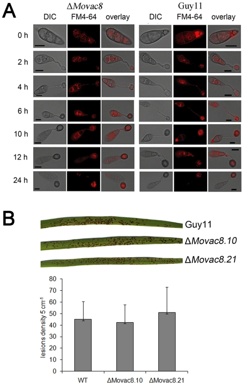
Figure 8 Phenotypic analysis of ΔMovac8 mutants. (A) MoVAC8 is not involved in vacuole movement from the conidium into the appressorium. FM4–64 was used to stain endosomes and vacuoles in conidia to visualize movement of vacuoles during appressorium development. (B) Rice blast infection assay of ΔMovac8 mutants on rice leaves. The virulence of ΔMovac8 is similar to that of strain Guy11. Lesion density represents the lesion number per 5 cm of infected leaf area on rice CO-39 seedlings (n>40).
Figure 8 Phenotypic analysis of ΔMovac8 mutants. (A) MoVAC8 is not involved in vacuole movement from the conidium into the appressorium. FM4–64 was used to stain endosomes and vacuoles in conidia to visualize movement of vacuoles during appressorium development. (B) Rice blast infection assay of ΔMovac8 mutants on rice leaves. The virulence of ΔMovac8 is similar to that of strain Guy11. Lesion density represents the lesion number per 5 cm of infected leaf area on rice CO-39 seedlings (n>40).CC BY · PubMed Central
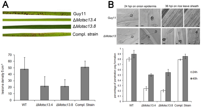
Figure 9 MoTSC13 is required for full virulence of M. oryzae. (A) Targeted deletion of MoTSC13 results in reduced pathogenicity on rice CO-39. Lesion density and size was severely reduced in ΔMotsc13 rice infections compared to Guy11. Full pathogenicity of ΔMotsc13 mutants was restored by introduction of MoTSC13:GFP (Compl.). Lesion density represents the lesion number per 5 cm of infected leaf area (n>40). (B) MoTSC13 is necessary for penetration peg formation and invasive hyphae expansion during in planta growth. Scale bar = 10 µm.
Figure 9 MoTSC13 is required for full virulence of M. oryzae. (A) Targeted deletion of MoTSC13 results in reduced pathogenicity on rice CO-39. Lesion density and size was severely reduced in ΔMotsc13 rice infections compared to Guy11. Full pathogenicity of ΔMotsc13 mutants was restored by introduction of MoTSC13:GFP (Compl.). Lesion density represents the lesion number per 5 cm of infected leaf area (n>40). (B) MoTSC13 is necessary for penetration peg formation and invasive hyphae expansion during in planta growth. Scale bar = 10 µm.CC BY · PubMed Central
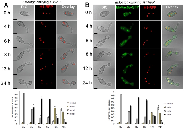
Figure 10 Macroautophagy is necessary for nuclear degeneration during appressorium development in M. oryzae. (A) The Macroautophagy gene MoATG1 is required for nuclear degeneration. Upper panel: time course live cell images showing nuclear division and nuclear degeneration during appressorium development in a M. oryzae ΔMoatg1 mutant. ΔMoatg1 conidia expressing H1:RFP were examined by epifluorescence microscopy at indicated time points. Lower panel: time series of bar charts showing the percentage of spores in ΔMoatg1 containing 0 to 4 nuclei (mean ± SD, n>100, triple replications). (B) The Macroautophagy core gene MoATG4 is required for nuclear degeneration. Upper panel: time course live cell images showing nuclear division and nuclear degeneration during appressorium development in M. oryzae ΔMoatg4 mutant. ΔMoatg4 conidia expressing H1:RFP were examined by epifluorescence microscopy at indicated time points during appressorium development. Lower panel: time series of bar charts showing the percentage of spores in ΔMoatg4 containing 0 to 4 nuclei (mean±SD, n>100, triple replications). Scale bar = 10 µm.
Figure 10 Macroautophagy is necessary for nuclear degeneration during appressorium development in M. oryzae. (A) The Macroautophagy gene MoATG1 is required for nuclear degeneration. Upper panel: time course live cell images showing nuclear division and nuclear degeneration during appressorium development in a M. oryzae ΔMoatg1 mutant. ΔMoatg1 conidia expressing H1:RFP were examined by epifluorescence microscopy at indicated time points. Lower panel: time series of bar charts showing the percentage of spores in ΔMoatg1 containing 0 to 4 nuclei (mean ± SD, n>100, triple replications). (B) The Macroautophagy core gene MoATG4 is required for nuclear degeneration. Upper panel: time course live cell images showing nuclear division and nuclear degeneration during appressorium development in M. oryzae ΔMoatg4 mutant. ΔMoatg4 conidia expressing H1:RFP were examined by epifluorescence microscopy at indicated time points during appressorium development. Lower panel: time series of bar charts showing the percentage of spores in ΔMoatg4 containing 0 to 4 nuclei (mean±SD, n>100, triple replications). Scale bar = 10 µm.CC BY · PubMed Central

Verification

Claims the paper makes

The claims and statistics of this paper haven’t been checked yet.

Run the check — free, no account needed. It reads the open-access text and checks the sources it cites.

A few free checks a day · papers already verified open instantly · results are public

Claims other papers make about this one

Each is checked against this paper’s own text: does it say what the citing paper says it does?

Self-check

Percentages, sums and reported differences re-computed from the paper’s own numbers.

The self-check needs the paper’s full text.

Citations

Cites

No references could be resolved yet.

Research works printed but not linked: 52

  1. RA Wilson, 2009, Under pressure: investigating the biology of plant infection by Magnaporthe oryzae., Nat Rev Microbiol, 7, 185, 10.1038/nrmicro2032 · DOI
  2. JE Hamer, 1988, A mechanism for surface attachment in spores of a plant pathogenic fungus., Science, 239, 288, 10.1126/science.239.4837.288 · DOI
  3. NJ Talbot, 2003, On the trail of a cereal killer: Exploring the biology of Magnaporthe grisea., Annu Rev Microbiol, 57, 177, 10.1146/annurev.micro.57.030502.090957 · DOI
  4. RA Dean, 1997, Signal pathways and appressorium morphogenesis., Annu Rev Phytopathol, 35, 211, 10.1146/annurev.phyto.35.1.211 · DOI
  5. JR Xu, 2000, Map kinases in fungal pathogens., Fungal Genet Biol, 31, 137, 10.1006/fgbi.2000.1237 · DOI
  6. JC de Jong, 1997, Glycerol generates turgor in rice blast., Nature, 389, 244, 10.1038/38418 · DOI
  7. C Veneault-Fourrey, 2006, Autophagic fungal cell death is necessary for infection by the rice blast fungus., Science, 312, 580, 10.1126/science.1124550 · DOI
  8. KP Dixon, 1999, Independent signaling pathways regulate cellular turgor during hyperosmotic stress and appressorium-mediated plant infection by Magnaporthe grisea., Plant Cell, 11, 2045, 10.1105/tpc.11.10.2045 · DOI
  9. E Thines, 2000, MAP kinase and protein kinase A-dependent mobilization of triacylglycerol and glycogen during appressorium turgor generation by Magnaporthe grisea., Plant Cell, 12, 1703
  10. DG Saunders, 2010, Cell cycle-mediated regulation of plant infection by the rice blast fungus., Plant Cell, 22, 497, 10.1105/tpc.109.072447 · DOI
  11. MJ Kershaw, 2009, Genome-wide functional analysis reveals that infection-associated fungal autophagy is necessary for rice blast disease., Proc Natl Acad Sci U S A, 106, 15967, 10.1073/pnas.0901477106 · DOI
  12. XH Liu, 2007, Involvement of a Magnaporthe grisea serine/threonine kinase gene, MgATG1, in appressorium turgor and pathogenesis., Eukaryot Cell, 6, 997, 10.1128/EC.00011-07 · DOI
  13. JP Lu, 2009, An autophagy gene, MgATG5, is required for cell differentiation and pathogenesis in Magnaporthe oryzae., Curr Genet, 55, 461, 10.1007/s00294-009-0259-5 · DOI
  14. TB Liu, 2010, The cysteine protease MoAtg4 interacts with MoAtg8 and is required for differentiation and pathogenesis in Magnaporthe oryzae., Autophagy, 6, 74, 10.4161/auto.6.1.10438 · DOI
  15. X Pan, 2000, Nucleus-vacuole junctions in Saccharomyces cerevisiae are formed through the direct interaction of Vac8p with Nvj1p., Mol Biol Cell, 11, 2445, 10.1091/mbc.11.7.2445 · DOI
  16. P Roberts, 2003, Piecemeal microautophagy of nucleus in Saccharomyces cerevisiae., Mol Biol Cell, 14, 129, 10.1091/mbc.E02-08-0483 · DOI · Paper page
  17. R Dawaliby, 2010, A Microautophagy of the Nucleus Coincides with a Vacuolar Diffusion Barrier at Nuclear-Vacuolar Junctions., Mol Biol Cell, 21, 4173, 10.1091/mbc.E09-09-0782 · DOI
  18. E Kvam, 2006, Nucleus-vacuole junctions in yeast: anatomy of a membrane contact site., Biochem Soc Trans, 34, 340, 10.1042/BST0340340 · DOI
  19. E Kvam, 2004, Nvj1p is the outer-nuclear-membrane receptor for oxysterol-binding protein homolog Osh1p in Saccharomyces cerevisiae., J Cell Sci, 117, 4959, 10.1242/jcs.01372 · DOI
  20. R Krick, 2008, Piecemeal microautophagy of the nucleus requires the core macroautophagy genes., Mol Biol Cell, 19, 4492, 10.1091/mbc.E08-04-0363 · DOI
  21. E Kvam, 2007, Nucleus-vacuole junctions and piecemeal microautophagy of the nucleus in S. cerevisiae., Autophagy, 3, 85, 10.4161/auto.3586 · DOI
  22. E Kvam, 2005, Targeting of Tsc13p to nucleus-vacuole junctions: a role for very-long-chain fatty acids in the biogenesis of microautophagic vesicles., Mol Biol Cell, 16, 3987, 10.1091/mbc.E05-04-0290 · DOI
  23. D Fleckenstein, 1998, Yel013p (Vac8p), an armadillo repeat protein related to plakoglobin and importin alpha is associated with the yeast vacuole membrane., J Cell Sci, 111, 3109, 10.1242/jcs.111.20.3109 · DOI
  24. YX Wang, 2001, Fusion of docked membranes requires the armadillo repeat protein Vac8p., J Biol Chem, 276, 35133, 10.1074/jbc.M103937200 · DOI
  25. F Tang, 2006, Vac8p, an armadillo repeat protein, coordinates vacuole inheritance with multiple vacuolar processes., Traffic, 7, 1368, 10.1111/j.1600-0854.2006.00458.x · DOI
  26. Y Peng, 2006, Palmitoylation plays a role in targeting Vac8p to specific membrane subdomains., Traffic, 7, 1378, 10.1111/j.1600-0854.2006.00472.x · DOI
  27. X Pan, 1998, YEB3/VAC8 encodes a myristylated armadillo protein of the Saccharomyces cerevisiae vacuolar membrane that functions in vacuole fusion and inheritance., J Cell Sci, 111, 2137, 10.1242/jcs.111.15.2137 · DOI
  28. M Peifer, 1994, A repeating amino acid motif shared by proteins with diverse cellular roles., Cell, 76, 789, 10.1016/0092-8674(94)90353-0 · DOI
  29. YX Wang, 1998, Vac8p, a vacuolar protein with armadillo repeats, functions in both vacuole inheritance and protein targeting from the cytoplasm to vacuole., J Cell Biol, 140, 1063, 10.1083/jcb.140.5.1063 · DOI
  30. YX Wang, 1996, Multiple classes of yeast mutants are defective in vacuole partitioning yet target vacuole proteins correctly., Mol Biol Cell, 7, 1375, 10.1091/mbc.7.9.1375 · DOI
  31. K Subramanian, 2006, Palmitoylation determines the function of Vac8 at the yeast vacuole., J Cell Sci, 119, 2477, 10.1242/jcs.02972 · DOI
  32. MD Resh, 1993, Interaction of tyrosine kinase oncoproteins with cellular membranes., Biochim Biophys Acta, 1155, 307
  33. MR Fry, 2006, Early and late molecular events of glucose-induced pexophagy in Pichia pastoris require Vac8., Autophagy, 2, 280
  34. M Oku, 2006, Role of Vac8 in formation of the vacuolar sequestering membrane during micropexophagy., Autophagy, 2, 272, 10.4161/auto.3135 · DOI
  35. V Veses, 2009, Vacuole inheritance regulates cell size and branching frequency of Candida albicans hyphae., Mol Microbiol, 71, 505, 10.1111/j.1365-2958.2008.06545.x · DOI
  36. CJ Barelle, 2006, Candida albicans VAC8 is required for vacuolar inheritance and normal hyphal branching., Eukaryot Cell, 5, 359, 10.1128/EC.5.2.359-367.2006 · DOI
  37. SD Kohlwein, 2001, Tsc13p is required for fatty acid elongation and localizes to a novel structure at the nuclear-vacuolar interface in Saccharomyces cerevisiae., Mol Cell Biol, 21, 109, 10.1128/MCB.21.1.109-125.2001 · DOI
  38. S Paul, 2007, A six-membrane-spanning topology for yeast and Arabidopsis Tsc13p, the enoyl reductases of the microsomal fatty acid elongating system., J Biol Chem, 282, 19237, 10.1074/jbc.M701774200 · DOI
  39. RW Weber, 2001, The vacuole as central element of the lytic system and sink for lipid droplets in maturing appressoria of Magnaporthe grisea., Protoplasma, 216, 101, 10.1007/BF02680137 · DOI
  40. K Gable, 2004, Functional characterization of the Arabidopsis thaliana orthologue of Tsc13p, the enoyl reductase of the yeast microsomal fatty acid elongating system., J Exp Bot, 55, 543, 10.1093/jxb/erh061 · DOI
  41. WQ Song, 2009, Characterization of two cotton cDNAs encoding trans-2-enoyl-CoA reductase reveals a putative novel NADPH-binding motif., J Exp Bot, 60, 1839, 10.1093/jxb/erp057 · DOI
  42. MD Resh, 1999, Fatty acylation of proteins: new insights into membrane targeting of myristoylated and palmitoylated proteins., Biochim Biophys Acta, 1451, 1, 10.1016/S0167-4889(99)00075-0 · DOI
  43. K Kuranda, 2006, Investigating the caffeine effects in the yeast Saccharomyces cerevisiae brings new insight into the connection between TOR, PKC and Ras/cAMP signalling pathways., Molec Microbiol, 61, 1147, 10.1111/j.1365-2958.2006.05300.x · DOI
  44. J-y Shoji, 2010, Macroautophagy-Mediated Degradation of Whole Nuclei in the Filamentous Fungus Aspergillus oryzae., PLoS ONE, 5, e15650, 10.1371/journal.pone.0015650 · DOI · Paper page
  45. H Leung, 1988, Magnaporthe grisea (Pyricularia grisea), the blast fungus., Adv Plant Pathol, 6, 175, 10.1016/B978-0-12-033706-4.50015-3 · DOI
  46. NJ Talbot, 1993, Identification and characterization of MPG1, a gene involved in pathogenicity from the rice blast fungus Magnaporthe grisea., Plant Cell, 5, 1575, 10.1105/tpc.5.11.1575 · DOI
  47. NL Catlett, 2003, Split-Marker Recombination for Efficient Targeted Deletion of Fungal Genes., Fungal Genet Newsl, 50, 9
  48. J Sweigard, 1997, A series of vectors for fungal transformation., Fungal Genet Newsl, 44, 52
  49. KR Oldenburg, 1997, Recombination-mediated PCR-directed plasmid construction in vivo in yeast., Nucleic Acids Res, 25, 451, 10.1093/nar/25.2.451 · DOI
  50. RJ Howard, 1991, Penetration of hard substrates by a fungus employing enormous turgor pressures., Proc Natl Acad Sci U S A, 88, 11281, 10.1073/pnas.88.24.11281 · DOI
  51. MJ Kershaw, 1998, Complementation of the mpg1 mutant phenotype in Magnaporthe grisea reveals functional relationships between fungal hydrophobins., EMBO J, 17, 3838, 10.1093/emboj/17.14.3838 · DOI
  52. MA Sheff, 2004, Optimized cassettes for fluorescent protein tagging in Saccharomyces cerevisiae., Yeast, 21, 661, 10.1002/yea.1130 · DOI

Cited by

Sorted by citations

Metadata

Authors
He, M., Kershaw, M. J., Soanes, D. M., Xia, Y., & Talbot, N. J.
Venue
PLoS ONE
Published
2012-03-20
Volume
7
Issue
3
Pages
e33270
ISSN
1932-6203
Cited by
40
References
0
Language
en

About these checks

Registry, retraction, Cites and statistics results are computed, not judged, with one exception: a printed reference that no index lists is read once by a model before it counts as a citation error. Claims are extracted by a model and kept only when their quote appears verbatim in the text; each cited source is then checked, with a model’s help, for whether it says what the claim relies on. Those verdicts can be wrong — read the claim’s quote and the cited source before relying on one.

Statistics and arithmetic are recomputed, not judged.

Confidence bands are shown only for fields calibrated on our annotated test set. None is calibrated yet, so no band is shown.

What counts as a claim ›How we measure confidence ›How we check this ›

Are you an author, or do you see a problem with this page? Request a review