

The DOI matches its Crossref record; 34 of the 34 references it cites were found in a registry or an index.
Co-Stimulation of PAFR and CD36 Is Required for oxLDL-Induced Human Macrophages Activation
Open accessAbstract
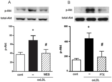
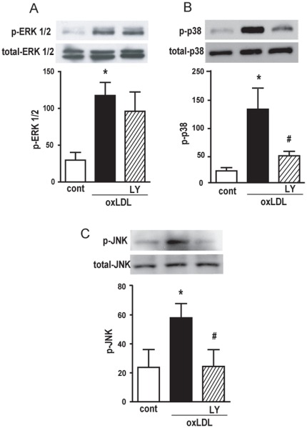
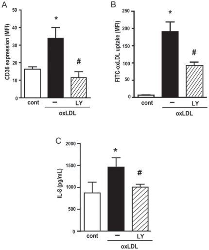
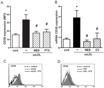
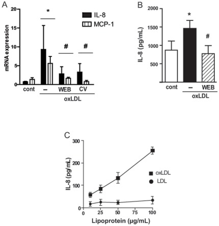
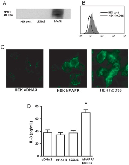
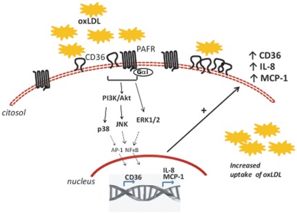
The oxidative process of LDL particles generates molecules which are structurally similar to platelet-activating factor (PAF), and some effects of oxidized LDL (oxLDL) have been shown to be dependent on PAF receptor (PAFR) activation. In a previous study, we showed that PAFR is required for upregulation of CD36 and oxLDL uptake. In the present study we analyzed the molecular mechanisms activated by oxLDL in human macrophages and the contribution of PAFR to this response. Human adherent monocytes/macrophages were stimulated with oxLDL. Uptake of oxLDL and CD36 expression were determined by flow cytometry; MAP kinases and Akt phosphorylation by Western blot; IL-8 and MCP-1 concentration by ELISA and mRNA expression by real-time PCR. To investigate the participation of the PI3K/Akt pathway, Gαi-coupled protein or PAFR, macrophages were treated with LY294002, pertussis toxin or with the PAFR antagonists WEB2170 and CV3988, respectively before addition of oxLDL. It was found that the addition of oxLDL to human monocytes/macrophages activates the PI3K/Akt pathway which in turn activates the MAPK (p38 and JNK). Phosphorylation of Akt requires the engagement of PAFR and a Gαi-coupled protein. The upregulation of CD36 protein and the uptake of oxLDL as well as the IL-8 production are dependent on PI3K/Akt pathway activation. The increased CD36 protein expression is dependent on PAFR and Gαi-coupled protein. Transfection studies using HEK 293t cells showed that oxLDL uptake occurs with either PAFR or CD36, but IL-8 production requires the co-transfection of both PAFR and CD36. These findings show that PAFR has a pivotal role in macrophages response to oxLDL and suggest that pharmacological intervention at the level of PAFR activation might be beneficial in atherosclerosis.
Overview
The claims and statistics of this paper haven’t been checked yet.
Run the check — free, no account needed. It reads the open-access text and checks the sources it cites.
Integrity signals
Registry
Cites
Statistics
Figures
Verification
Claims the paper makes
The claims and statistics of this paper haven’t been checked yet.
Run the check — free, no account needed. It reads the open-access text and checks the sources it cites.
Claims other papers make about this one
Each is checked against this paper’s own text: does it say what the citing paper says it does?
The source backs part of the claim, not all of it — or backs it for a different population, condition or comparison.
In the citing paper
“we performed WB to confirm that AKT phosphorylation was intermediate according to previous studies (P < 0.05 by t-test, Fig. 4A) [19].”
From Matrix stiffness modulates hepatic stellate cell activation into tumor-promoting myofibroblasts via E2F3-dependent signaling and regulates malignant progression (2021) · cites this paper as [19]
Experimental — accuracy not yet measured
Self-check
Percentages, sums and reported differences re-computed from the paper’s own numbers.
The self-check needs the paper’s full text.
Citations
Cites
Analysis of Relative Gene Expression Data Using Real-Time Quantitative PCR and the 2−ΔΔCT Method
Livak, K. J., & Schmittgen, T. D. (2001) · Methods · doi:10.1006/meth.2001.1262 · Cited by 180,860
Glass, C. K., & Witztum, J. L. (2001) · Cell · doi:10.1016/s0092-8674(01)00238-0 · Cited by 3,094
Oxidized LDL Regulates Macrophage Gene Expression through Ligand Activation of PPARγ
Nagy, L., Tontonoz, P., Alvarez, J. G. A., Chen, H.-W., & Evans, R. M. (1998) · Cell · doi:10.1016/s0092-8674(00)81574-3 · Cited by 1,861
Stewart, C. R., Stuart, L. M., Wilkinson, K., van Gils, J. M., Deng, J., Halle, A., Rayner, K. J., Boyer, L., Zhong, R., Frazier, W. A., et al. (2010) · Nature Immunology · doi:10.1038/ni.1836 · Cited by 1,513
INFLAMMATION AND ATHEROSCLEROSIS
Hansson, G. K., Robertson, A.-K. L., & Söderberg‐Nauclér, C. (2006) · Annual Review of Pathology Mechanisms of Disease · doi:10.1146/annurev.pathol.1.110304.100100 · Cited by 1,082
Multiple control of interleukin-8 gene expression
Hoffmann, E., Dittrich-Breiholz, O., Holtmann, H., & Kracht, M. (2002) · Journal of Leukocyte Biology · doi:10.1189/jlb.72.5.847 · Cited by 1,058
Febbraio, M., Podrez, E. A., Smith, J. D., Hajjar, D. P., Hazen, S. L., Hoff, H. F., Sharma, K., & Silverstein, R. L. (2000) · Journal of Clinical Investigation · doi:10.1172/jci9259 · Cited by 1,037
Triantafilou, M., Gamper, F. G. J., Haston, R. M., Mouratis, M. A., Morath, S., Härtung, T., & Triantafilou, K. (2006) · Journal of Biological Chemistry · doi:10.1074/jbc.m602794200 · Cited by 471
Nozaki, S., Kashiwagi, H., Yamashita, S., Nakagawa, T., Kostner, B., Tomiyama, Y., Nakata, A., Ishigami, M., Miyagawa, J.-I., & Kameda-Takemura, K. (1995) · Journal of Clinical Investigation · doi:10.1172/jci118231 · Cited by 340
Frémont, L., Belguendouz, L., & Delpal, S. (1999) · Life Sciences · doi:10.1016/s0024-3205(99)00209-x · Cited by 336
Cited by
Sorted by citations
Oxidized LDL Induces Alternative Macrophage Phenotype through Activation of CD36 and PAFR
Rios, F. J. O., Koga, M. M., Pecenin, M. F., Ferracini, M., Gidlund, M., & Jancar, S. (2013) · Mediators of Inflammation · doi:10.1155/2013/198193 · Cited by 233
Self-citationAre GPCRs Still a Source of New Targets?
Garland, S. L. (2013) · SLAS DISCOVERY · doi:10.1177/1087057113498418 · Cited by 151
Pisonero‐Vaquero, S., Martínez‐Ferreras, Á., García‐Mediavilla, M. V., Martínez‐Flórez, S., Fernández, A., Benet, M., Olcoz, J. L., Jover, R., González‐Gallego, J., & Sánchez‐Campos, S. (2015) · Molecular Nutrition & Food Research · doi:10.1002/mnfr.201400913 · Cited by 128
Role of PI3K in the Progression and Regression of Atherosclerosis
Zhao, Y., Qian, Y., Sun, Z., Shen, X., Cai, Y., Li, L., & Wang, Z. (2021) · Frontiers in Pharmacology · doi:10.3389/fphar.2021.632378 · Cited by 104
Isolation, synthesis and bioactivity studies of phomactin terpenoids
Kuroda, Y., Nicácio, K. D. J., Silva, I., Leger, P. R., Chang, S., Gubiani, J. R., Deflon, V. M., Nagashima, N., Rode, A., Blackford, K., et al. (2018) · Nature Chemistry · doi:10.1038/s41557-018-0084-x · Cited by 86
Self-citation
Metadata
- Authors
- Rios, F. J. O., Koga, M. M., Ferracini, M., & Jancar, S.
- Affiliations
- Universidade de São Paulo (BR)
- Corresponding author
- Rios, Francisco J. O.
- Venue
- PLoS ONE
- Publisher
- Public Library of Science (PLoS)
- Published
- 2012-05-03
- Volume
- 7
- Issue
- 5
- Pages
- e36632
- ISSN
- 1932-6203 (ISSN-L)
- PMC
- PMC3343035
- OpenAlex
- W2056458080
- Cited by
- 51
- References
- 34
- Citations by year
- 2024: 2 · 2023: 5 · 2021: 4 · 2020: 3 · 2019: 3 · 2018: 2 · 2017: 2 · 2016: 6 · 2015: 2 · 2014: 7 · 2013: 11 · 2012: 3
- FWCI
- 3.22
- Type
- journal article
- Language
- en
- Open access
- gold
- doi.org
- Locations
- PLoS ONE (publishedVersion, cc-by)
- Europe PMC (PubMed Central) (publishedVersion, cc-by)
- PubMed Central (publishedVersion, cc-by)
- PubMed (publishedVersion)
- Portuguese National Funding Agency for Science, Research and Technology (RCAAP Project by FCT) (publishedVersion)
- DOAJ (DOAJ: Directory of Open Access Journals) (submittedVersion, cc-by-sa)
- Funders
- Fundação de Amparo à Pesquisa do Estado de São Paulo (06/03982-5)
- Coordenação de Aperfeiçoamento de Pessoal de Nível Superior
- Conselho Nacional de Desenvolvimento Científico e Tecnológico (2006/03982-5)
- Topics
- Life Sciences › Immunology and Microbiology › Immunology › Atherosclerosis and Cardiovascular Diseases
- Life Sciences › Biochemistry, Genetics and Molecular Biology › Biochemistry › Cholesterol and Lipid Metabolism
- Life Sciences › Biochemistry, Genetics and Molecular Biology › Molecular Biology › Sphingolipid Metabolism and Signaling
- Keywords
- platelet-activating factor receptor, CD36, oxidized LDL, macrophage activation, PI3K/Akt signaling, MAPK signaling, interleukin-8, MCP-1, atherosclerosis, macrophage foam cell formation
- MeSH terms
- Chromones, Enzyme Inhibitors, Humans, Lipoproteins, LDL, Macrophage Activation, Macrophages, Models, Biological, Morpholines, Platelet Membrane Glycoproteins, Signal Transduction, Up-Regulation, Interleukin-8, Chemokine CCL2, CD36 Antigens, GTP-Binding Protein alpha Subunits, Gi-Go, Phosphatidylinositol 3-Kinases, Receptors, G-Protein-Coupled, Proto-Oncogene Proteins c-akt, HEK293 Cells
About these checks
Registry, retraction, Cites and statistics results are computed, not judged, with one exception: a printed reference that no index lists is read once by a model before it counts as a citation error. Claims are extracted by a model and kept only when their quote appears verbatim in the text; each cited source is then checked, with a model’s help, for whether it says what the claim relies on. Those verdicts can be wrong — read the claim’s quote and the cited source before relying on one.
Statistics and arithmetic are recomputed, not judged.
Confidence bands are shown only for fields calibrated on our annotated test set. None is calibrated yet, so no band is shown.
What counts as a claim ›How we measure confidence ›How we check this ›
Are you an author, or do you see a problem with this page? Request a review
How to cite
Cite CoChat for the checks on this page, and the paper for its own findings.
- This page
- CoChat. (2026). Citation check: Co-Stimulation of PAFR and CD36 Is Required for oxLDL-Induced Human Macrophages Activation. https://app.cochat.ai/papers/10.1371/journal.pone.0036632
- The paper
- Rios, F. J. O., Koga, M. M., Ferracini, M., & Jancar, S. (2012). Co-Stimulation of PAFR and CD36 Is Required for oxLDL-Induced Human Macrophages Activation. PLoS ONE. https://doi.org/10.1371/journal.pone.0036632
