

The DOI matches its Crossref record; 43 of the 43 references it cites were found in a registry or an index.
Stimulated Human Mast Cells Secrete Mitochondrial Components That Have Autocrine and Paracrine Inflammatory Actions
Open accessAbstract
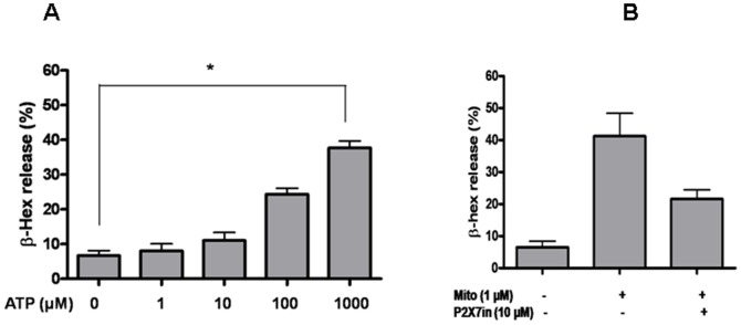
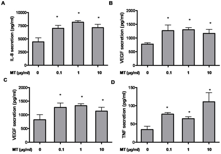
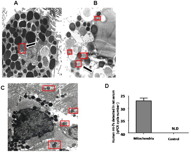
Mast cells are hematopoietically-derived tissue immune cells that participate in acquired and innate immunity, as well as in inflammation through release of many chemokines and cytokines, especially in response to the pro-inflammatory peptide substance P (SP). Inflammation is critical in the pathogenesis of many diseases, but the trigger(s) is often unknown. We investigated if mast cell stimulation leads to secretion of mitochondrial components and whether these could elicit autocrine and/or paracrine inflammatory effects. Here we show that human LAD2 mast cells stimulated by IgE/anti-IgE or by the SP led to secretion of mitochondrial particles, mitochondrial (mt) mtDNA and ATP without cell death. Mitochondria purified from LAD2 cells and, when mitochondria added to mast cells trigger degranulation and release of histamine, PGD(2), IL-8, TNF, and IL-1β. This stimulatory effect is partially inhibited by an ATP receptor antagonist and by DNAse. These results suggest that the mitochondrial protein fraction may also contribute. Purified mitochondria also stimulate IL-8 and vascular endothelial growth factor (VEGF) release from cultured human keratinocytes, and VEGF release from primary human microvascular endothelial cells. In order to investigate if mitochondrial components could be secreted in vivo, we injected rats intraperiotoneally (ip) with compound 48/80, which mimicks the action of SP. Peritoneal mast cells degranulated and mitochondrial particles were documented by transimission electron microscopy outside the cells. We also wished to investigate if mitochondrial components secreted locally could reach the systemic circulation. Administration ip of mtDNA isolated from LAD2 cells in rats was detected in their serum within 4 hr, indicating that extravascular mtDNA could enter the systemic circulation. Secretion of mitochondrial components from stimulated live mast cells may act as "autopathogens" contributing to the pathogenesis of inflammatory diseases and may be used as targets for novel treatments.
Overview
The claims and statistics of this paper haven’t been checked yet.
Run the check — free, no account needed. It reads the open-access text and checks the sources it cites.
Integrity signals
Registry
Cites
Statistics
Figures
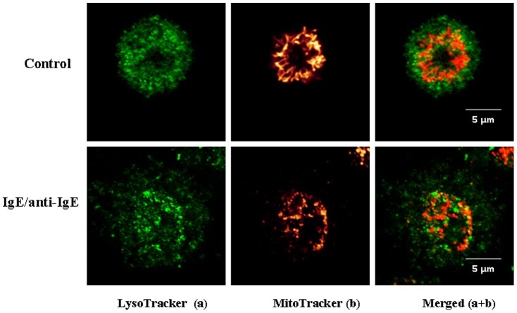
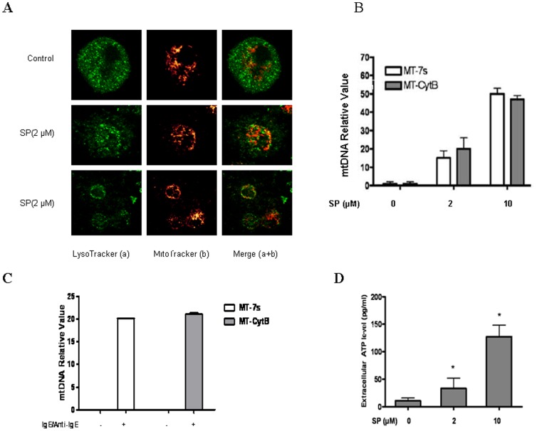
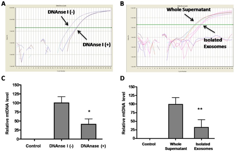
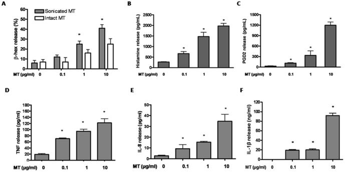
Verification
Claims the paper makes
The claims and statistics of this paper haven’t been checked yet.
Run the check — free, no account needed. It reads the open-access text and checks the sources it cites.
Claims other papers make about this one
Each is checked against this paper’s own text: does it say what the citing paper says it does?
The source backs part of the claim, not all of it — or backs it for a different population, condition or comparison.
In the citing paper
“Different cellular lineages (10–18) may extrude their mitochondria upon activation.”
From Autoantibodies in Systemic Lupus Erythematosus Target Mitochondrial RNA (2019) · cites this paper as [16]
Experimental — accuracy not yet measuredThe cited source says what the claim attributes to it.
In the citing paper
“Various cellular lineages are capable of extruding their mitochondria. Activated mast cells10, T-cells11, eosinophils12, hepatocytes13, neutrophils14–16 and platelets17,18, in addition to damaged organs or tissues7,13,19,20, release extracellular mitochondria.”
From Anti-mitochondrial autoantibodies in systemic lupus erythematosus and their association with disease manifestations (2019) · cites this paper as [10]
Self-check
Percentages, sums and reported differences re-computed from the paper’s own numbers.
The self-check needs the paper’s full text.
Citations
Cites
Circulating mitochondrial DAMPs cause inflammatory responses to injury
Zhang, Q., Raoof, M., Chen, Y., Sumi, Y., Sursal, T., Junger, W., Brohi, K., Itagaki, K., & Hauser, C. J. (2010) · Nature · doi:10.1038/nature08780 · Cited by 3,812
Yang, Y., Li, S., Yang, Q., Shi, Y., Zheng, M., Liu, Y., Chen, F., Song, G., Xu, H., Wan, T., et al. (2014) · Cellular Physiology and Biochemistry · doi:10.1159/000358696 · Cited by 3,263
Wallace, D. C. (2005) · Annual Review of Genetics · doi:10.1146/annurev.genet.39.110304.095751 · Cited by 3,185
DAMPs, PAMPs and alarmins: all we need to know about danger
Bianchi, M. E. (2007) · Journal of Leukocyte Biology · doi:10.1189/jlb.0306164 · Cited by 2,794
Mitochondria: Dynamic Organelles in Disease, Aging, and Development
Chan, D. C. (2006) · Cell · doi:10.1016/j.cell.2006.06.010 · Cited by 1,987
Mast cells in the development of adaptive immune responses
Galli, S. J., Nakae, S., & Tsai, M. M. (2005) · Nature Immunology · doi:10.1038/ni1158 · Cited by 1,280
Mast cells as a source of both preformed and immunologically inducible TNF-α/cachectin
Gordon, J., & Galli, S. J. (1990) · Nature · doi:10.1038/346274a0 · Cited by 984
Mast cell-orchestrated immunity to pathogens
Abraham, S. N., & John, A. L. S. (2010) · Nature reviews. Immunology · doi:10.1038/nri2782 · Cited by 959
Mitochondrial fission in apoptosis
Youle, R. J., & Karbowski, M. (2005) · Nature Reviews Molecular Cell Biology · doi:10.1038/nrm1697 · Cited by 773
Theoharides, T. C., Alysandratos, K., Angelidou, A., Delivanis, D.-A., Sismanopoulos, N., Zhang, B., Asadi, S., Vasiadi, M., Weng, Z., Miniati, A., et al. (2010) · Biochimica et Biophysica Acta (BBA) - Molecular Basis of Disease · doi:10.1016/j.bbadis.2010.12.014 · Cited by 772
Self-citation
Research works printed but not linked: 1
- B Zhang, 2012, Preformed, but not de novo synthesized, TNF secretion from human mast cells is regulated by mitochondria, Int Arch Allergy Immunol
Cited by
Sorted by citations
Haraszti, R. A., Didiot, M., Sapp, E., Leszyk, J., Shaffer, S. A., Rockwell, H. E., Gao, F., Narain, N. R., DiFiglia, M., Kiebish, M. A., et al. (2016) · Journal of Extracellular Vesicles · doi:10.3402/jev.v5.32570 · Cited by 746
Boudreau, L. H., Duchez, A.-C., Cloutier, N., Soulet, D., Martin, N., Bollinger, J., Paré, A., Rousseau, M., Naika, G. S., Lévesque, T., et al. (2014) · Blood · doi:10.1182/blood-2014-05-573543 · Cited by 703
Petra, A. I., Panagiotidou, S., Hatziagelaki, E., Stewart, J. M., Conti, P., & Theoharides, T. C. (2015) · Clinical Therapeutics · doi:10.1016/j.clinthera.2015.04.002 · Cited by 592
Self-citationMast Cells, Mastocytosis, and Related Disorders
Theoharides, T. C., Valent, P., & Akin, C. (2015) · New England Journal of Medicine · doi:10.1056/nejmra1409760 · Cited by 518
Self-citationKey role of CRF in the skin stress response system
Slominski, A. T., Zmijewski, M. A., Zbytek, B., Tobin, D. J., Theoharides, T. C., & Rivier, J. (2013) · Endocrine Reviews · doi:10.1210/er.2012-1092 · Cited by 400
Self-citation
Metadata
- Authors
- Zhang, B., Asadi, S., Weng, Z., Sismanopoulos, N., & Theoharides, T. C.
- Affiliations
- Tufts University (US)
- Corresponding author
- Theoharides, Theoharis C.
- Venue
- PLoS ONE
- Publisher
- Public Library of Science (PLoS)
- Published
- 2012-12-17
- Volume
- 7
- Issue
- 12
- Pages
- e49767
- ISSN
- 1932-6203 (ISSN-L)
- PMC
- PMC3524249
- OpenAlex
- W2169711067
- Cited by
- 121
- References
- 43
- Citations by year
- 2026: 3 · 2025: 8 · 2024: 10 · 2023: 6 · 2022: 7 · 2021: 7 · 2020: 7 · 2019: 10 · 2018: 10 · 2017: 11 · 2016: 11 · 2015: 13 · 2014: 7 · 2013: 11
- FWCI
- 4.27
- Type
- journal article
- Language
- en
- Open access
- gold
- doi.org
- Locations
- PLoS ONE (publishedVersion, cc-by)
- Europe PMC (PubMed Central) (publishedVersion, other-oa)
- PubMed Central (publishedVersion, cc-by)
- PubMed (publishedVersion)
- DOAJ (DOAJ: Directory of Open Access Journals) (submittedVersion, cc-by-sa)
- Figshare (submittedVersion, cc-by)
- Funders
- National Institutes of Health
- National Institute of Arthritis and Musculoskeletal and Skin Diseases (R01AR047652)
- Topics
- Life Sciences › Immunology and Microbiology › Immunology › Mast cells and histamine
- Life Sciences › Biochemistry, Genetics and Molecular Biology › Molecular Biology › Mitochondrial Function and Pathology
- Life Sciences › Immunology and Microbiology › Immunology › Neutrophil, Myeloperoxidase and Oxidative Mechanisms
- Keywords
- mast cells, mitochondrial DNA, ATP, substance P, mast cell degranulation, LAD2 cells, histamine release, prostanoids, interleukin-8, TNF-α, IL-1β, vascular endothelial growth factor, keratinocytes, microvascular endothelial cells, compound 48/80, systemic circulation
- MeSH terms
- Animals, p-Methoxy-N-methylphenethylamine, DNA, Mitochondrial, Extracellular Space, Humans, Inflammation, Male, Mast Cells, Mitochondria, Cell Degranulation, Cytokines, Autocrine Communication, Paracrine Communication, Rats, Exosomes, Receptors, Purinergic P2X7, Inflammasomes
About these checks
Registry, retraction, Cites and statistics results are computed, not judged, with one exception: a printed reference that no index lists is read once by a model before it counts as a citation error. Claims are extracted by a model and kept only when their quote appears verbatim in the text; each cited source is then checked, with a model’s help, for whether it says what the claim relies on. Those verdicts can be wrong — read the claim’s quote and the cited source before relying on one.
Statistics and arithmetic are recomputed, not judged.
Confidence bands are shown only for fields calibrated on our annotated test set. None is calibrated yet, so no band is shown.
What counts as a claim ›How we measure confidence ›How we check this ›
Are you an author, or do you see a problem with this page? Request a review
