

The DOI matches its Crossref record; 2 of the 2 references it cites were found in a registry or an index.
Inter- and Intra-Patient Heterogeneity of Response and Progression to Targeted Therapy in Metastatic Melanoma
Overview
The claims and statistics of this paper haven’t been checked yet.
Run the check — free, no account needed. It reads the open-access text and checks the sources it cites.
Integrity signals
Registry
Cites
Statistics
Figures
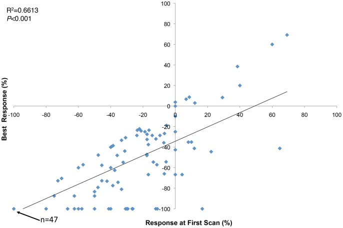
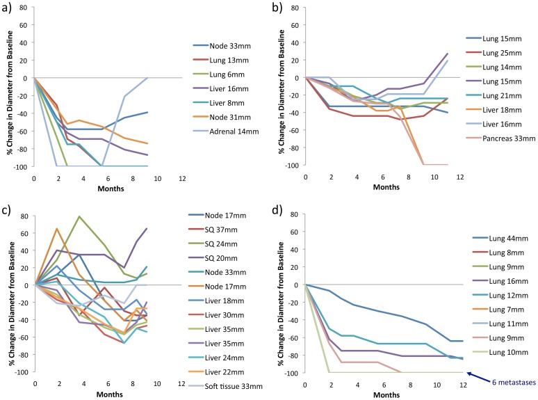
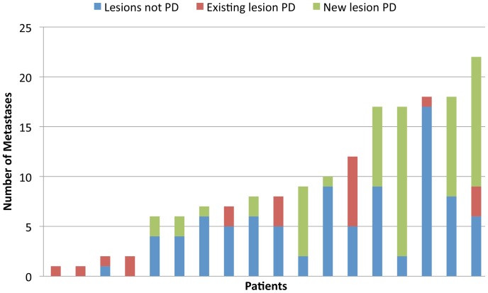
Verification
Claims the paper makes
The claims and statistics of this paper haven’t been checked yet.
Run the check — free, no account needed. It reads the open-access text and checks the sources it cites.
Claims other papers make about this one
Each is checked against this paper’s own text: does it say what the citing paper says it does?
In the citing paper
“Tumor heterogeneity—both within a single tumor (intra-tumoral) and between patients (inter-patient)—affects how cancers respond to therapy [21].”
From Advances in green-synthesized magnetic nanoparticles for targeted cancer therapy: mechanisms, applications, and future perspectives (2026) · cites this paper as [21]
Self-check
Percentages, sums and reported differences re-computed from the paper’s own numbers.
The self-check needs the paper’s full text.
Citations
Cites
Combined BRAF and MEK inhibition in melanoma with BRAF V600 mutations
Flaherty, K. T., Infante, J. R., Daud, A., Gonzalez, R., Kefford, R. F., Sosman, J., Hamid, O., Schuchter, L., Cebon, J., Ibrahim, N., et al. (2012) · New England Journal of Medicine · doi:10.1056/nejmoa1210093 · Cited by 2,810
Self-citationFrederick, D. T., Piris, A., Cogdill, A. P., Cooper, Z. A., Lezcano, C., Ferrone, C. R., Mitra, D., Boni, A., Newton, L. P., Liu, C., et al. (2013) · Clinical Cancer Research · doi:10.1158/1078-0432.ccr-12-1630 · Cited by 944
Research works printed but not linked: 20
- CE Meacham, 2013, Tumour heterogeneity and cancer cell plasticity, Nature, 501, 328, 10.1038/nature12624 · DOI
- B Vogelstein, 2013, Cancer genome landscapes, Science, 339, 1546, 10.1126/science.1235122 · DOI · Paper page
- M Yancovitz, 2012, Intra- and inter-tumor heterogeneity of BRAF(V600E))mutations in primary and metastatic melanoma, PLoS ONE, 7, e29336, 10.1371/journal.pone.0029336 · DOI
- M Takata, 2000, Clonal heterogeneity in sporadic melanomas as revealed by loss-of-heterozygosity analysis, Int J Cancer, 85, 492, 10.1002/(SICI)1097-0215(20000215)85:4<492::AID-IJC8>3.0.CO;2-# · DOI
- J Lin, 2011, Polyclonality of BRAF mutations in primary melanoma and the selection of mutant alleles during progression, British Journal of Cancer, 104, 464, 10.1038/sj.bjc.6606072 · DOI
- TM Katona, 2007, Genetically heterogeneous and clonally unrelated metastases may arise in patients with cutaneous melanoma, Am J Surg Pathol, 31, 1029, 10.1097/PAS.0b013e31802b3488 · DOI
- JS Wilmott, 2012, Intratumoral Molecular Heterogeneity in a BRAF-Mutant, BRAF Inhibitor-Resistant Melanoma: A Case Illustrating the Challenges for Personalized Medicine, Molecular Cancer Therapeutics, 11, 2704, 10.1158/1535-7163.MCT-12-0530 · DOI
- MR Junttila, 2013, Influence of tumour micro-environment heterogeneity on therapeutic response, Nature, 501, 346, 10.1038/nature12626 · DOI · Paper page
- PB Chapman, 2011, Improved survival with vemurafenib in melanoma with BRAF V600E mutation, New England Journal of Medicine, 364, 2507, 10.1056/NEJMoa1103782 · DOI · Paper page
- A Hauschild, 2012, Dabrafenib in BRAF-mutated metastatic melanoma: a multicentre, open-label, phase 3 randomised controlled trial, Lancet, 380, 358, 10.1016/S0140-6736(12)60868-X · DOI
- Kim KB, Flaherty KT, Chapman PB, Sosman JA, Ribas A, <etal>et al</etal>.. (2011) Pattern and outcome of disease progression in phase I study of vemurafenib in patients with metastatic melanoma (MM). Journal of Clinical Oncology 29: (abstract 8519). · DOI
- Azer MF, Menzies AM, Haydu LE, Kefford RF, Long GV (2013) Patterns of Response and Progression in Patients with BRAF-mutant Melanoma Metastatic to the Brain treated with Dabrafenib. Cancer: in press. · DOI
- EA Eisenhauer, 2009, New response evaluation criteria in solid tumours: revised RECIST guideline (version 1.1), European Journal of Cancer, 45, 228, 10.1016/j.ejca.2008.10.026 · DOI · Paper page
- GA McArthur, 2012, Marked, homogeneous, and early [18F]fluorodeoxyglucose-positron emission tomography responses to vemurafenib in BRAF-mutant advanced melanoma, Journal of Clinical Oncology, 30, 1628, 10.1200/JCO.2011.39.1938 · DOI
- MS Carlino, 2013, (18)F-labelled fluorodeoxyglucose-positron emission tomography (FDG-PET) heterogeneity of response is prognostic in dabrafenib treated BRAF mutant metastatic melanoma, European Journal of Cancer, 49, 395, 10.1016/j.ejca.2012.08.018 · DOI
- JS Wilmott, 2012, Selective BRAF inhibitors induce marked T-cell infiltration into human metastatic melanoma, Clinical Cancer Research, 18, 1386, 10.1158/1078-0432.CCR-11-2479 · DOI
- Chan M, Haydu L, Menzies AM, Azer MWF, Klein O, <etal>et al</etal>.. (2013) Clinical characteristics and survival of BRAF-mutant metastatic melanoma patients treated with BRAF inhibitor dabrafenib or vemurafenib beyond disease progression. Journal of Clinical Oncology 31: (abstract 9062). · DOI
- Ackerman A, Klein O, McDermott DF, Lawrence DP, Gunturi A, <etal>et al</etal>.. (2013) Outcomes of patients with metastatic melanoma treated with immunotherapy prior to or after BRAF inhibitors. Cancer: in press. · DOI
- PA Ascierto, 2012, Sequencing of BRAF inhibitors and ipilimumab in patients with metastatic melanoma: a possible algorithm for clinical use, Journal of Translational Medicine, 10, 107, 10.1186/1479-5876-10-107 · DOI
- MS Carlino, 2013, Antiproliferative effects of continued mitogen-activated protein kinase pathway inhibition following acquired resistance to BRAF and/or MEK inhibition in melanoma, Molecular Cancer Therapeutics, 12, 1332, 10.1158/1535-7163.MCT-13-0011 · DOI
Cited by
Sorted by citations
Long, G. V., Fung, C., Menzies, A. M., Pupo, G. M., Carlino, M. S., Hyman, J., Shahheydari, H., Tembe, V., Thompson, J. F., Saw, R. P., et al. (2014) · Nature Communications · doi:10.1038/ncomms6694 · Cited by 375
Self-citationA Convergence-Based Framework for Cancer Drug Resistance
Konieczkowski, D. J., Johannessen, C. M., & Garraway, L. A. (2018) · Cancer Cell · doi:10.1016/j.ccell.2018.03.025 · Cited by 258
Barish, S., Ochs, M. F., Sontag, E. D., & Gevertz, J. L. (2017) · Proceedings of the National Academy of Sciences · doi:10.1073/pnas.1703355114 · Cited by 48
El-khawaga, A. M., Youssef, O. I., El-dydamoni, O. A., Ragab, W. M., & Mohammed, R. A. E. (2026) · European Journal of Medical Research · doi:10.1186/s40001-025-03804-9 · Cited by 14
Metadata
- Authors
- Menzies, A. M., Haydu, L. E., Carlino, M. S., Azer, M. W. F., Carr, P. J. A., Kefford, R. F., & Long, G. V.
- Venue
- PLoS ONE
- Publisher
- Public Library of Science (PLoS)
- Published
- 2014-01-06
- Volume
- 9
- Issue
- 1
- Pages
- e85004
- ISSN
- 1932-6203
- PMC
- PMC3882277
- Cited by
- 71
- References
- 2
- Language
- en
About these checks
Registry, retraction, Cites and statistics results are computed, not judged, with one exception: a printed reference that no index lists is read once by a model before it counts as a citation error. Claims are extracted by a model and kept only when their quote appears verbatim in the text; each cited source is then checked, with a model’s help, for whether it says what the claim relies on. Those verdicts can be wrong — read the claim’s quote and the cited source before relying on one.
Statistics and arithmetic are recomputed, not judged.
Confidence bands are shown only for fields calibrated on our annotated test set. None is calibrated yet, so no band is shown.
What counts as a claim ›How we measure confidence ›How we check this ›
Are you an author, or do you see a problem with this page? Request a review
