

The DOI matches its Crossref record; 75 of the 75 references it cites were found in a registry or an index.
Quantitative Profiling of Colorectal Cancer-Associated Bacteria Reveals Associations between Fusobacterium spp., Enterotoxigenic Bacteroides fragilis (ETBF) and Clinicopathological Features of Colorectal Cancer
Open accessAbstract
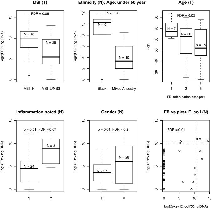
Various studies have presented clinical or in vitro evidence linking bacteria to colorectal cancer, but these bacteria have not previously been concurrently quantified by qPCR in a single cohort. We quantify these bacteria (Fusobacterium spp., Streptococcus gallolyticus, Enterococcus faecalis, Enterotoxigenic Bacteroides fragilis (ETBF), Enteropathogenic Escherichia coli (EPEC), and afaC- or pks-positive E. coli) in paired tumour and normal tissue samples from 55 colorectal cancer patients. We further investigate the relationship between a) the presence and b) the level of colonisation of each bacterial species with site and stage of disease, age, gender, ethnicity and MSI-status. With the exception of S. gallolyticus, we detected all bacteria profiled here in both tumour and normal samples at varying frequencies. ETBF (FDR = 0.001 and 0.002 for normal and tumour samples) and afaC-positive E. coli (FDR = 0.03, normal samples) were significantly enriched in the colon compared to the rectum. ETBF (FDR = 0.04 and 0.002 for normal and tumour samples, respectively) and Fusobacterium spp. (FDR = 0.03 tumour samples) levels were significantly higher in late stage (III/IV) colorectal cancers. Fusobacterium was by far the most common bacteria detected, occurring in 82% and 81% of paired tumour and normal samples. Fusobacterium was also the only bacterium that was significantly higher in tumour compared to normal samples (p = 6e-5). We also identified significant associations between high-level colonisation by Fusobacterium and MSI-H (FDR = 0.05), age (FDR = 0.03) or pks-positive E. coli (FDR = 0.01). Furthermore, we exclusively identified atypical EPEC in our cohort, which has not been previously reported in association with colorectal cancer. By quantifying colorectal cancer-associated bacteria across a single cohort, we uncovered inter- and intra-individual patterns of colonization not previously recognized, as well as important associations with clinicopathological features, especially in the case of Fusobacterium and ETBF.
Overview
The claims and statistics of this paper haven’t been checked yet.
Run the check — free, no account needed. It reads the open-access text and checks the sources it cites.
Integrity signals
Registry
Cites
Statistics
Figures
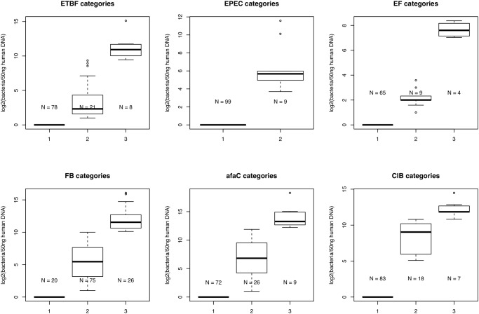
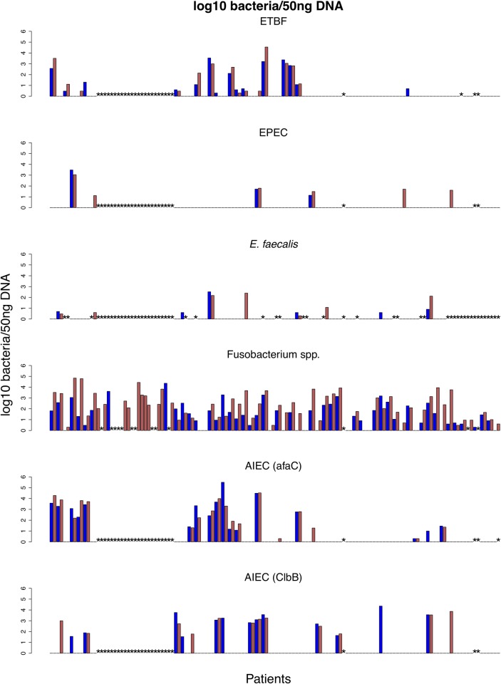
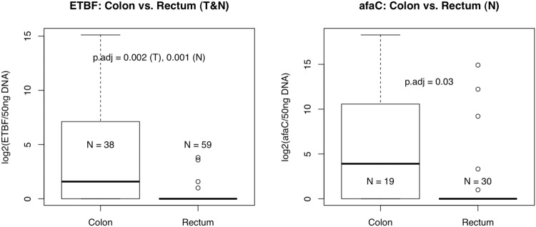
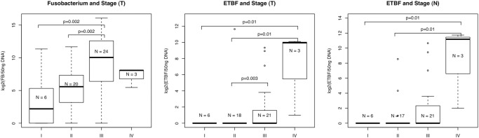
Verification
Claims the paper makes
The claims and statistics of this paper haven’t been checked yet.
Run the check — free, no account needed. It reads the open-access text and checks the sources it cites.
Claims other papers make about this one
Each is checked against this paper’s own text: does it say what the citing paper says it does?
In the citing paper
“Among them, 2 found significant enrichment of ETBF in tumor tissues compared to tissues from controls or adjacent normal tissues40, 43.”
From Influence of the Gut Microbiome, Diet, and Environment on Risk of Colorectal Cancer (2020) · cites this paper as [43]
Self-check
Percentages, sums and reported differences re-computed from the paper’s own numbers.
The self-check needs the paper’s full text.
Citations
Cites
Primer-BLAST: A tool to design target-specific primers for polymerase chain reaction
Ye, J., Coulouris, G., Zaretskaya, I., Cutcutache, I., Rozen, S., & Madden, T. (2012) · BMC Bioinformatics · doi:10.1186/1471-2105-13-134 · Cited by 6,369
Human papillomavirus and cervical cancer
SCHIFFMAN, M. H., Castle, P. E., Jerónimo, J., Rodríguez, A. C., & Wacholder, S. (2007) · The Lancet · doi:10.1016/s0140-6736(07)61416-0 · Cited by 3,135
Kostic, A. D., Chun, E., Robertson, L., Glickman, J. N., Gallini, C. A., Michaud, M., Clancy, T. E., Chung, D. C., Lochhead, P., Hold, G. L., et al. (2013) · Cell Host & Microbe · doi:10.1016/j.chom.2013.07.007 · Cited by 2,718
‘Touchdown’ PCR to circumvent spurious priming during gene amplification
Don, R. H., Cox, P. T., Wainwright, B. J., Baker, K. N., & Mattick, J. S. (1991) · Nucleic Acids Research · doi:10.1093/nar/19.14.4008 · Cited by 2,586
Rubinstein, M. R., Wang, X., Liu, W., Hao, Y., Cai, G., & Han, Y. W. (2013) · Cell Host & Microbe · doi:10.1016/j.chom.2013.07.012 · Cited by 2,476
Intestinal inflammation targets cancer-inducing activity of the microbiota
Arthur, J. C., Perez-Chanona, E., Mühlbauer, M., Tomkovich, S., Uronis, J. M., Fan, T.-J., Campbell, B. J., Abujamel, T., Dogan, B., Rogers, A. B., et al. (2012) · Science · doi:10.1126/science.1224820 · Cited by 2,263
Fusobacterium nucleatum infection is prevalent in human colorectal carcinoma
Castellarin, M., Warren, R. L., Freeman, J. D., Dreolini, L., Krzywinski, M., Strauss, J., Barnes, R., Watson, P., Allen-Vercoe, E., Moore, R. A., et al. (2012) · Genome Research · doi:10.1101/gr.126516.111 · Cited by 2,043
Genomic analysis identifies association of Fusobacterium with colorectal carcinoma
Kostic, A. D., Gevers, D., Pedamallu, C. S., Michaud, M., Duke, F., Earl, A. M., Ojesina, A. I., Jung, J., Bass, A. J., Tabernero, J., et al. (2012) · Genome Research · doi:10.1101/gr.126573.111 · Cited by 1,960
Wu, S., Rhee, K.-J., Albesiano, E., Rabizadeh, S., Wu, X., Yen, H.-R., Huso, D. L., Brancati, F. L., Wick, E., McAllister, F., et al. (2009) · Nature Medicine · doi:10.1038/nm.2015 · Cited by 1,812
Větrovský, T., & Baldrián, P. (2013) · PLoS ONE · doi:10.1371/journal.pone.0057923 · Cited by 1,201
Research works printed but not linked: 3
- E Buc, 2013, High Prevalence of Mucosa-Associated E. coli Producing Cyclomodulin and Genotoxin in Colon Cancer, PLoS One, 8
- HM Martin, 2004, Enhanced Escherichia coli adherence and invasion in Crohn’s disease and colon cancer, Gastroenterology, 127, 80
- J Dolezel, 2003, Nuclear DNA content and genome size of trout and human, Cytometry A, 51, 127
Cited by
Sorted by citations
Gut microbiota imbalance and colorectal cancer
Gagnière, J. (2016) · World Journal of Gastroenterology · doi:10.3748/wjg.v22.i2.501 · Cited by 890
The Intestinal Microbiota in Colorectal Cancer.
Tilg, H., Adolph, T. E., Gerner, R. R., & Moschen, A. R. (2018) · Cancer Cell · doi:10.1016/j.ccell.2018.03.004 · Cited by 777
Influence of the Gut Microbiome, Diet, and Environment on Risk of Colorectal Cancer
Song, M., Chan, A. T., & Sun, J. (2020) · Gastroenterology · doi:10.1053/j.gastro.2019.06.048 · Cited by 752
The inflammatory pathogenesis of colorectal cancer
Schmitt, M., & Greten, F. R. (2021) · Nature reviews. Immunology · doi:10.1038/s41577-021-00534-x · Cited by 840
Gut Microbiota, Inflammation, and Colorectal Cancer
Brennan, C. A., & Garrett, W. S. (2016) · Annual Review of Microbiology · doi:10.1146/annurev-micro-102215-095513 · Cited by 586
Metadata
- Authors
- Viljoen, K. S., Dakshinamurthy, A., Goldberg, P., & Blackburn, J. M.
- Affiliations
- University of Cape Town (ZA)
- Groote Schuur Hospital (ZA)
- Corresponding author
- Blackburn, Jonathan M.
- Venue
- PLoS ONE
- Publisher
- Public Library of Science (PLoS)
- Published
- 2015-03-09
- Volume
- 10
- Issue
- 3
- Pages
- e0119462
- ISSN
- 1932-6203 (ISSN-L)
- PMC
- PMC4353626
- OpenAlex
- W2086248022
- Cited by
- 256
- References
- 74
- Citations by year
- 2026: 14 · 2025: 26 · 2024: 25 · 2023: 32 · 2022: 29 · 2021: 34 · 2020: 21 · 2019: 23 · 2018: 21 · 2017: 16 · 2016: 12 · 2015: 1
- FWCI
- 7.24
- Type
- journal article
- Language
- en
- Open access
- gold
- doi.org
- Licence
- Locations
- PLoS ONE (publishedVersion, cc-by)
- Europe PMC (PubMed Central) (publishedVersion, cc-by)
- PubMed Central (publishedVersion, cc-by)
- PubMed (publishedVersion)
- DOAJ (DOAJ: Directory of Open Access Journals) (submittedVersion, cc-by-sa)
- Figshare (submittedVersion, cc-by)
- Open University of Cape Town (University of Cape Town) (submittedVersion, cc-by)
- Funders
- National Research Foundation
- Cancer Association of South Africa
- Deutscher Akademischer Austauschdienst
- Harry Crossley Foundation
- University of Cape Town
- Topics
- Life Sciences › Biochemistry, Genetics and Molecular Biology › Molecular Biology › Gut microbiota and health
- Life Sciences › Immunology and Microbiology › Microbiology › Escherichia coli research studies
- Health Sciences › Dentistry › Periodontics › Oral microbiology and periodontitis research
- Keywords
- colorectal cancer, Fusobacterium, enterotoxigenic Bacteroides fragilis, Streptococcus bovis, Enterococcus faecalis, enteropathogenic Escherichia coli, tumor microbiome, tumor stage, microsatellite instability, clinicopathological features
- MeSH terms
- Adenocarcinoma, Adult, Bacterial Infections, Bacteroides fragilis, Colon, Female, Fusobacterium, Genes, Bacterial, Humans, Male, Middle Aged, Neoplasm Staging, Rectum, Colorectal Neoplasms, Case-Control Studies, Enteropathogenic Escherichia coli, Bacterial Load
About these checks
Registry, retraction, Cites and statistics results are computed, not judged, with one exception: a printed reference that no index lists is read once by a model before it counts as a citation error. Claims are extracted by a model and kept only when their quote appears verbatim in the text; each cited source is then checked, with a model’s help, for whether it says what the claim relies on. Those verdicts can be wrong — read the claim’s quote and the cited source before relying on one.
Statistics and arithmetic are recomputed, not judged.
Confidence bands are shown only for fields calibrated on our annotated test set. None is calibrated yet, so no band is shown.
What counts as a claim ›How we measure confidence ›How we check this ›
Are you an author, or do you see a problem with this page? Request a review
