

The DOI matches its Crossref record; 37 of the 37 references it cites were found in a registry or an index.
Hypoxia Pretreatment of Bone Marrow Mesenchymal Stem Cells Facilitates Angiogenesis by Improving the Function of Endothelial Cells in Diabetic Rats with Lower Ischemia
Open accessAbstract
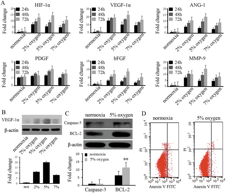
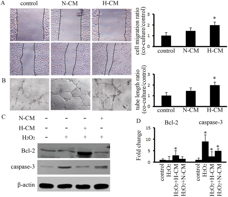
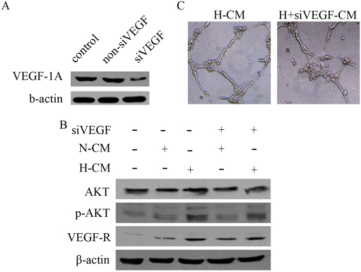
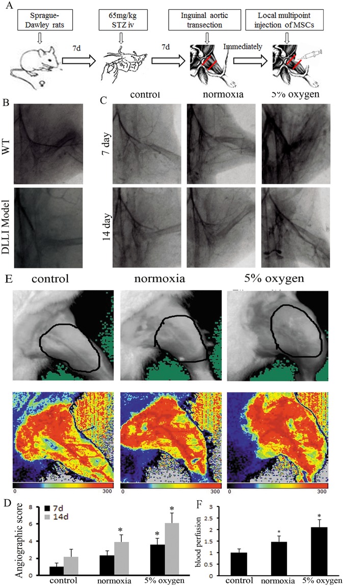
Endothelial dysfunction induced by unordered metabolism results in vascular reconstruction challenges in diabetic lower limb ischemia (DLLI). Mesenchymal stem cells (MSCs) are multipotent secretory cells that are suitable for clinical DLLI treatment, but their use has been hampered by poor survival after injection. Hypoxia can significantly enhance the capacity of MSCs to secrete angiogenic factors. We investigated transient hypoxia pretreatment of MSCs to facilitate revascularization in DLLI. Rat bone marrow MSCs (BM-MSCs) were cultured at different oxygen concentrations for varying time periods. The results indicated that transient pretreatment (5% O2, 48 h) not only increased the expression of VEGF-1α, ANG, HIF-1α and MMP-9 in BM-MSCs as assessed by real-time RT-PCR, but also increased the expression of Bcl-2 as determined by western blotting. The transplantation of pretreated BM-MSCs into rats with DLLI demonstrated accelerated vascular reconstruction when assayed by angiography and immunohistochemistry. CM-Dil-labeled tracer experiments indicated that the survival of BM-MSCs was significantly improved, with approximately 5% of the injected cells remaining alive at 14 days. The expression levels of VEGF-1α, MMP-9 and VEGF-R were significantly increased, and the expression of pAKT was up-regulated in ischemic muscle. Double immunofluorescence studies confirmed that the pretreated BM-MSCs promoted the proliferation and inhibited the apoptosis of endothelial cells. In vitro, pretreated BM-MSCs increased the migratory and tube forming capacity of endothelial cells (ECs). Hypoxia pretreatment of BM-MSCs significantly improved angiogenesis in response to tissue ischemia by ameliorating endothelial cell dysfunction and is a promising therapeutic treatment for DLLI.
Overview
The claims and statistics of this paper haven’t been checked yet.
Run the check — free, no account needed. It reads the open-access text and checks the sources it cites.
Integrity signals
Registry
Cites
Statistics
Figures
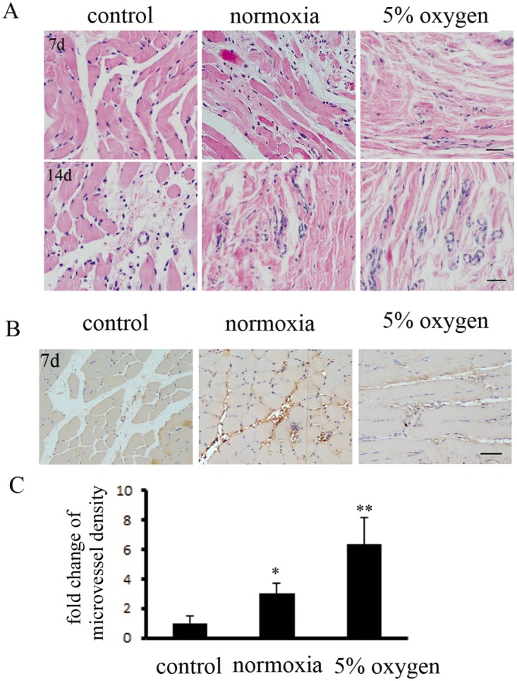
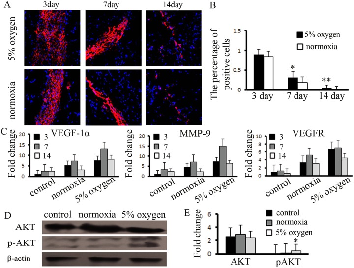
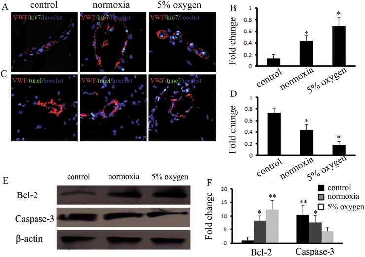
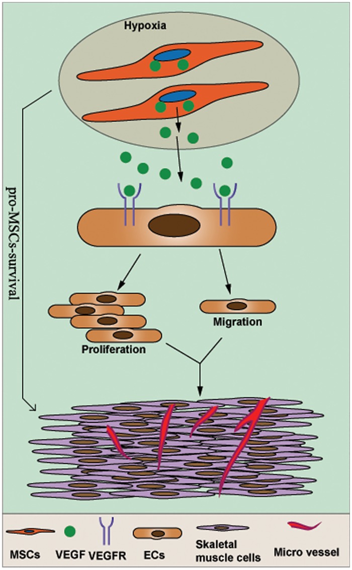
Verification
Claims the paper makes
The claims and statistics of this paper haven’t been checked yet.
Run the check — free, no account needed. It reads the open-access text and checks the sources it cites.
Claims other papers make about this one
Each is checked against this paper’s own text: does it say what the citing paper says it does?
The cited source says what the claim attributes to it.
In the citing paper
“Upregulation of the VEGF gene was evident in all hypoxia cultures, which is consistent with other studies [53–56].”
From Short-term physiological hypoxia potentiates the therapeutic function of mesenchymal stem cells (2018) · cites this paper as [56]
Self-check
Percentages, sums and reported differences re-computed from the paper’s own numbers.
The self-check needs the paper’s full text.
Citations
Cites
Multilineage potential of adult human mesenchymal stem cells
Pittenger, M. F., Mackay, A. M., Beck, S. C., Jaiswal, R. K., Douglas, R., Mosca, J. D., Moorman, M. A., Simonetti, D. W., Craig, S., & Marshak, D. R. (1999) · Science · doi:10.1126/science.284.5411.143 · Cited by 21,052
Wild, S., Roglic, G., Green, A., Sicree, R., & King, H. (2004) · Diabetes Care · doi:10.2337/diacare.27.5.1047 · Cited by 15,297
Angiogenesis in cancer and other diseases
Carmeliet, P., & Jain, R. K. (2000) · Nature · doi:10.1038/35025220 · Cited by 9,070
Nitric Oxide and Peroxynitrite in Health and Disease
Pacher, P., Beckman, J. S., & Liaudet, L. (2007) · Physiological Reviews · doi:10.1152/physrev.00029.2006 · Cited by 6,390
Mechanisms of angiogenesis and arteriogenesis
Carmeliet, P. (2000) · Nature Medicine · doi:10.1038/74651 · Cited by 4,235
Oxidative Stress and Stress-Activated Signaling Pathways: A Unifying Hypothesis of Type 2 Diabetes
Evans, J. L., Goldfine, I. D., Maddux, B. A., & Grodsky, G. M. (2002) · Endocrine Reviews · doi:10.1210/er.2001-0039 · Cited by 2,232
Pathways to Diabetic Limb Amputation: Basis for Prevention
Pecoraro, R. E., Reiber, G. E., & Burgess, E. M. (1990) · Diabetes Care · doi:10.2337/diacare.13.5.513 · Cited by 1,585
Kinnaird, T., Stabile, E., Burnett, M. S., Lee, C. W., Barr, S., Fuchs, S., & Epstein, S. E. (2004) · Circulation Research · doi:10.1161/01.res.0000118601.37875.ac · Cited by 1,415
Lumeij, J. T. (1997) · Elsevier eBooks · doi:10.1016/b978-012396305-5/50031-2 · Cited by 1,185
Central nervous system pericytes in health and disease
Winkler, E. A., Bell, R. D., & Zlokovic, B. V. (2011) · Nature Neuroscience · doi:10.1038/nn.2946 · Cited by 962
Cited by
Sorted by citations
Mesenchymal stromal cell secretome: influencing therapeutic potential by cellular pre-conditioning
Ferreira, J. R., Teixeira, G. Q., Santos, S. G., Barbosa, M. A., Almeida-Porada, G., & Gonçalves, R. M. (2018) · Frontiers in Immunology · doi:10.3389/fimmu.2018.02837 · Cited by 572
Macro‐ and microvascular endothelial dysfunction in diabetes
Shi, Y., & Vanhoutte, P. M. (2017) · Journal of Diabetes · doi:10.1111/1753-0407.12521 · Cited by 562
Preconditioning influences mesenchymal stem cell properties in vitro and in vivo
Hu, C., & Li, L. (2018) · Journal of Cellular and Molecular Medicine · doi:10.1111/jcmm.13492 · Cited by 452
The challenges and promises of allogeneic mesenchymal stem cells for use as a cell-based therapy
Zhang, J., Huang, X., Wang, H., Liu, X., Zhang, T., Wang, Y., & Hu, D. (2015) · Stem Cell Research & Therapy · doi:10.1186/s13287-015-0240-9 · Cited by 311
Mesenchymal Stem Cells for Chronic Wound Healing: Current Status of Preclinical and Clinical Studies
Huang, Y., Gou, M., Da, L., Zhang, W., & Xie, H. (2020) · Tissue Engineering Part B Reviews · doi:10.1089/ten.teb.2019.0351 · Cited by 213
Short-term physiological hypoxia potentiates the therapeutic function of mesenchymal stem cells
Antebi, B., Rodriguez, L. A., Walker, K. P., Asher, A. M., Kamucheka, R. M., Alvarado, L., Mohammadipoor, A., & Cancio, L. C. (2018) · Stem Cell Research & Therapy · doi:10.1186/s13287-018-1007-x · Cited by 137
Liu, Y. F., Chen, J., Liang, H., Cai, Y., Li, X., Li, Y., Zhou, L., Shan, L., & Wang, H. (2022) · Stem Cell Research & Therapy · doi:10.1186/s13287-022-02927-8 · Cited by 123
Rivera‐Cruz, C. M., Shearer, J. J., Neto, M. F., & Figueiredo, M. L. (2017) · Stem Cells International · doi:10.1155/2017/4015039 · Cited by 117
Babajani, A., Soltani, P., Jamshidi, E., Farjoo, M. H., & Niknejad, H. (2020) · Frontiers in Bioengineering and Biotechnology · doi:10.3389/fbioe.2020.00748 · Cited by 99
Wang, X., Shen, K., Wang, J., Liu, K., Wu, G., Li, Y., Luo, L., Zheng, Z., & Hu, D. (2020) · Free Radical Biology and Medicine · doi:10.1016/j.freeradbiomed.2020.07.023 · Cited by 99
Kuss, M. A., Harms, R., Wu, S., Wang, Y., Untrauer, J. B., Carlson, M. A., & Duan, B. (2017) · RSC Advances · doi:10.1039/c7ra04372d · Cited by 81
Zhao, J., Yu, G., Cai, M., Lei, X., Yang, Y., Wang, Q., & Zhai, X. (2018) · Stem Cell Research & Therapy · doi:10.1186/s13287-018-0785-5 · Cited by 80
Potential role of hydrogen sulfide in diabetes-impaired angiogenesis and ischemic tissue repair
Cheng, Z. A., & Kishore, R. (2020) · Redox Biology · doi:10.1016/j.redox.2020.101704 · Cited by 81
Yuan, Y., Shi, M., Li, L., Liu, J., Chen, B., Chen, Y., An, X., Liu, S., Luo, R., Long, D., et al. (2016) · Clinical Science · doi:10.1042/cs20160235 · Cited by 70
Self-citationHan, K. H., Kim, A., Kim, M., Kim, D., Go, H., & Kim, D. (2015) · Cell Biology International · doi:10.1002/cbin.10519 · Cited by 67
Wobma, H., Kanai, M., Ma, S. P., Shih, Y., Li, H. W., Duran‐Struuck, R., WINCHESTER, R. J., Goeta, S., Brown, L. M., & Vunjak‐Novakovic, G. (2018) · Journal of Immunology and Regenerative Medicine · doi:10.1016/j.regen.2018.01.001 · Cited by 66
Zhilai, Z., Biling, M., Sujun, Q., Chao, D., Benchao, S., Huang, S., Shun, Y., & Zhang, H. (2016) · Brain Research · doi:10.1016/j.brainres.2016.04.025 · Cited by 65
Kahrizi, M. S., Mousavi, E., Khosravi, A., Rahnama, S., Salehi, A., Nasrabadi, N., Ebrahimzadeh, F., & Jamali, S. (2023) · Stem Cell Research & Therapy · doi:10.1186/s13287-023-03374-9 · Cited by 61
Jin, C., Yang, Y., Shen, L., Ding, W., Chen, X., Wu, E., Cai, K., & Wang, G. (2016) · Cellular and Molecular Neurobiology · doi:10.1007/s10571-016-0445-1 · Cited by 53
Emerging roles of mesenchymal stem cell therapy in patients with critical limb ischemia
Shirbaghaee, Z., Hassani, M., Keshel, S. H., & Soleimani, M. (2022) · Stem Cell Research & Therapy · doi:10.1186/s13287-022-03148-9 · Cited by 51
CD73 + extracellular vesicles inhibit angiogenesis through adenosine A 2B receptor signalling
Angioni, R., Liboni, C., Herkenne, S., Sánchez‐Rodríguez, R., Borile, G., Marcuzzi, E., Calì, B., Muraca, M., & Viola, A. (2020) · Journal of Extracellular Vesicles · doi:10.1080/20013078.2020.1757900 · Cited by 49
Hao, D., He, C., Ma, B., Lankford, L., Reynaga, L., Farmer, D. L., Guo, F., & Wang, A. (2019) · Stem Cells International · doi:10.1155/2019/9695239 · Cited by 44
Rodriguez, L., Mohammadipoor, A., Alvarado, L., Kamucheka, R. M., Asher, A. M., Cancio, L. C., & Antebi, B. (2019) · Cells · doi:10.3390/cells8050462 · Cited by 44
Calcat-i-Cervera, S., Sanz‐Nogués, C., & O’Brien, T. (2021) · Frontiers in Medicine · doi:10.3389/fmed.2021.728496 · Cited by 42
Rovere, M., Reverberi, D., Arnaldi, P., Palamà, M. E. F., & Gentili, C. (2023) · Frontiers in Bioengineering and Biotechnology · doi:10.3389/fbioe.2023.1297644 · Cited by 39
Chai, Y. C., Mendes, L. F., van Gastel, N., Carmeliet, G., & Luyten, F. P. (2018) · Acta Biomaterialia · doi:10.1016/j.actbio.2018.03.048 · Cited by 37
Xu, T., Lv, Z., Chen, Q., Guo, M., Wang, X., & Huang, F. (2018) · Biomedicine & Pharmacotherapy · doi:10.1016/j.biopha.2018.06.129 · Cited by 37
Phelps, J., Hart, D. A., Mitha, A. P., Duncan, N. A., & Sen, A. (2023) · Stem Cell Research & Therapy · doi:10.1186/s13287-023-03439-9 · Cited by 36
Cozens, D., Grahame, E., Sutherland, E., Taylor, G., Berry, C. C., & Davies, R. L. (2018) · Scientific Reports · doi:10.1038/s41598-017-19079-y · Cited by 36
Enhancement of the efficacy of mesenchymal stem cells in the treatment of ischemic diseases
Luo, R., Lu, Y., Liu, J., Cheng, J., & Chen, Y. (2018) · Biomedicine & Pharmacotherapy · doi:10.1016/j.biopha.2018.11.068 · Cited by 36
Self-citationLee, S., & Joe, Y. A. (2018) · Biochemical and Biophysical Research Communications · doi:10.1016/j.bbrc.2018.05.086 · Cited by 35
Mesenchymal Stem Cells in Cardiac Repair: Effects on Myocytes, Vasculature, and Fibroblasts
White, S., & Chong, J. J. H. (2020) · Clinical Therapeutics · doi:10.1016/j.clinthera.2020.08.010 · Cited by 35
Rekittke, N. E., Ang, M., Rawat, D., Khatri, R., & Linn, T. (2016) · Stem Cells International · doi:10.1155/2016/3764681 · Cited by 34
Zhu, F., Wei, C., Wu, H., Shuai, B., Yu, T., Gao, F., Yuan, Y., Zuo, D., Liu, X., Zhang, L., et al. (2022) · International Immunopharmacology · doi:10.1016/j.intimp.2022.109426 · Cited by 35
Esquivel‐Ruiz, S., González‐Rodríguez, P., Lorente, J. A., Pérez‐Vizcaíno, F., Herrero, R., & Moreno, L. (2021) · Frontiers in Physiology · doi:10.3389/fphys.2021.752287 · Cited by 33
Advancements in diabetic foot ulcer research: Focus on mesenchymal stem cells and their exosomes
Wu, S., Zhou, Z., Li, Y., & Jiang, J. (2024) · Heliyon · doi:10.1016/j.heliyon.2024.e37031 · Cited by 30
Zhāng, Q., Wan, X.-X., Hu, X., Zhao, W., Ban, X.-X., Huang, Y., Yan, W., & Xiong, K. (2021) · Frontiers in Cell and Developmental Biology · doi:10.3389/fcell.2021.809656 · Cited by 28
Neo, S. H., Her, Z., Othman, R. B., Tee, C. A., Ong, L. C., Wang, Y., Tan, I., Tan, J., Yang, Y., Yang, Z., et al. (2023) · Stem Cell Research & Therapy · doi:10.1186/s13287-023-03481-7 · Cited by 26
Yin, C.-P., Liang, Y., Zhang, J., Ruan, G., Li, Z.-A., Pang, R., & Pan, X. (2016) · Stem Cells International · doi:10.1155/2016/9742034 · Cited by 26
Khasawneh, R. R., Abu‐El‐Rub, E., Serhan, A. O., Serhan, B. O., & Abu-El-Rub, H. (2019) · The Journal of Physiological Sciences · doi:10.1007/s12576-019-00720-6 · Cited by 24
Han, K.-H., Kim, A.-K., Kim, M.-H., Kim, D.-H., Go, H.-N., Kang, D., Chang, J. W., Choi, S. W., Kang, K.-S., & Kim, D.-I. (2017) · Tissue and Cell · doi:10.1016/j.tice.2017.09.006 · Cited by 20
First Clinical Experiences Using Preconditioning Approaches to Improve MSC-Based Therapies
Le, B., Cressman, A., Morales, D. L. S., & Fierro, F. A. (2024) · Current Stem Cell Reports · doi:10.1007/s40778-023-00232-5 · Cited by 20
Han, T. T. Y., & Flynn, L. E. (2020) · Journal of Tissue Engineering and Regenerative Medicine · doi:10.1002/term.3133 · Cited by 20
Wei, S., Chen, T., Kao, F., Hsu, Y.-J., & Chen, Y. (2022) · Journal of Tissue Engineering · doi:10.1177/20417314221084096 · Cited by 19
Biological mechanisms and applied prospects of mesenchymal stem cells in premature ovarian failure
Shi, L., Zhang, Z., Deng, M., Zheng, F., Liu, W., & Ye, S. (2022) · Medicine · doi:10.1097/md.0000000000030013 · Cited by 20
Jiang, Q., Yu, T., Huang, K., Lu, J., Zhang, H., & Hu, S. (2016) · PLoS ONE · doi:10.1371/journal.pone.0146074 · Cited by 18
Zhao, Y., Zhang, M., Lu, G., Huang, B., Wang, D., Shao, Y., & Lu, M. (2020) · Frontiers in Cell and Developmental Biology · doi:10.3389/fcell.2020.580131 · Cited by 19
Self-citationCooper, T. T., Sherman, S. E., Bell, G. I., Dayarathna, T. K., McRae, D. M., Ma, J., Lagugné‐Labarthet, F., Pasternak, S., Lajoie, G., & Hess, D. A. (2021) · Stem Cells and Development · doi:10.1089/scd.2020.0206 · Cited by 19
Adipose‐derived stem cells: A candidate for liver regeneration
Yang, D., Wang, Z. Q., Deng, J., Liao, J. Y., Wang, X., Xie, J., Deng, M., & Lü, M. H. (2015) · Journal of Digestive Diseases · doi:10.1111/1751-2980.12268 · Cited by 17
Shen, C., Chen, B., Gu, J., Ning, J., Chen, L., Zeng, J., Yi, B., & Lu, K. (2016) · Experimental Cell Research · doi:10.1016/j.yexcr.2016.04.013 · Cited by 17
Subbarayan, R., Barathidasan, R., Raja, S. T. K., Gnanamani, A., Kuruvilla, S., Shanthi, P., & Rao, S. R. (2019) · Journal of Cellular Biochemistry · doi:10.1002/jcb.29452 · Cited by 17
The Angiogenic Paracrine Potential of Mesenchymal Stem Cells
Rezaie, J., Heidarzadeh, M., Hassanpour, M., Amini, H., Shokrollahi, E., Ahmadi, M., & Rahbarghazi, R. (2019) · IntechOpen eBooks · doi:10.5772/intechopen.84433 · Cited by 17
Peng, X., Liang, B., Wang, H., Hou, J., & Yuan, Q. (2022) · Cell and Tissue Research · doi:10.1007/s00441-021-03562-0 · Cited by 16
Taheri, M., Tehrani, H. A., Dehghani, S., Rajabzadeh, A., Alibolandi, M., Zamani, N., Arefian, E., & Ramezani, M. (2024) · Cytokine & Growth Factor Reviews · doi:10.1016/j.cytogfr.2024.01.004 · Cited by 17
Wang, J., Xiang, B., Deng, J., Lin, H., Freed, D. H., Arora, R. C., & Tian, G. (2017) · Journal of Huazhong University of Science and Technology [Medical Sciences] · doi:10.1007/s11596-017-1766-0 · Cited by 16
Santos, R. D. A., Asensi, K. D., de Barros, J. H. O., de Menezes, R. C. S., Cordeiro, I. R., Brito, J. M., Kasai-Brunswick, T. H., & Goldenberg, R. C. D. S. (2020) · International Journal of Molecular Sciences · doi:10.3390/ijms21249563 · Cited by 15
Subbarayan, R., Girija, D. M., Raja, S. T. K., Krishnamoorthy, A., Srinivasan, D., Shrestha, R., Srivastava, N., & Rao, S. R. (2024) · Molecular Biology Reports · doi:10.1007/s11033-024-09503-8 · Cited by 14
Hirose, S., Tabata, Y., Sone, K., Takahashi, N., Yoshino, D., & Funamoto, K. (2021) · Cell Adhesion & Migration · doi:10.1080/19336918.2021.1978368 · Cited by 12
Hypoxia in Mesenchymal Stem Cell
Widowati, W., Rihibiha, D. D., Khiong, K., Widodo, M. A., Sumitro, S. B., & Bachtiar, I. (2017) · InTech eBooks · doi:10.5772/65510 · Cited by 11
Lee, S. M., Jun, D. W., Kang, H. T., Oh, J., Saeed, W. K., & Ahn, S. B. (2021) · International Journal of Stem Cells · doi:10.15283/ijsc20096 · Cited by 10
Vu, N. B., Phi, L. T., Dao, T. T.-T., Le, H. T.-N., Ta, V. T., & Pham, P. V. (2016) · Biomedical Research and Therapy · doi:10.7603/s40730-016-0046-0 · Cited by 10
Zhao, P., Zhang, Y., Zhang, G., Niu, H., Li, J. J., Hao, R., Xie, M., Chu, S., Li, X., & Li, G. (2025) · Journal of Hypertension · doi:10.1097/hjh.0000000000004022 · Cited by 9
Ciavarella, C., Fittipaldi, S., & Pasquinelli, G. (2016) · Methods in molecular biology · doi:10.1007/7651_2016_350 · Cited by 8
Pham, P. V., Vu, N. B., Dao, T. T.-T., Le, H. T.-N., Phi, L. T., Huynh, O. T., Truong, M. T.-H., Nguyen, O. T.-K., & Phan, N. K. (2017) · Cytotechnology · doi:10.1007/s10616-017-0095-2 · Cited by 6
Weldearegay, Y. B., Brogaard, L., Rautenschlein, S., Meens, J., Valentin‐Weigand, P., & Schaaf, D. (2025) · Frontiers in Cellular and Infection Microbiology · doi:10.3389/fcimb.2025.1565513 · Cited by 5
He, Y. Z., Ma, M., Yan, Y., Chen, C., Luo, H., & Lei, W. (2020) · Journal of Cellular and Molecular Medicine · doi:10.1111/jcmm.15598 · Cited by 5
Fuchs, B., Birt, A., Moellhoff, N., Kuhlmann, C., Giunta, R. E., & Wiggenhauser, P. S. (2023) · Medicina · doi:10.3390/medicina59040706 · Cited by 5
Update on Mesenchymal and Induced Pluripotent Stem Cells
Al‐Anazi, K. A. (2019) · IntechOpen eBooks · doi:10.5772/intechopen.77857 · Cited by 5
Unlocking MSC Potential: Metabolic Reprogramming via Synthetic Biology Approaches
Trufanova, N. A., Trufanov, O., & Petrenko, O. (2025) · SynBio · doi:10.3390/synbio3030013 · Cited by 4
Li, Z., Sun, Y., Wu, J., Zhang, W., Li, T., Tang, S., & Ding, G. (2026) · International Journal of Nanomedicine · doi:10.2147/ijn.s584494 · Cited by 4
Eltrombopag increases the hematopoietic supporting ability of mesenchymal stem/stromal cells
Muntión, S., Preciado, S., Sánchez-Luis, E., Corchete, L. A., Díez‐Campelo, M., Osugui, L., Martí-Chillón, G. J., Vídriales, M., Navarro‐Bailón, A., Rivas, J. D. L., et al. (2022) · Therapeutic Advances in Hematology · doi:10.1177/20406207221142137 · Cited by 4
Metabolic conditioning enhances human bmMSC therapy of doxorubicin-induced heart failure
Jacques, V., Benaouadi, S., Descamps, J.-G., Reina, N., Espagnolle, N., Marsal, D. J., Sainte–Marie, Y., Boudet, A., Carla, P., Farge, T., et al. (2024) · Stem Cells · doi:10.1093/stmcls/sxae050 · Cited by 3
Cell therapy of critical limb ischemia: A review of preclinical and clinical research in China
Liao, L. (2020) · Vascular Investigation and Therapy · doi:10.4103/vit.vit_7_20 · Cited by 3
Boulos, A. N., Elagawany, A. M., Awwad, A., Fouad, G., & El-Rahman, A. A. (2020) · The Egyptian Journal of Histology · doi:10.21608/ejh.2020.24878.1257 · Cited by 3
Orassay, A., Yerzhigit, N., Ganina, A., Chuvakova, E., Lookin, O. N., & Baigenzhin, A. K. (2026) · International Journal of Molecular Sciences · doi:10.3390/ijms27104323 · Cited by 2
Quantitative Expression of Key Cancer Markers in the AS-30D Hepatocarcinoma Model
Briones-Orta, M. A., Delgado‐Coello, B., Gutiérrez‐Vidal, R., Sosa‐Garrocho, M., Macías‐Silva, M., & Mas‐Oliva, J. (2021) · Frontiers in Oncology · doi:10.3389/fonc.2021.670292 · Cited by 2
Gofron, M., Mrozikiewicz‐Rakowska, B., Sieńko, D., & Czupryniak, L. (2020) · Clinical Diabetology · doi:10.5603/dk.2020.0065 · Cited by 2
Hypoxia: A Potent Regulator of Angiogenesis Through Extracellular Matrix Remodelling
Karavasili, K., & Koolwijk, P. (2022) · Biology of extracellular matrix · doi:10.1007/978-3-031-19616-4_8 · Cited by 2
Hypoxia Effects in Intervertebral Disc-Derived Stem Cells and Discus Secretomes: An in vitro Study
Romaniyanto, Mahyudin, F., Prakoeswa, C. R. S., Notobroto, H. B., Tinduh, D., Ausrin, R., Rantam, F. A., Suroto, H., Utomo, D. N., & Rhatomy, S. (2022) · Stem Cells and Cloning Advances and Applications · doi:10.2147/sccaa.s363951 · Cited by 2
Karimi, M., Zandsalimi, K., Jafary, H., Akbari, N., & Heidari, B. (2025) · Research Square · doi:10.21203/rs.3.rs-7136519/v1 · Cited by 2
Asadi-Yousefabad, S.-L., Nammian, P., Sheikhha, M. H., Tabei, K., Daneshi, S., Nikukar, H., Lotfi, M., & Razban, V. (2022) · Cell and Tissue Banking · doi:10.1007/s10561-022-10007-7 · Cited by 1
Бобылева, П. И., Rudimova, Y. V., & Buravkova, L. B. (2023) · Bulletin of Experimental Biology and Medicine · doi:10.1007/s10517-023-05870-7 · Cited by 1
The Inflammatory Environment and Its Effects on Mesenchymal Stem/Stromal Cells
Németh, K. (2016) · Elsevier eBooks · doi:10.1016/b978-0-12-803102-5.00018-5
Yuen, J. S. K., Barrick, B. M., DiCindio, H., Pietropinto, J. A., & Kaplan, D. L. (2023) · ACS Biomaterials Science & Engineering · doi:10.1021/acsbiomaterials.3c00358
Neo, S. H., Her, Z., Othman, R. B., Tee, C. A., Ong, L. C., Wang, Y., Tan, I., Tan, J., Yang, Y., Yang, Z., et al. (2023) · Figshare · doi:10.6084/m9.figshare.24164987
Neo, S. H., Her, Z., Othman, R. B., Tee, C. A., Ong, L. C., Wang, Y., Tan, I., Tan, J., Yang, Y., Yang, Z., et al. (2023) · Figshare · doi:10.6084/m9.figshare.24164990
Neo, S. H., Her, Z., Othman, R. B., Tee, C. A., Ong, L. C., Wang, Y., Tan, I., Tan, J., Yang, Y., Yang, Z., et al. (2023) · Figshare · doi:10.6084/m9.figshare.24164972
Neo, S. H., Her, Z., Othman, R. B., Tee, C. A., Ong, L. C., Wang, Y., Tan, I., Tan, J., Yang, Y., Yang, Z., et al. (2023) · Figshare · doi:10.6084/m9.figshare.24164984
Neo, S. H., Her, Z., Othman, R. B., Tee, C. A., Ong, L. C., Wang, Y., Tan, I., Tan, J., Yang, Y., Yang, Z., et al. (2023) · Figshare · doi:10.6084/m9.figshare.24164975
Phelps, J., Hart, D. A., Mitha, A. P., Duncan, N. A., & Sen, A. (2024) · Figshare · doi:10.6084/m9.figshare.26612204
Phelps, J., Hart, D. A., Mitha, A. P., Duncan, N. A., & Sen, A. (2024) · Figshare · doi:10.6084/m9.figshare.26612195
Neo, S. H., Her, Z., Othman, R. B., Tee, C. A., Ong, L. C., Wang, Y., Tan, I., Tan, J., Yang, Y., Yang, Z., et al. (2023) · Figshare · doi:10.6084/m9.figshare.24164969
Neo, S. H., Her, Z., Othman, R. B., Tee, C. A., Ong, L. C., Wang, Y., Tan, I., Tan, J., Yang, Y., Yang, Z., et al. (2023) · Figshare · doi:10.6084/m9.figshare.24164978
Phelps, J., Hart, D. A., Mitha, A. P., Duncan, N. A., & Sen, A. (2024) · Figshare · doi:10.6084/m9.figshare.26612201
Phelps, J., Hart, D. A., Mitha, A. P., Duncan, N. A., & Sen, A. (2024) · Figshare · doi:10.6084/m9.figshare.26612207
Phelps, J., Hart, D. A., Mitha, A. P., Duncan, N. A., & Sen, A. (2024) · Figshare · doi:10.6084/m9.figshare.26612198
Liu, Y., Chen, J., Liang, H., 蔡月琴, Li, X., Yan, L., Zhou, L., Shan, L., & Wang, H. (2022) · Figshare · doi:10.6084/m9.figshare.20098331
Neo, S. H., Her, Z., Othman, R. B., Tee, C. A., Ong, L. C., Wang, Y., Tan, I., Tan, J., Yang, Y., Yang, Z., et al. (2023) · Figshare · doi:10.6084/m9.figshare.24164966
Phelps, J., Hart, D. A., Mitha, A. P., Duncan, N. A., & Sen, A. (2024) · Figshare · doi:10.6084/m9.figshare.26612192
Basu, R., Madison, M. K., Sualeh, A. H., Clark, T. S., Stashevsky, J., Aridi, H. D., Zhang, N., Anekal, S., Murphy, M. P., Miller, S. J., et al. (2026) · Bioengineering & Translational Medicine · doi:10.1002/btm2.70119
Metadata
- Authors
- Liu, J., Hao, H., Xia, L., Ti, D., Huang, H., Dong, L., Tong, C., Hou, Q., Zhao, Y., Liu, H., Fu, X., & Han, W.
- Affiliations
- Chinese PLA General Hospital (CN)
- Corresponding author
- Fu, Xiaobing
- Han, Weidong
- ORCID
- Venue
- PLOS ONE
- Publisher
- Public Library of Science (PLoS)
- Published
- 2015-05-21
- Volume
- 10
- Issue
- 5
- Pages
- e0126715
- ISSN
- 1932-6203 (ISSN-L)
- PMC
- PMC4440823
- OpenAlex
- W1506316383
- Cited by
- 101
- References
- 37
- Citations by year
- 2026: 4 · 2025: 4 · 2024: 12 · 2023: 13 · 2022: 11 · 2021: 7 · 2020: 14 · 2019: 6 · 2018: 10 · 2017: 7 · 2016: 10 · 2015: 3
- FWCI
- 5.60
- Type
- journal article
- Language
- en
- Open access
- gold
- doi.org
- Licence
- Locations
- PLoS ONE (publishedVersion, cc-by)
- Europe PMC (PubMed Central) (publishedVersion, cc-by)
- PubMed Central (publishedVersion, cc-by)
- PubMed (publishedVersion)
- DOAJ (DOAJ: Directory of Open Access Journals) (submittedVersion, cc-by-sa)
- Figshare (submittedVersion, cc-by)
- Funders
- National Natural Science Foundation of China (2012CB518105, 81121004, 81230041, 2013AA020105, 2012AA020502, 2012CB518103)
- Topics
- Life Sciences › Biochemistry, Genetics and Molecular Biology › Cell Biology › Mesenchymal stem cell research
- Life Sciences › Biochemistry, Genetics and Molecular Biology › Molecular Biology › Angiogenesis and VEGF in Cancer
- Health Sciences › Medicine › Surgery › Tissue Engineering and Regenerative Medicine
- Keywords
- bone marrow mesenchymal stem cells, angiogenesis, endothelial dysfunction, revascularization, HIF-1α, MMP-9, Bcl-2, cell survival after transplantation, Akt signaling, angiogenic factors
- MeSH terms
- Angiography, Animals, Hypoxia, Cell Survival, Cells, Cultured, Diabetes Mellitus, Experimental, Disease Models, Animal, Humans, Ischemia, Male, Neovascularization, Pathologic, Ultrasonography, Signal Transduction, Apoptosis, Lower Extremity, Vascular Endothelial Growth Factors, Endothelial Cells, Angiogenesis Inducing Agents, Mesenchymal Stem Cell Transplantation, Proto-Oncogene Proteins c-akt, Rats, Mesenchymal Stem Cells, Human Umbilical Vein Endothelial Cells
About these checks
Registry, retraction, Cites and statistics results are computed, not judged, with one exception: a printed reference that no index lists is read once by a model before it counts as a citation error. Claims are extracted by a model and kept only when their quote appears verbatim in the text; each cited source is then checked, with a model’s help, for whether it says what the claim relies on. Those verdicts can be wrong — read the claim’s quote and the cited source before relying on one.
Statistics and arithmetic are recomputed, not judged.
Confidence bands are shown only for fields calibrated on our annotated test set. None is calibrated yet, so no band is shown.
What counts as a claim ›How we measure confidence ›How we check this ›
Are you an author, or do you see a problem with this page? Request a review
