

The DOI matches its Crossref record; 21 of the 21 references it cites were found in a registry or an index.
Autophagosome proteins LC3A, LC3B and LC3C have distinct subcellular distribution kinetics and expression in cancer cell lines.
Open accessAbstract
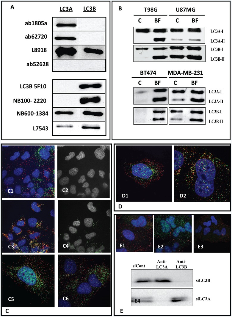
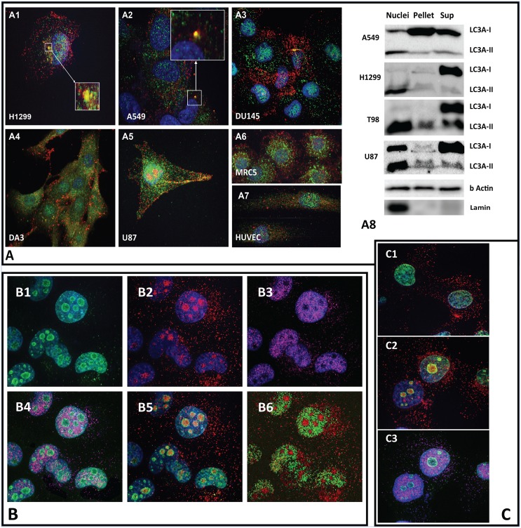
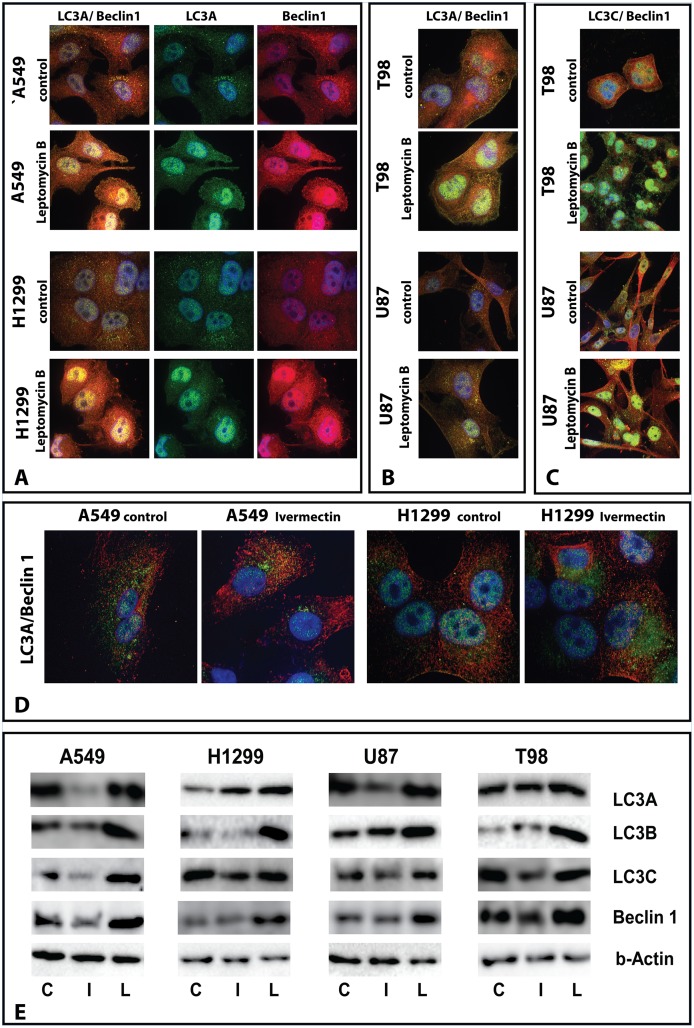
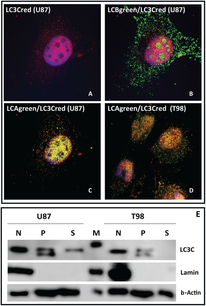
LC3s (MAP1-LC3A, B and C) are structural proteins of autophagosomal membranes, widely used as biomarkers of autophagy. Whether these three LC3 proteins have a similar biological role in autophagy remains obscure. We examine in parallel the subcellular expression patterns of the three LC3 proteins in a panel of human cancer cell lines, as well as in normal MRC5 fibroblasts and HUVEC, using confocal microscopy and western blot analysis of cell fractions. In the cytoplasm, there was a minimal co-localization between LC3A, B and C staining, suggesting that the relevant autophagosomes are formed by only one out of the three LC3 proteins. LC3A showed a perinuclear and nuclear localization, while LC3B was equally distributed throughout the cytoplasm and localized in the nucleolar regions. LC3C was located in the cytoplasm and strongly in the nuclei (excluding nucleoli), where it extensively co-localized with the LC3A and the Beclin-1 autophagy initiating protein. Beclin 1 is known to contain a nuclear trafficking signal. Blocking nuclear export function by Leptomycin B resulted in nuclear accumulation of all LC3 and Beclin-1 proteins, while Ivermectin that blocks nuclear import showed reduction of accumulation, but not in all cell lines. Since endogenous LC3 proteins are used as major markers of autophagy in clinical studies and cell lines, it is essential to check the specificity of the antibodies used, as the kinetics of these molecules are not identical and may have distinct biological roles. The distinct subcellular expression patterns of LC3s provide a basis for further studies.
Overview
The claims and statistics of this paper haven’t been checked yet.
Run the check — free, no account needed. It reads the open-access text and checks the sources it cites.
Integrity signals
Registry
Cites
Statistics
Figures
Verification
Claims the paper makes
The claims and statistics of this paper haven’t been checked yet.
Run the check — free, no account needed. It reads the open-access text and checks the sources it cites.
Claims other papers make about this one
Each is checked against this paper’s own text: does it say what the citing paper says it does?
In the citing paper
“some studies suggest isoform specificity in selective autophagy (Shpilka et al., 2011; Maruyama et al., 2014; Koukourakis et al., 2015).”
From Microtubule-associated protein 1 light chain 3B, (LC3B) is necessary to maintain lipid-mediated homeostasis in the retinal pigment epithelium (2018) · cites this paper as Koukourakis et al., 2015
In the citing paper
“Specifying individual LC3 isoform is necessary as the isoforms have different expression patterns and biology (Cann et al., 2008; Koukourakis et al., 2015).”
From Microtubule-associated protein 1 light chain 3B, (LC3B) is necessary to maintain lipid-mediated homeostasis in the retinal pigment epithelium (2018) · cites this paper as Koukourakis et al., 2015
Self-check
Percentages, sums and reported differences re-computed from the paper’s own numbers.
The self-check needs the paper’s full text.
Citations
Cites
LC3, a mammalian homologue of yeast Apg8p, is localized in autophagosome membranes after processing.
Kabeya, Y., Mizushima, N., Ueno, T., Yamamoto, A., Kirisako, T., Noda, T., Kominami, E., Ohsumi, Y., & Yoshimori, T. (2000) · The EMBO Journal · doi:10.1093/emboj/19.21.5720 · Cited by 6,605
Cui, C., Cui, J., Jin, F., Cui, Y., Li, R., Jiang, X., Tian, Y., Wang, R. C., Jiang, P., & Gao, J. (2017) · Cellular Physiology and Biochemistry · doi:10.1159/000479571 · Cited by 5,214
Tissue fractionation studies. 6. Intracellular distribution patterns of enzymes in rat-liver tissue
Duve, C. D., Pressman, B. C., Gianetto, R., Wattiaux, R., & Appelmans, F. (1955) · Biochemical Journal · doi:10.1042/bj0600604 · Cited by 4,176
Autophagy as a Regulated Pathway of Cellular Degradation
Klionsky, D. J., & Emr, S. D. (2000) · Science · doi:10.1126/science.290.5497.1717 · Cited by 3,630
Antimicrobial Autophagy: A Conserved Innate Immune Response in Drosophila
Moy, R. H., & Cherry, S. R. (2013) · Journal of Innate Immunity · doi:10.1159/000350326 · Cited by 3,551
A ubiquitin-like system mediates protein lipidation
Ichimura, Y., Kirisako, T., Takao, T., Satomi, Y., Shimonishi, Y., Ishihara, N., Mizushima, N., Tanida, I., Kominami, E., Ohsumi, M., et al. (2000) · Nature · doi:10.1038/35044114 · Cited by 2,139
Exportin 1 (Crm1p) Is an Essential Nuclear Export Factor
Stade, K., Ford, C. S., Guthrie, C., & Weis, K. (1997) · Cell · doi:10.1016/s0092-8674(00)80370-0 · Cited by 1,145
Kudo, N. R., Matsumori, N., Taoka, H., Fujiwara, D., Schreiner, E. P., Wolff, B., Yoshida, M., & Horinouchi, S. (1999) · Proceedings of the National Academy of Sciences · doi:10.1073/pnas.96.16.9112 · Cited by 1,033
Autophagy: assays and artifacts
Barth, S., Glick, D. R., & Macleod, K. F. (2010) · The Journal of Pathology · doi:10.1002/path.2694 · Cited by 819
Wagstaff, K. M., Sivakumaran, H., Heaton, S. M., Harrich, D., & Jans, D. A. (2012) · Biochemical Journal · doi:10.1042/bj20120150 · Cited by 682
He, H., Dang, Y., Dai, F., Guo, Z., Wu, J., She, X., Pei, Y., Chen, Y., Ling, W., Wu, C., et al. (2003) · Journal of Biological Chemistry · doi:10.1074/jbc.m303800200 · Cited by 273
Liang, X., Yu, J., Brown, K., & Levine, B. (2001) · PubMed · Cited by 267
Tanida, I., Ueno, T., & Kominami, E. (2004) · Journal of Biological Chemistry · doi:10.1074/jbc.m407016200 · Cited by 263
Defective autophagy in neurons and astrocytes from mice deficient in PI(3,5)P2
Ferguson, C. J., Lenk, G. M., & Meisler, M. H. (2009) · Human Molecular Genetics · doi:10.1093/hmg/ddp460 · Cited by 202
Molecular cloning and characterization of rat LC3A and LC3B—Two novel markers of autophagosome
Wu, J., Dang, Y., Su, W., Liu, C., Ma, H., Shan, Y., Pei, Y., Wan, B., Guo, J., & Yu, L. (2005) · Biochemical and Biophysical Research Communications · doi:10.1016/j.bbrc.2005.10.211 · Cited by 180
Aoki, H., Kondo, Y., Aldape, K., Yamamoto, A., Iwado, E., Yokoyama, T., Hollingsworth, E., Kobayashi, R., Hess, K. R., Shinojima, N., et al. (2008) · Autophagy · doi:10.4161/auto.5668 · Cited by 126
Karim, M. R., Kanazawa, T., Daigaku, Y., Fujimura, S., Miotto, G., & Kadowaki, M. (2007) · Autophagy · doi:10.4161/auto.4615 · Cited by 120
Nucleocytoplasmic Distribution and Dynamics of the Autophagosome Marker EGFP-LC3
Drake, K. R., Kang, M., & Kenworthy, A. K. (2010) · PLoS ONE · doi:10.1371/journal.pone.0009806 · Cited by 97
Bai, H., Inoue, J., Kawano, T., & Inazawa, J. (2012) · Oncogene · doi:10.1038/onc.2011.613 · Cited by 92
"Autophagic flux" in normal mouse tissues: Focus on endogenous LC3A processing
Zois, C. E., Giatromanolaki, A. N., Sivridis, E., Papaiakovou, M., Kainulainen, H., & Koukourakis, M. I. (2011) · Autophagy · doi:10.4161/auto.7.11.16664 · Cited by 63
He, W., Hu, C. X., Hou, J., Fan, L., Xu, Y., Liu, M.-H., Yan, S. Y., Chen, G. Q., & Huang, Y. (2014) · PLoS ONE · doi:10.1371/journal.pone.0113089 · Cited by 14
Cited by
Sorted by citations
Guidelines for the use and interpretation of assays for monitoring autophagy (4th edition) 1
Klionsky, D. J., Abdel-Aziz, A. K., Abdelfatah, S., Abdellatif, M., Abdoli, A., Abel, S., Abeliovich, H., Abildgaard, M. H., Abudu, Y. P., Acevedo-Arozena, A., et al. (2021) · Autophagy · doi:10.1080/15548627.2020.1797280 · Cited by 2,897
Self-citationKandemır, F. M., Küçükler, S., Çağlayan, C., Gür, C., Batil, A. A., & Gülçın, İ. (2017) · Journal of Food Biochemistry · doi:10.1111/jfbc.12398 · Cited by 141
Kandemır, F. M., Küçükler, S., Eldutar, E., Çağlayan, C., & Gülçın, İ. (2017) · Scientia Pharmaceutica · doi:10.3390/scipharm85010004 · Cited by 132
Maiti, P., Scott, J., Sengupta, D., Gharaibeh, A., & Dunbar, G. L. (2019) · International Journal of Molecular Sciences · doi:10.3390/ijms20020399 · Cited by 118
Ji, Y., Li, L., Ma, Y., Li, W., Li, L., Zhu, H., Wu, M., & Zhou, J. (2019) · The Journal of Nutritional Biochemistry · doi:10.1016/j.jnutbio.2019.03.018 · Cited by 105
Metadata
- Authors
- Koukourakis, M. I., Kalamida, D., Giatromanolaki, A., Zois, C. E., Sivridis, E., Pouliliou, S., Mitrakas, A., Gatter, K. C., & Harris, A. L.
- Affiliations
- Democritus University of Thrace (GR)
- John Radcliffe Hospital (GB)
- University of Oxford (GB)
- Corresponding author
- Koukourakis, Michael I.
- ORCID
- Venue
- PLoS ONE
- Publisher
- Public Library of Science (PLoS)
- Published
- 2015-09-17
- Volume
- 10
- Issue
- 9
- Pages
- e0137675
- ISSN
- 1932-6203 (ISSN-L)
- PMC
- PMC4574774
- OpenAlex
- W2300762239
- Cited by
- 183
- References
- 21
- Citations by year
- 2026: 13 · 2025: 12 · 2024: 27 · 2023: 16 · 2022: 16 · 2021: 17 · 2020: 19 · 2019: 24 · 2018: 13 · 2017: 14 · 2016: 11 · 2015: 1
- FWCI
- 10.15
- Type
- journal article
- Language
- en
- Open access
- gold
- doi.org
- Licence
- Locations
- PLoS ONE (publishedVersion, cc-by)
- Europe PMC (PubMed Central) (publishedVersion, cc-by)
- PubMed Central (publishedVersion, cc-by)
- PubMed (publishedVersion)
- DOAJ (DOAJ: Directory of Open Access Journals) (submittedVersion)
- Funders
- Cancer Research UK (11359, 16466)
- National Institute for Health and Care Research (NF-SI-0611-10163)
- Topics
- Life Sciences › Biochemistry, Genetics and Molecular Biology › Cell Biology › Autophagy in Disease and Therapy
- Life Sciences › Biochemistry, Genetics and Molecular Biology › Molecular Biology › Ubiquitin and proteasome pathways
- Life Sciences › Biochemistry, Genetics and Molecular Biology › Cell Biology › Cellular transport and secretion
- Keywords
- LC3A, LC3B, LC3C, autophagosomes, autophagy, subcellular localization, nuclear localization, Beclin-1, nuclear export, nuclear import, leptomycin B, ivermectin, antibody specificity, cancer cell lines
- MeSH terms
- Beclin-1, Antibodies, Autophagy, Fatty Acids, Unsaturated, Gene Expression Regulation, Humans, Ivermectin, Membrane Proteins, Microtubule-Associated Proteins, Neoplasms, Macrolides, Active Transport, Cell Nucleus, RNA Interference, RNA, Small Interfering, Cell Line, Tumor, Apoptosis Regulatory Proteins, Human Umbilical Vein Endothelial Cells
About these checks
Registry, retraction, Cites and statistics results are computed, not judged, with one exception: a printed reference that no index lists is read once by a model before it counts as a citation error. Claims are extracted by a model and kept only when their quote appears verbatim in the text; each cited source is then checked, with a model’s help, for whether it says what the claim relies on. Those verdicts can be wrong — read the claim’s quote and the cited source before relying on one.
Statistics and arithmetic are recomputed, not judged.
Confidence bands are shown only for fields calibrated on our annotated test set. None is calibrated yet, so no band is shown.
What counts as a claim ›How we measure confidence ›How we check this ›
Are you an author, or do you see a problem with this page? Request a review
