

The DOI matches its Crossref record; 110 of the 111 references it cites were found in a registry or an index.
A publicly available virtual cohort of four-chamber heart meshes for cardiac electro-mechanics simulations
Open accessAbstract
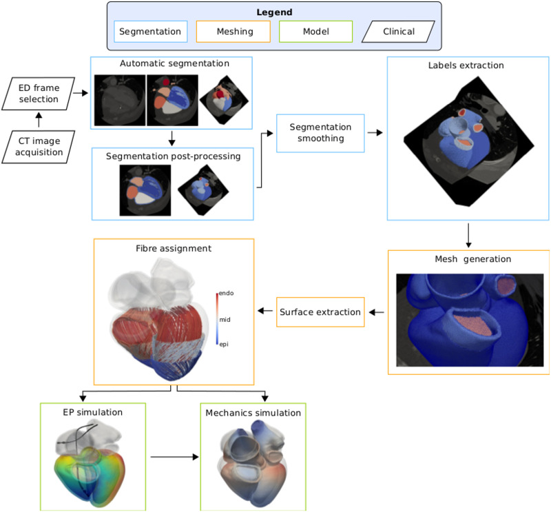
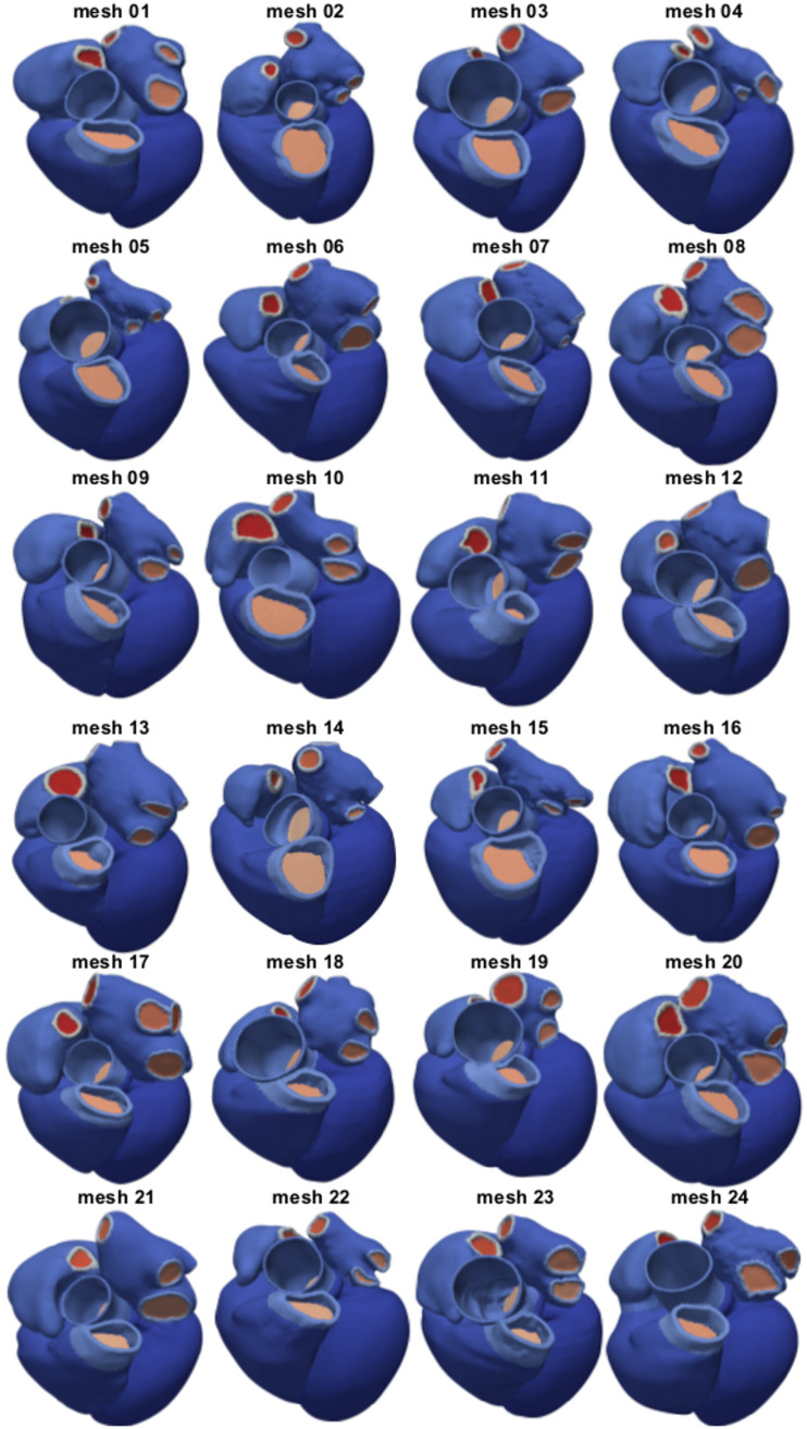
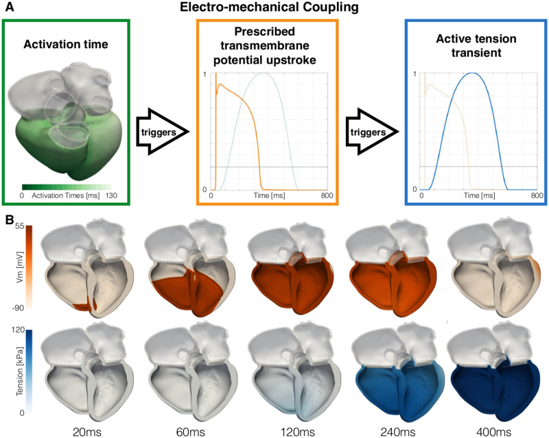
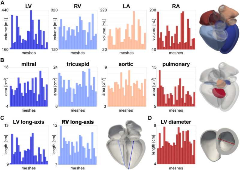
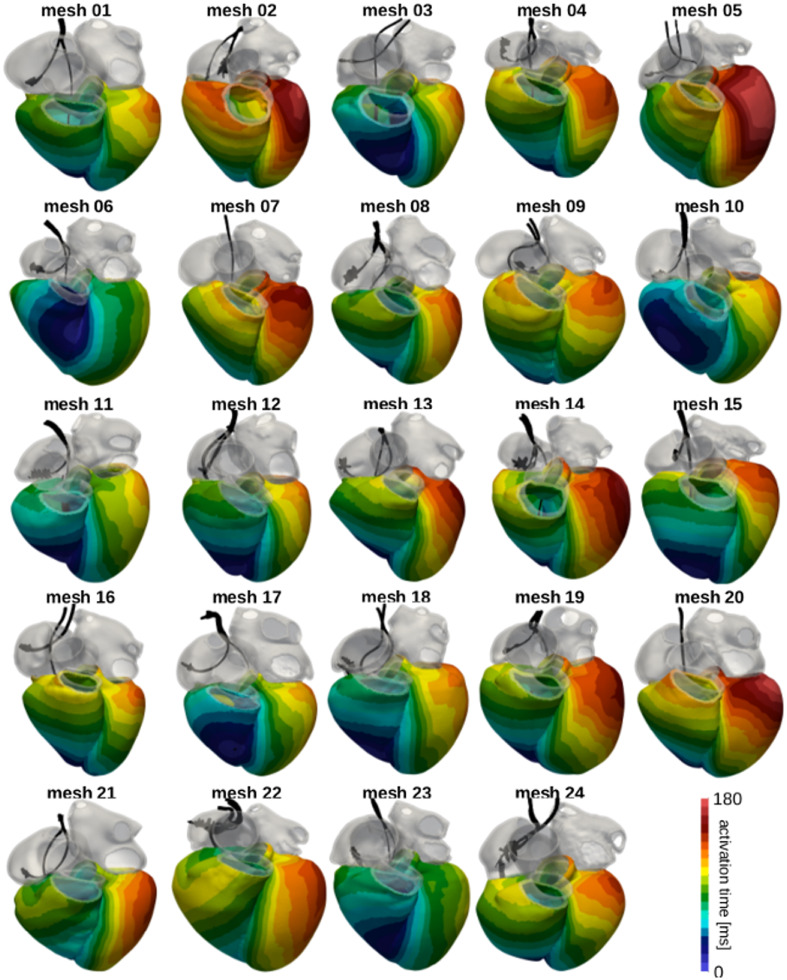
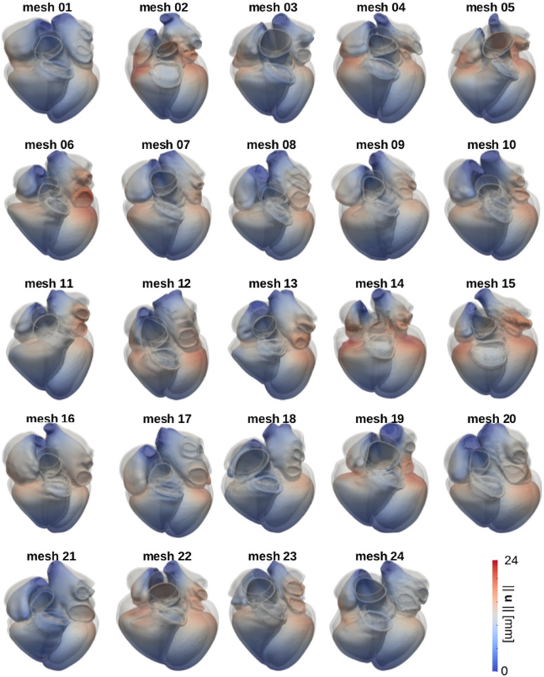
Computational models of the heart are increasingly being used in the development of devices, patient diagnosis and therapy guidance. While software techniques have been developed for simulating single hearts, there remain significant challenges in simulating cohorts of virtual hearts from multiple patients. To facilitate the development of new simulation and model analysis techniques by groups without direct access to medical data, image analysis techniques and meshing tools, we have created the first publicly available virtual cohort of twenty-four four-chamber hearts. Our cohort was built from heart failure patients, age 67±14 years. We segmented four-chamber heart geometries from end-diastolic (ED) CT images and generated linear tetrahedral meshes with an average edge length of 1.1±0.2mm. Ventricular fibres were added in the ventricles with a rule-based method with an orientation of -60° and 80° at the epicardium and endocardium, respectively. We additionally refined the meshes to an average edge length of 0.39±0.10mm to show that all given meshes can be resampled to achieve an arbitrary desired resolution. We ran simulations for ventricular electrical activation and free mechanical contraction on all 1.1mm-resolution meshes to ensure that our meshes are suitable for electro-mechanical simulations. Simulations for electrical activation resulted in a total activation time of 149±16ms. Free mechanical contractions gave an average left ventricular (LV) and right ventricular (RV) ejection fraction (EF) of 35±1% and 30±2%, respectively, and a LV and RV stroke volume (SV) of 95±28mL and 65±11mL, respectively. By making the cohort publicly available, we hope to facilitate large cohort computational studies and to promote the development of cardiac computational electro-mechanics for clinical applications.
Overview
The claims and statistics of this paper haven’t been checked yet.
Run the check — free, no account needed. It reads the open-access text and checks the sources it cites.
Integrity signals
Registry
Cites
Statistics
Figures
Verification
Claims the paper makes
The claims and statistics of this paper haven’t been checked yet.
Run the check — free, no account needed. It reads the open-access text and checks the sources it cites.
Claims other papers make about this one
Each is checked against this paper’s own text: does it say what the citing paper says it does?
The cited source says what the claim attributes to it.
In the citing paper
“For example, imaging data from 24 subjects was used in [40] to develop 24 four-chamber heart models.”
From Credibility assessment of in silico clinical trials for medical devices (2024) · cites this paper as [40]
The cited source says what the claim attributes to it.
In the citing paper
“Each of the 24 anatomical models represent a specific patient, although tissue material properties were defined from the literature.”
From Credibility assessment of in silico clinical trials for medical devices (2024) · cites this paper as [40]
Self-check
Percentages, sums and reported differences re-computed from the paper’s own numbers.
The self-check needs the paper’s full text.
Citations
Cites
Cardiac Resynchronization in Chronic Heart Failure
Abraham, W. T., Fisher, W. G., Smith, A. L., Delurgio, D. B., Leon, A. R., Loh, E., Kocovic, D. Z., Packer, M., Clavell, A. L., Hayes, D. L., et al. (2002) · New England Journal of Medicine · doi:10.1056/nejmoa013168 · Cited by 4,648
CARDIAC RESYNCHRONIZATION IN CHRONIC HEART FAILURE
Braham, W. I. T. A., Isher, W. E. G. F., Mith, L. S., Elurgio, D. A. B. D., Eon, A. N. R. L., Oh, E., Ocovic, D. U. Z. K., Acker, M. G., Lavell, L. C., Ayes, D. A. L. H., et al. (2002) · Cited by 3,808
Young, J. B., Abraham, W. T., & Smith, A. L. (2003) · ACC Current Journal Review · doi:10.1016/j.accreview.2003.08.087 · Cited by 1,670
Linde, C. M., Abraham, W. T., Gold, M. R., Sutton, M. S. J., Ghio, S., & Daubert, J. (2008) · Journal of the American College of Cardiology · doi:10.1016/j.jacc.2008.08.027 · Cited by 1,269
Alternans and spiral breakup in a human ventricular tissue model
ten Tusscher, K., & Panfilov, A. V. (2006) · American Journal of Physiology-Heart and Circulatory Physiology · doi:10.1152/ajpheart.00109.2006 · Cited by 1,126
Linde, C. M., Leclercq, C., Rex, S., Garrigue, S., Lavergne, T., Cazeau, S., McKenna, W. J., Fitzgerald, M., Deharo, J., Alonso, C., et al. (2002) · Journal of the American College of Cardiology · doi:10.1016/s0735-1097(02)01932-0 · Cited by 965
Thermodynamic relations for high elastic materials
Flory, P. J. (1961) · Transactions of the Faraday Society · doi:10.1039/tf9615700829 · Cited by 958
Maceira, A. M., Prasad, S. K., Khan, M. N., & Pennell, D. J. (2006) · Journal of Cardiovascular Magnetic Resonance · doi:10.1080/10976640600572889 · Cited by 851
Left Ventricular Remodeling in Heart Failure
Konstam, M. A., Kramer, D. G., Patel, A. R., Maron, M. S., & Udelson, J. E. (2011) · JACC. Cardiovascular imaging · doi:10.1016/j.jcmg.2010.10.008 · Cited by 808
Anatomy, echocardiography, and normal right ventricular dimensions
Ho, S. Y., & Nihoyannopoulos, P. (2006) · Heart · doi:10.1136/hrt.2005.077875 · Cited by 628
Cited by
Sorted by citations
The openCARP simulation environment for cardiac electrophysiology
Plank, G., Loewe, A., Neic, A., Augustin, C. M., Huang, Y.-L., Gsell, M. A. F., Karabelas, E., Nothstein, M., Prassl, A. J., Sánchez, J., et al. (2021) · Computer Methods and Programs in Biomedicine · doi:10.1016/j.cmpb.2021.106223 · Cited by 277
Self-citationScaling digital twins from the artisanal to the industrial
Niederer, S., Sacks, M. S., Girolami, M., & Willcox, K. E. (2021) · Nature Computational Science · doi:10.1038/s43588-021-00072-5 · Cited by 231
Self-citationTrayanova, N. A., Lyon, A., Shade, J. K., & Heijman, J. (2023) · Physiological Reviews · doi:10.1152/physrev.00017.2023 · Cited by 129
Fedele, M., Piersanti, R., Regazzoni, F., Salvador, M., Africa, P. C., Bucelli, M., Zingaro, A., Dede’, L., & Quarteroni, A. M. (2023) · Computer Methods in Applied Mechanics and Engineering · doi:10.1016/j.cma.2023.115983 · Cited by 126
Electro-Mechanical Whole-Heart Digital Twins: A Fully Coupled Multi-Physics Approach
Gerach, T., Schuler, S., Fröhlich, J., Lindner, L. P., Kovacheva, E. L., Moss, R., Wülfers, E. M., Seemann, G., Wieners, C., & Loewe, A. (2021) · Mathematics · doi:10.3390/math9111247 · Cited by 118
Metadata
- Authors
- Strocchi, M., Augustin, C. M., Gsell, M. A. F., Karabelas, E., Neic, A., Gillette, K., Razeghi, O., Prassl, A. J., Vigmond, E. J., Behar, J. M., Gould, J., Sidhu, B., Rinaldi, C. A., Bishop, M. J., Plank, G., & Niederer, S. A.
- Affiliations
- King's College London (GB)
- Medical University of Graz (AT)
- Université de Bordeaux (FR)
- Corresponding author
- Strocchi, Marina
- ORCID
- Venue
- PLoS ONE
- Publisher
- Public Library of Science (PLoS)
- Published
- 2020-06-26
- Volume
- 15
- Issue
- 6
- Pages
- e0235145
- ISSN
- 1932-6203 (ISSN-L)
- PMC
- PMC7319311
- OpenAlex
- W3037817333
- Cited by
- 112
- References
- 111
- Citations by year
- 2026: 13 · 2025: 17 · 2024: 18 · 2023: 24 · 2022: 21 · 2021: 15 · 2020: 3
- FWCI
- 9.75
- Type
- journal article
- Language
- en
- Open access
- gold
- doi.org
- Licence
- Locations
- PLoS ONE (publishedVersion, cc-by)
- PubMed Central (publishedVersion, cc-by)
- PubMed (publishedVersion)
- Research Portal (King's College London) (publishedVersion, other-oa)
- HAL (submittedVersion)
- RePEc: Research Papers in Economics (submittedVersion)
- DOAJ (DOAJ: Directory of Open Access Journals) (submittedVersion, cc-by-sa)
- Oskar-Bordeaux (Universite de Bordeaux) (submittedVersion)
- Unipub UB Graz (Universität Graz) (submittedVersion, cc-by)
- Funders
- Engineering and Physical Sciences Research Council (EP/M012492/1, NS/A000049/1, EP/L015226/1 and EP/P01268X/1)
- British Heart Foundation (PG/15/91/31812 and PG/13/37/30280)
- Kings Health Partners London National Institute for Health Research (NIHR) Biomedical Research Centre
- H2020 Marie Skłodowska-Curie Actions (750835)
- Austrian Science Fund (PI2760-B30)
- Topics
- Health Sciences › Medicine › Cardiology and Cardiovascular Medicine › Cardiac electrophysiology and arrhythmias
- Health Sciences › Medicine › Cardiology and Cardiovascular Medicine › Cardiac Arrhythmias and Treatments
- Health Sciences › Medicine › Cardiology and Cardiovascular Medicine › Cardiac pacing and defibrillation studies
- Keywords
- cardiac electromechanics, heart failure, tetrahedral meshes, ejection fraction, stroke volume
- MeSH terms
- Adult, Aged, Aged, 80 and over, Computer Simulation, Female, Heart Failure, Heart Ventricles, Humans, Male, Middle Aged, Surgical Mesh, Cohort Studies, Ventricular Function, Left, Ventricular Function, Right
About these checks
Registry, retraction, Cites and statistics results are computed, not judged, with one exception: a printed reference that no index lists is read once by a model before it counts as a citation error. Claims are extracted by a model and kept only when their quote appears verbatim in the text; each cited source is then checked, with a model’s help, for whether it says what the claim relies on. Those verdicts can be wrong — read the claim’s quote and the cited source before relying on one.
Statistics and arithmetic are recomputed, not judged.
Confidence bands are shown only for fields calibrated on our annotated test set. None is calibrated yet, so no band is shown.
What counts as a claim ›How we measure confidence ›How we check this ›
Are you an author, or do you see a problem with this page? Request a review
