

The DOI matches its Crossref record; 110 of the 111 references it cites were found in a registry or an index.
A publicly available virtual cohort of four-chamber heart meshes for cardiac electro-mechanics simulations
Open accessAbstract
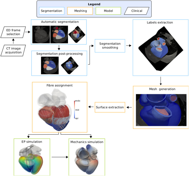
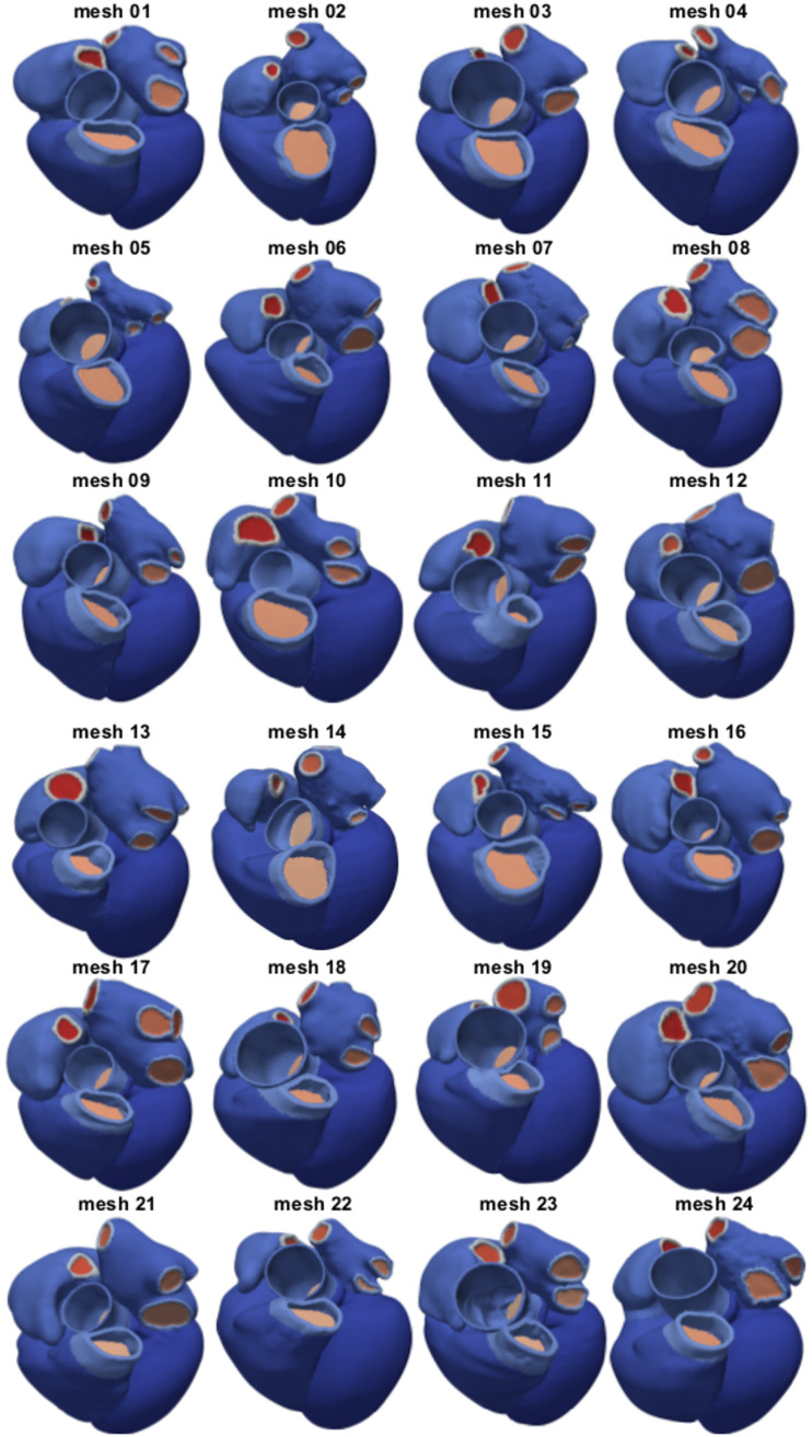
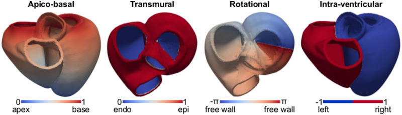
Computational models of the heart are increasingly being used in the development of devices, patient diagnosis and therapy guidance. While software techniques have been developed for simulating single hearts, there remain significant challenges in simulating cohorts of virtual hearts from multiple patients. To facilitate the development of new simulation and model analysis techniques by groups without direct access to medical data, image analysis techniques and meshing tools, we have created the first publicly available virtual cohort of twenty-four four-chamber hearts. Our cohort was built from heart failure patients, age 67±14 years. We segmented four-chamber heart geometries from end-diastolic (ED) CT images and generated linear tetrahedral meshes with an average edge length of 1.1±0.2mm. Ventricular fibres were added in the ventricles with a rule-based method with an orientation of -60° and 80° at the epicardium and endocardium, respectively. We additionally refined the meshes to an average edge length of 0.39±0.10mm to show that all given meshes can be resampled to achieve an arbitrary desired resolution. We ran simulations for ventricular electrical activation and free mechanical contraction on all 1.1mm-resolution meshes to ensure that our meshes are suitable for electro-mechanical simulations. Simulations for electrical activation resulted in a total activation time of 149±16ms. Free mechanical contractions gave an average left ventricular (LV) and right ventricular (RV) ejection fraction (EF) of 35±1% and 30±2%, respectively, and a LV and RV stroke volume (SV) of 95±28mL and 65±11mL, respectively. By making the cohort publicly available, we hope to facilitate large cohort computational studies and to promote the development of cardiac computational electro-mechanics for clinical applications.
Overview
The claims and statistics of this paper haven’t been checked yet.
Run the check — free, no account needed. It reads the open-access text and checks the sources it cites.
Integrity signals
Registry
Cites
Statistics
Figures
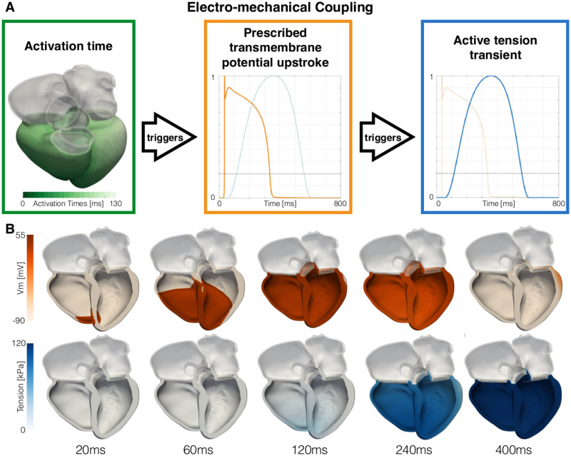
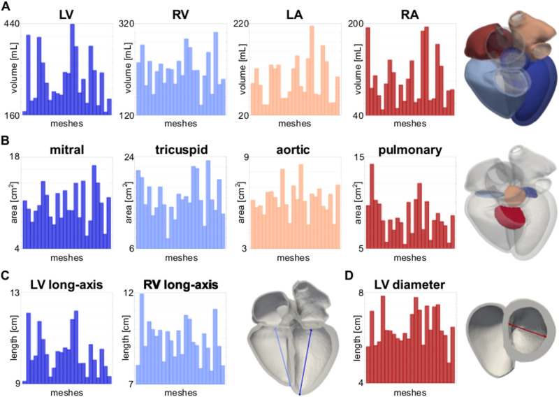
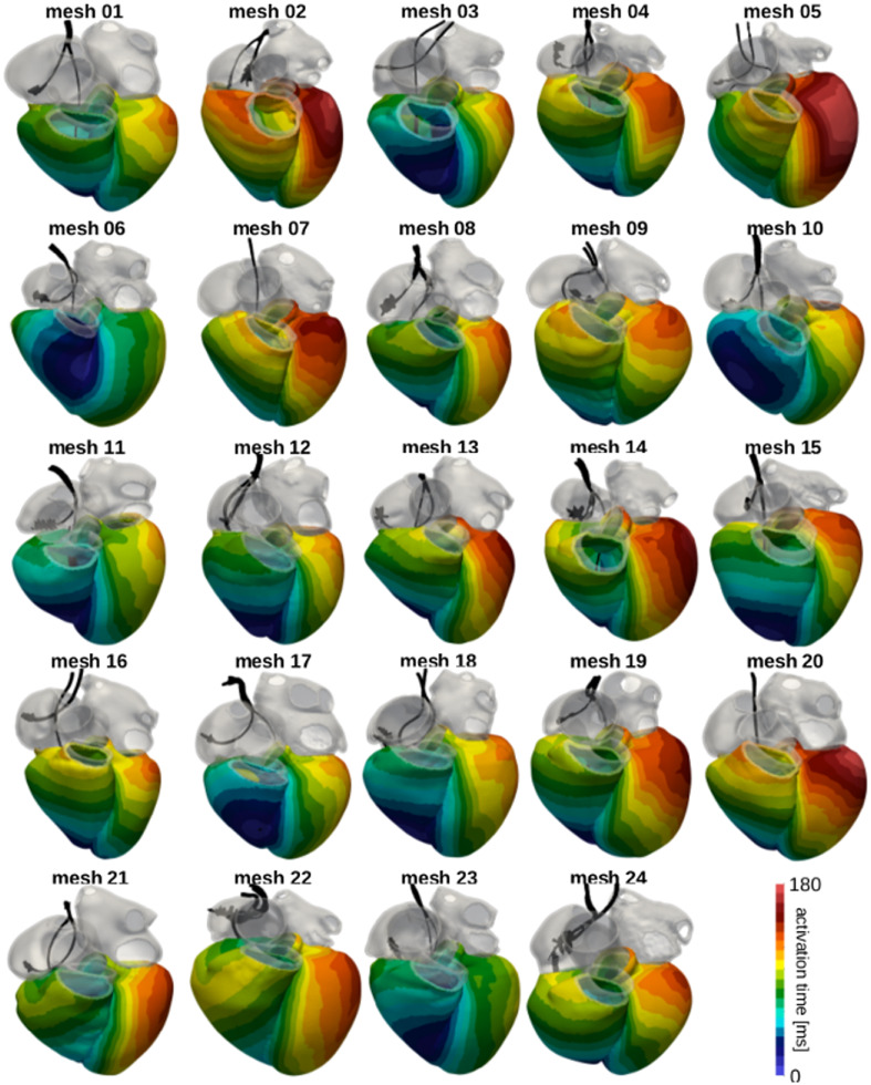
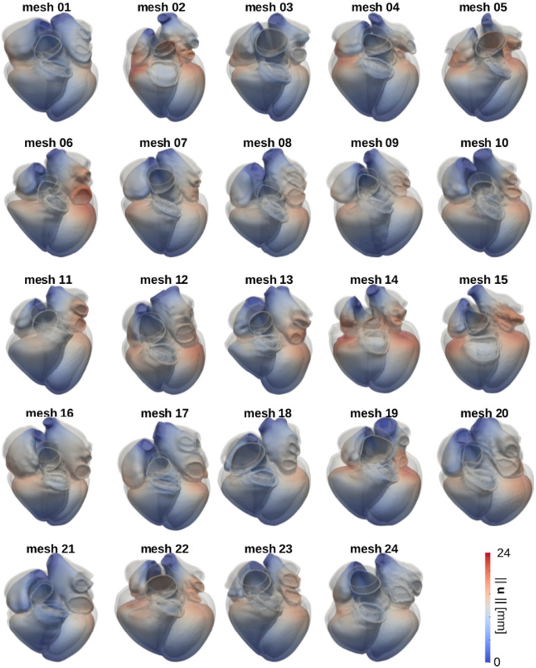
Verification
Claims the paper makes
The claims and statistics of this paper haven’t been checked yet.
Run the check — free, no account needed. It reads the open-access text and checks the sources it cites.
Claims other papers make about this one
Each is checked against this paper’s own text: does it say what the citing paper says it does?
The cited source says what the claim attributes to it.
In the citing paper
“For example, imaging data from 24 subjects was used in [40] to develop 24 four-chamber heart models.”
From Credibility assessment of in silico clinical trials for medical devices (2024) · cites this paper as [40]
The cited source says what the claim attributes to it.
In the citing paper
“Each of the 24 anatomical models represent a specific patient, although tissue material properties were defined from the literature.”
From Credibility assessment of in silico clinical trials for medical devices (2024) · cites this paper as [40]
Self-check
Percentages, sums and reported differences re-computed from the paper’s own numbers.
The self-check needs the paper’s full text.
Citations
Cites
Cardiac Resynchronization in Chronic Heart Failure
Abraham, W. T., Fisher, W. G., Smith, A. L., Delurgio, D. B., Leon, A. R., Loh, E., Kocovic, D. Z., Packer, M., Clavell, A. L., Hayes, D. L., et al. (2002) · New England Journal of Medicine · doi:10.1056/nejmoa013168 · Cited by 4,648
CARDIAC RESYNCHRONIZATION IN CHRONIC HEART FAILURE
Braham, W. I. T. A., Isher, W. E. G. F., Mith, L. S., Elurgio, D. A. B. D., Eon, A. N. R. L., Oh, E., Ocovic, D. U. Z. K., Acker, M. G., Lavell, L. C., Ayes, D. A. L. H., et al. (2002) · Cited by 3,808
Young, J. B., Abraham, W. T., & Smith, A. L. (2003) · ACC Current Journal Review · doi:10.1016/j.accreview.2003.08.087 · Cited by 1,670
Linde, C. M., Abraham, W. T., Gold, M. R., Sutton, M. S. J., Ghio, S., & Daubert, J. (2008) · Journal of the American College of Cardiology · doi:10.1016/j.jacc.2008.08.027 · Cited by 1,269
Alternans and spiral breakup in a human ventricular tissue model
ten Tusscher, K., & Panfilov, A. V. (2006) · American Journal of Physiology-Heart and Circulatory Physiology · doi:10.1152/ajpheart.00109.2006 · Cited by 1,126
Linde, C. M., Leclercq, C., Rex, S., Garrigue, S., Lavergne, T., Cazeau, S., McKenna, W. J., Fitzgerald, M., Deharo, J., Alonso, C., et al. (2002) · Journal of the American College of Cardiology · doi:10.1016/s0735-1097(02)01932-0 · Cited by 965
Thermodynamic relations for high elastic materials
Flory, P. J. (1961) · Transactions of the Faraday Society · doi:10.1039/tf9615700829 · Cited by 958
Maceira, A. M., Prasad, S. K., Khan, M. N., & Pennell, D. J. (2006) · Journal of Cardiovascular Magnetic Resonance · doi:10.1080/10976640600572889 · Cited by 851
Left Ventricular Remodeling in Heart Failure
Konstam, M. A., Kramer, D. G., Patel, A. R., Maron, M. S., & Udelson, J. E. (2011) · JACC. Cardiovascular imaging · doi:10.1016/j.jcmg.2010.10.008 · Cited by 808
Anatomy, echocardiography, and normal right ventricular dimensions
Ho, S. Y., & Nihoyannopoulos, P. (2006) · Heart · doi:10.1136/hrt.2005.077875 · Cited by 628
Passive Material Properties of Intact Ventricular Myocardium Determined From a Cylindrical Model
Guccione, J. M., McCulloch, A. D., & Waldman, L. K. (1991) · Journal of Biomechanical Engineering · doi:10.1115/1.2894084 · Cited by 624
Petersen, S. E., Aung, N., Sanghvi, M. M., Zemrak, F., Fung, K., Paiva, J. M., Francis, J. M., Khanji, M. Y., Lukaschuk, E. I., Lee, A. M., et al. (2016) · Journal of Cardiovascular Magnetic Resonance · doi:10.1186/s12968-017-0327-9 · Cited by 601
Maceira, A. M., Prasad, S. K., Khan, M. A., & Pennell, D. J. (2006) · European Heart Journal · doi:10.1093/eurheartj/ehl336 · Cited by 565
Bayer, J. D., Blake, R. C., Plank, G., & Trayanova, N. A. (2012) · Annals of Biomedical Engineering · doi:10.1007/s10439-012-0593-5 · Cited by 557
Self-citationZheng, Y., Barbu, A., Georgescu, B., Scheuering, M., & Comaniciu, D. (2008) · IEEE Transactions on Medical Imaging · doi:10.1109/tmi.2008.2004421 · Cited by 546
Lewandowski, A. J., Augustine, D. X., Lamata, P., Davis, E., Lazdam, M., Francis, J. M., McCormick, K. P., Wilkinson, A. R., Singhal, A., Lucas, A., et al. (2012) · Circulation · doi:10.1161/circulationaha.112.126920 · Cited by 469
Njoku, A., Kannabhiran, M. K., Arora, R., Reddy, P. C., Gopinathannair, R., Lakkireddy, D., & Dominic, P. S. (2017) · EP Europace · doi:10.1093/europace/eux013 · Cited by 420
The Living Heart Project: A robust and integrative simulator for human heart function
Baillargeon, B., Rebelo, N., Fox, D. D., Taylor, R. L., & Kuhl, E. (2014) · European Journal of Mechanics - A/Solids · doi:10.1016/j.euromechsol.2014.04.001 · Cited by 370
Echocardiography-Guided Left Ventricular Lead Placement for Cardiac Resynchronization Therapy
Saba, S., Marek, J. J., SCHWARTZMAN, D. S., Jain, S. K., ADELSTEIN, E. C., White, P., Oyenuga, O. A., Onishi, T., Soman, P., & Gorcsan, J. (2013) · Circulation Heart Failure · doi:10.1161/circheartfailure.112.000078 · Cited by 365
Solvers for the cardiac bidomain equations
Vigmond, E. J., dos Santos, R. W., Prassl, A. J., Deo, M., & Plank, G. (2007) · Progress in Biophysics and Molecular Biology · doi:10.1016/j.pbiomolbio.2007.07.012 · Cited by 345
Self-citationPrakosa, A., Arevalo, H., Deng, D., Boyle, P. M., Nikolov, P. P., Ashikaga, H., Blauer, J. J. E., Ghafoori, E., Park, C. J., Blake, R., et al. (2018) · Nature Biomedical Engineering · doi:10.1038/s41551-018-0282-2 · Cited by 299
A clinical appraisal of left atrial function
Stefanadis, C. (2001) · European Heart Journal · doi:10.1053/euhj.1999.2581 · Cited by 282
Knupp, P. (2000) · International Journal for Numerical Methods in Engineering · doi:10.1002/(sici)1097-0207(20000720)48:8 · Cited by 253
Computational techniques for solving the bidomain equations in three dimensions
Vigmond, E. J., Aguel, F., & Trayanova, N. A. (2002) · IEEE Transactions on Biomedical Engineering · doi:10.1109/tbme.2002.804597 · Cited by 228
Self-citationVentricular arrhythmias and the His–Purkinje system
Haı̈ssaguerre, M., Vigmond, E. J., Stuyvers, B. D. M. Y., Hocini, M., & Bernus, O. (2016) · Nature Reviews Cardiology · doi:10.1038/nrcardio.2015.193 · Cited by 225
Self-citationKnupp, P. (2000) · International Journal for Numerical Methods in Engineering · doi:10.1002/(sici)1097-0207(20000530)48:3 · Cited by 222
MRI-based finite-element analysis of left ventricular aneurysm
Walker, J. C., Ratcliffe, M. B., Zhang, P., Wallace, A. W., Fata, B., Hsu, E. W., Saloner, D., & Guccione, J. M. (2005) · American Journal of Physiology-Heart and Circulatory Physiology · doi:10.1152/ajpheart.01226.2004 · Cited by 205
Inhomogeneous Transmural Conduction During Early Ischaemia in Patients with Coronary Artery Disease
Taggart, P. I., Sutton, P. M., Tobias Opthof, A., Coronel, R., Trimlett, R., Pugsley, W. B., & Kallis, P. (2000) · Journal of Molecular and Cellular Cardiology · doi:10.1006/jmcc.2000.1105 · Cited by 200
Neic, A., Campos, F. O., Prassl, A. J., Niederer, S., Bishop, M. J., Vigmond, E. J., & Plank, G. (2017) · Journal of Computational Physics · doi:10.1016/j.jcp.2017.06.020 · Cited by 192
Self-citationStatistical shape model of atria, ventricles and epicardium from short- and long-axis MR images
Lötjönen, J. M. P. (2004) · Medical Image Analysis · doi:10.1016/j.media.2004.06.013 · Cited by 180
Coupling multi-physics models to cardiac mechanics
Nordsletten, D., Niederer, S., Nash, M. P., Hunter, P. J., & Smith, N. P. (2009) · Progress in Biophysics and Molecular Biology · doi:10.1016/j.pbiomolbio.2009.11.001 · Cited by 177
Self-citationBishop, M. J., Plank, G., Burton, R. A. B., Schneider, J. E., Gavaghan, D. J., Grau, V., & Kohl, P. K. (2009) · American Journal of Physiology-Heart and Circulatory Physiology · doi:10.1152/ajpheart.00606.2009 · Cited by 171
Self-citationLand, S., Park‐Holohan, S., Smith, N. P., dos Remedios, C. G., Kentish, J. C., & Niederer, S. (2017) · Journal of Molecular and Cellular Cardiology · doi:10.1016/j.yjmcc.2017.03.008 · Cited by 169
Self-citationGlobal Left Atrial Failure in Heart Failure
Triposkiadis, F. K., Pieske, B. M., Butler, J., Parissis, J., Giamouzis, G., Skoularigis, J. S., Brutsaert, D. L., & Boudoulas, H. K. (2016) · European Journal of Heart Failure · doi:10.1002/ejhf.645 · Cited by 161
Length-dependent tension in the failing heart and the efficacy of cardiac resynchronization therapy
Niederer, S., Plank, G., Chinchapatnam, P. P., Ginks, M., Lamata, P., Rhode, K., Rinaldi, C. A., Razavi, R., & Smith, N. P. (2010) · Cardiovascular Research · doi:10.1093/cvr/cvq318 · Cited by 159
Self-citationPlank, G., Burton, R. A. B., Hales, P. W., Bishop, M. J., Mansoori, T., Bernabéu, M. O., Garny, A., Prassl, A. J., Bollensdorff, C., Mason, F. E., et al. (2009) · Philosophical Transactions of the Royal Society A Mathematical Physical and Engineering Sciences · doi:10.1098/rsta.2009.0056 · Cited by 158
Self-citationAugustin, C. M., Neic, A., Liebmann, M., Prassl, A. J., Niederer, S., Haase, G., & Plank, G. (2015) · Journal of Computational Physics · doi:10.1016/j.jcp.2015.10.045 · Cited by 154
Self-citationMorris, D. A., Gailani, M., Pérez, A. V., Blaschke, F., Dietz, R., Haverkamp, W., & Özcelik, C. (2011) · Journal of the American Society of Echocardiography · doi:10.1016/j.echo.2011.02.004 · Cited by 151
Nearly isochoric elastic deformations: Application to rubberlike solids
Ogden, R. W. (1978) · Journal of the Mechanics and Physics of Solids · doi:10.1016/0022-5096(78)90012-1 · Cited by 146
Beinart, R., Abbara, S., Blum, A. R., Ferencik, M., Heist, E. K., Ruskin, J. N., & MANSOUR, M. C. (2011) · Journal of Cardiovascular Electrophysiology · doi:10.1111/j.1540-8167.2011.02100.x · Cited by 134
Pfaller, M. R., Hörmann, J. M., Weigl, M., Nagler, A., Chabiniok, R., Bertoglio, C., & Wall, W. A. (2018) · Biomechanics and Modeling in Mechanobiology · doi:10.1007/s10237-018-1098-4 · Cited by 133
Bayer, J. D., Prassl, A. J., Pashaei, A., Gómez, J. F., Frontera, A., Neic, A., Plank, G., & Vigmond, E. J. (2018) · Medical Image Analysis · doi:10.1016/j.media.2018.01.005 · Cited by 131
Self-citationSimulating Human Cardiac Electrophysiology on Clinical Time-Scales
Niederer, S., Mitchell, L., Smith, N. P., & Plank, G. (2011) · Frontiers in Physiology · doi:10.3389/fphys.2011.00014 · Cited by 128
Self-citationLand, S., Gurev, V., Arens, S., Augustin, C. M., Baron, L., Blake, R., Bradley, C. P., Castro, S., Crozier, A., Favino, M., et al. (2015) · Proceedings of the Royal Society A Mathematical Physical and Engineering Sciences · doi:10.1098/rspa.2015.0641 · Cited by 128
Self-citationImage‐based models of cardiac structure in health and disease
Vadakkumpadan, F., Arevalo, H., Prassl, A. J., Chen, J., Kickinger, F., Kohl, P. K., Plank, G., & Trayanova, N. A. (2010) · WIREs Systems Biology and Medicine · doi:10.1002/wsbm.76 · Cited by 124
Self-citationLeft Atrial Volume and Geometry in Healthy Aging
Aurigemma, G. P., Gottdiener, J. S., Arnold, A. M., Chinali, M., Hill, J. C., & Kitzman, D. W. (2009) · Circulation Cardiovascular Imaging · doi:10.1161/circimaging.108.826602 · Cited by 121
Generating Purkinje networks in the human heart
Costabal, F. S., Hurtado, D. E., & Ellen Kuhl, A. (2015) · Journal of Biomechanics · doi:10.1016/j.jbiomech.2015.12.025 · Cited by 121
Left Ventricular Diameter and Risk Stratification for Sudden Cardiac Death
Narayanan, K., Reinier, K., Teodorescu, C., Uy‐Evanado, A., Aleong, R. G., Chugh, H. S., Nichols, G. A., Gunson, K., London, B., Jui, J., et al. (2014) · Journal of the American Heart Association · doi:10.1161/jaha.114.001193 · Cited by 120
Roney, C. H., Pashaei, A., Meo, M., Dubois, R., Boyle, P. M., Trayanova, N. A., Cochet, H., Niederer, S., & Vigmond, E. J. (2019) · Medical Image Analysis · doi:10.1016/j.media.2019.04.004 · Cited by 120
Self-citationMaceira, A. M., Cosín-Sales, J., Roughton, M., Prasad, S. K., & Pennell, D. J. (2013) · Journal of Cardiovascular Magnetic Resonance · doi:10.1186/1532-429x-15-29 · Cited by 114
Ferrer-Albero, A., Sebastián, R., Sánchez‐Quintana, D., Matas, J. F. R., Godoy, E. J., Martínez-Mateu, L., & Sáiz, J. (2015) · PLoS ONE · doi:10.1371/journal.pone.0141573 · Cited by 110
Parallel imaging with nonlinear reconstruction using variational penalties
Knöll, F., Clason, C., Bredies, K., Uecker, M., & Stollberger, R. (2011) · Magnetic Resonance in Medicine · doi:10.1002/mrm.22964 · Cited by 104
Boyden, P. A., Hirose, M., & Dun, W. (2009) · Heart Rhythm · doi:10.1016/j.hrthm.2009.09.017 · Cited by 100
Strocchi, M., Gsell, M. A. F., Augustin, C. M., Razeghi, O., Roney, C. H., Prassl, A. J., Vigmond, E. J., Behar, J. M., Gould, J., Rinaldi, C. A., et al. (2020) · Journal of Biomechanics · doi:10.1016/j.jbiomech.2020.109645 · Cited by 100
Self-citationMedrano-Gracia, P., Cowan, B. R., Ambale-Venkatesh, B., Bluemke, D. A., Eng, J., Finn, J. P., Fonseca, C. G., Lima, J. A., Suinesiaputra, A., & Young, A. A. (2014) · Journal of Cardiovascular Magnetic Resonance · doi:10.1186/s12968-014-0056-2 · Cited by 98
Fritz, T., Wieners, C., Seemann, G., Steen, H., & Dössel, O. (2013) · Biomechanics and Modeling in Mechanobiology · doi:10.1007/s10237-013-0523-y · Cited by 95
Mousavi, N., Tan, T. C., Ali, M. U., Halpern, E. F., Wang, L., & Scherrer‐Crosbie, M. (2015) · European Heart Journal - Cardiovascular Imaging · doi:10.1093/ehjci/jev113 · Cited by 95
A bilayer model of human atria: mathematical background, construction, and assessment
Labarthe, S., Bayer, J. D., Coudière, Y., Henry, J., Cochet, H., Jaı̈s, P., & Vigmond, E. J. (2014) · EP Europace · doi:10.1093/europace/euu256 · Cited by 92
Self-citationA High-Resolution Atlas and Statistical Model of the Human Heart From Multislice CT
Hoogendoorn, C., Duchateau, N., Sánchez‐Quintana, D., Whitmarsh, T., Sukno, F. M., Craene, M. D., Lekadir, K., & Frangi, A. F. (2012) · IEEE Transactions on Medical Imaging · doi:10.1109/tmi.2012.2230015 · Cited by 91
Effect of trabeculae and papillary muscles on the hemodynamics of the left ventricle
Vedula, V., Seo, J. H., Lardo, A. C., & Mittal, R. (2015) · Theoretical and Computational Fluid Dynamics · doi:10.1007/s00162-015-0349-6 · Cited by 90
Towards Personalized Cardiology: Multi-Scale Modeling of the Failing Heart
Kayvanpour, E., Mansi, T., Sedaghat‐Hamedani, F., Amr, A., Neumann, D., Georgescu, B., Seegerer, P., Kamen, A., Haas, J., Frese, K., et al. (2015) · PLoS ONE · doi:10.1371/journal.pone.0134869 · Cited by 87
Matsukubo, H., Matsuura, T., Endo, N., Asayama, J., & WATANABE, T. (1977) · Circulation · doi:10.1161/01.cir.56.2.278 · Cited by 85
Quantification of Full Left Ventricular Metrics via Deep Regression Learning With Contour-Guidance
Wang, W., Wang, Y., Wu, Y., Lin, T., Li, S., & Chen, B. (2019) · IEEE Access · doi:10.1109/access.2019.2907564 · Cited by 83
Linde, C., Leclercq, C., & Rex, S. (2002) · ACC Current Journal Review · doi:10.1016/s1062-1458(02)00962-5 · Cited by 79
Modeling our understanding of the His-Purkinje system
Vigmond, E. J., & Stuyvers, B. D. M. Y. (2015) · Progress in Biophysics and Molecular Biology · doi:10.1016/j.pbiomolbio.2015.12.013 · Cited by 74
Self-citationConstruction of a Statistical Model for Cardiac Motion Analysis Using Nonrigid Image Registration
Chandrashekara, R., Rao, A. W., Sanchez-Ortiz, G. I., Mohiaddin, R. H., & Rueckert, D. (2003) · Lecture notes in computer science · doi:10.1007/978-3-540-45087-0_50 · Cited by 73
The Role of Purkinje-Myocardial Coupling during Ventricular Arrhythmia: A Modeling Study
Behradfar, E., Nygren, A., & Vigmond, E. J. (2014) · PLoS ONE · doi:10.1371/journal.pone.0088000 · Cited by 72
Self-citationDuchateau, N., Craene, M. D., Piella, G., Silva, E., Doltra, A., Sitges, M., Bijnens, B. H., & Frangi, A. F. (2011) · Medical Image Analysis · doi:10.1016/j.media.2010.12.006 · Cited by 71
Personalized computational modeling of left atrial geometry and transmural myofiber architecture
Fastl, T. E., Tobon‐Gomez, C., Crozier, A., Whitaker, J., Rajani, R., McCarthy, K. P., Sánchez‐Quintana, D., Ho, S. Y., O'Neill, M. D., Plank, G., et al. (2018) · Medical Image Analysis · doi:10.1016/j.media.2018.04.001 · Cited by 67
Self-citationImage-Based Personalization of Cardiac Anatomy for Coupled Electromechanical Modeling
Crozier, A., Augustin, C. M., Neic, A., Prassl, A. J., Höller, M., Fastl, T. E., Hennemuth, A., Bredies, K., Kühne, T., Bishop, M. J., et al. (2015) · Annals of Biomedical Engineering · doi:10.1007/s10439-015-1474-5 · Cited by 65
Self-citationAn automatic service for the personalization of ventricular cardiac meshes
Lamata, P., Sinclair, M., Kerfoot, E., Lee, A., Crozier, A., Blazevic, B., Land, S., Lewandowski, A. J., Barber, D. C., Niederer, S., et al. (2013) · Journal of The Royal Society Interface · doi:10.1098/rsif.2013.1023 · Cited by 60
Self-citationIntra- and interventricular asynchrony of electromechanics in the ventricularly paced heart
Kerckhoffs, R. C. P., Bovendeerd, P. H. M., Prinzen, F. W., Smits, K. C., & Arts, T. G. J. (2003) · Journal of Engineering Mathematics · doi:10.1023/b:engi.0000007972.73874.da · Cited by 59
Influence of atrial contraction dynamics on cardiac function
Land, S., & Niederer, S. (2017) · International Journal for Numerical Methods in Biomedical Engineering · doi:10.1002/cnm.2931 · Cited by 56
Self-citationA Fast Iterative Method for Solving the Eikonal Equation on Tetrahedral Domains
Fu, Z., Kirby, R. M., & Whitaker, R. (2013) · SIAM Journal on Scientific Computing · doi:10.1137/120881956 · Cited by 54
The Importance of Purkinje Activation in Long Duration Ventricular Fibrillation
Huang, J. Y., Dosdall, D. J., Cheng, K., Li, L., Rogers, J. M., & Ideker, R. E. (2014) · Journal of the American Heart Association · doi:10.1161/jaha.113.000495 · Cited by 53
Strik, M., Regoli, F. D., Auricchio, A., & Prinzen, F. W. (2012) · Journal of Cardiovascular Translational Research · doi:10.1007/s12265-012-9351-1 · Cited by 52
Crozier, A., Blazevic, B., Lamata, P., Plank, G., Ginks, M., Duckett, S. G., Sohal, M., Shetty, A., Rinaldi, C. A., Razavi, R., et al. (2015) · Journal of Molecular and Cellular Cardiology · doi:10.1016/j.yjmcc.2015.10.026 · Cited by 50
Self-citationOkada, J., Washio, T., Nakagawa, M., WATANABE, M., Kadooka, Y., Kariya, T., Yamashita, H., Yamada, Y., Momomura, S., Nagai, R., et al. (2017) · Journal of Molecular and Cellular Cardiology · doi:10.1016/j.yjmcc.2017.05.006 · Cited by 48
Roney, C. H., Bayer, J. D., Cochet, H., Meo, M., Dubois, R., Jaı̈s, P., & Vigmond, E. J. (2018) · PLoS Computational Biology · doi:10.1371/journal.pcbi.1006166 · Cited by 48
Self-citationLyon, A., Bueno‐Orovio, A., Zacur, E., Ariga, R., Grau, V., Neubauer, S., Watkins, H. C., Rodríguez, B., & Mincholé, A. (2018) · EP Europace · doi:10.1093/europace/euy226 · Cited by 48
Sacco, F., Paun, B., Lehmkuhl, O., Iles, T. L., Iaizzo, P. A., Houzeaux, G., Vázquez, M., Butakoff, C., & Aguado‐Sierra, J. (2018) · Frontiers in Physiology · doi:10.3389/fphys.2018.00458 · Cited by 46
Bishop, M. J., & Plank, G. (2012) · The Journal of Physiology · doi:10.1113/jphysiol.2012.229062 · Cited by 45
Self-citationPatient-specific modeling of left ventricular electromechanics as a driver for haemodynamic analysis
Augustin, C. M., Crozier, A., Neic, A., Prassl, A. J., Karabelas, E., da Silva, T. F., Fernandes, J. F., Campos, F. O., Kühne, T., & Plank, G. (2016) · EP Europace · doi:10.1093/europace/euw369 · Cited by 45
Self-citationLee, A., Nguyên, U. C., Razeghi, O., Gould, J., Sidhu, B. S., Sieniewicz, B., Behar, J. M., Mafi-Rad, M., Plank, G., Prinzen, F. W., et al. (2019) · Medical Image Analysis · doi:10.1016/j.media.2019.06.017 · Cited by 44
Self-citationImproved identifiability of myocardial material parameters by an energy-based cost function
Nasopoulou, A., Shetty, A., Lee, J., Nordsletten, D., Rinaldi, C. A., Lamata, P., & Niederer, S. (2017) · Biomechanics and Modeling in Mechanobiology · doi:10.1007/s10237-016-0865-3 · Cited by 40
Self-citationKerckhoffs, R. C. P., Omens, J. H., & McCulloch, A. D. (2012) · EP Europace · doi:10.1093/europace/eus274 · Cited by 38
Bishop, M. J., Hales, P. W., Plank, G., Gavaghan, D. J., Scheider, J., & Grau, V. (2009) · Lecture notes in computer science · doi:10.1007/978-3-642-01932-6_10 · Cited by 38
Self-citationTobon‐Gomez, C., Duchateau, N., Sebastián, R., Marchesseau, S., Cámara, Ó., Donal, E., Craene, M. D., Pashaei, A., Relan, J., Steghofer, M., et al. (2013) · Medical & Biological Engineering & Computing · doi:10.1007/s11517-013-1044-7 · Cited by 34
Mean Thoracic Aortic Wall Thickness Determination by Cine MRI with Steady-State Free Precession
Mensel, B., Kühn, J., Schneider, T., Quadrat, A., & Hegenscheid, K. (2013) · Academic Radiology · doi:10.1016/j.acra.2013.03.014 · Cited by 33
Imaging technologies for cardiac fiber and heart failure: a review
Watson, S. R., Dormer, J. D., & Fei, B. (2018) · Heart Failure Reviews · doi:10.1007/s10741-018-9684-1 · Cited by 33
Gsell, M. A. F., Augustin, C. M., Prassl, A. J., Karabelas, E., Fernandes, J. F., Kelm, M., Goubergrits, L., Kühne, T., & Plank, G. (2018) · International Journal for Numerical Methods in Biomedical Engineering · doi:10.1002/cnm.3147 · Cited by 30
Self-citationValidation of an arterial constitutive model accounting for collagen content and crosslinking
Tian, L., Wang, Z., Liu, Y., Eickhoff, J. C., Eliceiri, K. W., & Chesler, N. C. (2015) · Acta Biomaterialia · doi:10.1016/j.actbio.2015.11.058 · Cited by 29
In vivo porcine left atrial wall stress: Computational model
Martino, E. S. D., Bellini, C. M., & SCHWARTZMAN, D. S. (2011) · Journal of Biomechanics · doi:10.1016/j.jbiomech.2011.08.023 · Cited by 25
Quality Metrics for High Order Meshes: Analysis of the Mechanical Simulation of the Heart Beat
Lamata, P., Roy, I., Blazevic, B., Crozier, A., Land, S., Niederer, S., Hose, D. R., & Smith, N. P. (2012) · IEEE Transactions on Medical Imaging · doi:10.1109/tmi.2012.2231094 · Cited by 23
Self-citationIvanov, A. V., Mohamed, A., Asfour, A. M. S., Ho, J., Khan, S., Chenn, O., Klem, I., Ramasubbu, K., Brener, S. J., & Heitner, J. F. (2017) · PLoS ONE · doi:10.1371/journal.pone.0173245 · Cited by 21
Orthogonal decomposition of left ventricular remodeling in myocardial infarction
Zhang, X., Medrano−Gracia, P., Ambale‐Venkatesh, B., Bluemke, D. A., Cowan, B. R., Finn, J. P., Kadish, A. H., Lee, D. C., Lima, J. A. C., Young, A. A., et al. (2017) · GigaScience · doi:10.1093/gigascience/gix005 · Cited by 17
García-Bustos, V., Sebastián, R., Izquierdo, M., Ríos‐Navarro, C., Bodı́, V., Chorro, F. J., & Ruiz‐Saurí, A. (2019) · PLoS ONE · doi:10.1371/journal.pone.0212096 · Cited by 17
Classification of left ventricular size: diameter or volume with contrast echocardiography?
Gibson, P. H., Becher, H. H., & Choy, J. B. (2014) · Open Heart · doi:10.1136/openhrt-2014-000147 · Cited by 16
A framework for biomechanics simulations using four-chamber cardiac models
Jafari, A., Pszczolkowski, E., & Krishnamurthy, A. (2019) · Journal of Biomechanics · doi:10.1016/j.jbiomech.2019.05.019 · Cited by 14
Hyde, E., Behar, J. M., Crozier, A., Claridge, S., Jackson, T., Sohal, M., Gill, J. S., O'Neill, M. D., Razavi, R., Niederer, S., et al. (2016) · Pacing and Clinical Electrophysiology · doi:10.1111/pace.12854 · Cited by 12
Self-citationDistributed Data Management Service for VPH Applications
Koulouzis, S., Belloum, A. S. Z., Bubak, M., Lamata, P., Nolte, D., Vasyunin, D., & de Laat, C. T. A. M. (2015) · IEEE Internet Computing · doi:10.1109/mic.2015.71 · Cited by 10
Continuous Spatio-temporal Atlases of the Asymptomatic and Infarcted Hearts
Medrano−Gracia, P., Cowan, B. R., Bluemke, D. A., Finn, J. P., Kadish, A. H., Lee, D. C., Lima, J. A. C., Suinesiaputra, A., & Young, A. A. (2014) · Lecture notes in computer science · doi:10.1007/978-3-642-54268-8_17 · Cited by 8
Cardiac resynchronization in chronic heart failure
Abraham, W. T., Fisher, W. G., & Smith, A. L. (2002) · ACC Current Journal Review · doi:10.1016/s1062-1458(02)00961-3 · Cited by 8
Identification of the material parameters of an aortic wall
Horný, L., Žitný, R., Chlup, H., & Macková, H. (2007) · Cited by 7
The Cardiac Atlas Project: Towards a Map of the Heart
Backhaus, M., Chung, J. D., Cowan, B. R., Fonseca, C. G., Tao, W., & Young, A. A. (2010) · Springer eBooks · doi:10.1007/978-1-4419-6691-9_7 · Cited by 4
Young, J. B. (2003) · JAMA · doi:10.1001/jama.289.20.2685 · Cited by 4
Strocchi, M., Augustin, C. M., Gsell, M. A. F., Karabelas, E., Neic, A., Gillette, K. K., Razeghi, O., Prassl, A. J., Vigmond, E. J., Behar, J. M., et al. (2020) · Figshare · doi:10.5281/zenodo.3890033 · Cited by 2
Self-citationRevealing Differences in Anatomical Remodelling of the Systemic Right Ventricle
Zacur, E., Wong, J., Razavi, R., Geva, T., Greil, G. F., & Lamata, P. (2015) · Lecture notes in computer science · doi:10.1007/978-3-319-20309-6_12 · Cited by 1
Papillary muscles contraction does not change ventricular wall mechanics
Gurev, V., Korte, J. C., Hafez, O. M., Fattebert, J., Richards, D. F., & Rice, J. J. (2015) · Computing in cardiology · doi:10.1109/cic.2015.7408654 · Cited by 1
Martinez, M. E., Walton, R. D., Haı̈ssaguerre, M., Hocini, M., & Bernus, O. (2018) · Archives of Cardiovascular Diseases Supplements · doi:10.1016/j.acvdsp.2018.02.036 · Cited by 1
Cited by
Sorted by citations
The openCARP simulation environment for cardiac electrophysiology
Plank, G., Loewe, A., Neic, A., Augustin, C. M., Huang, Y.-L., Gsell, M. A. F., Karabelas, E., Nothstein, M., Prassl, A. J., Sánchez, J., et al. (2021) · Computer Methods and Programs in Biomedicine · doi:10.1016/j.cmpb.2021.106223 · Cited by 277
Self-citationScaling digital twins from the artisanal to the industrial
Niederer, S., Sacks, M. S., Girolami, M., & Willcox, K. E. (2021) · Nature Computational Science · doi:10.1038/s43588-021-00072-5 · Cited by 231
Self-citationTrayanova, N. A., Lyon, A., Shade, J. K., & Heijman, J. (2023) · Physiological Reviews · doi:10.1152/physrev.00017.2023 · Cited by 129
Fedele, M., Piersanti, R., Regazzoni, F., Salvador, M., Africa, P. C., Bucelli, M., Zingaro, A., Dede’, L., & Quarteroni, A. M. (2023) · Computer Methods in Applied Mechanics and Engineering · doi:10.1016/j.cma.2023.115983 · Cited by 126
Electro-Mechanical Whole-Heart Digital Twins: A Fully Coupled Multi-Physics Approach
Gerach, T., Schuler, S., Fröhlich, J., Lindner, L. P., Kovacheva, E. L., Moss, R., Wülfers, E. M., Seemann, G., Wieners, C., & Loewe, A. (2021) · Mathematics · doi:10.3390/math9111247 · Cited by 118
Metadata
- Authors
- Strocchi, M., Augustin, C. M., Gsell, M. A. F., Karabelas, E., Neic, A., Gillette, K., Razeghi, O., Prassl, A. J., Vigmond, E. J., Behar, J. M., Gould, J., Sidhu, B., Rinaldi, C. A., Bishop, M. J., Plank, G., & Niederer, S. A.
- Affiliations
- King's College London (GB)
- Medical University of Graz (AT)
- Université de Bordeaux (FR)
- Corresponding author
- Strocchi, Marina
- ORCID
- Venue
- PLoS ONE
- Publisher
- Public Library of Science (PLoS)
- Published
- 2020-06-26
- Volume
- 15
- Issue
- 6
- Pages
- e0235145
- ISSN
- 1932-6203 (ISSN-L)
- PMC
- PMC7319311
- OpenAlex
- W3037817333
- Cited by
- 112
- References
- 111
- Citations by year
- 2026: 13 · 2025: 17 · 2024: 18 · 2023: 24 · 2022: 21 · 2021: 15 · 2020: 3
- FWCI
- 9.75
- Type
- journal article
- Language
- en
- Open access
- gold
- doi.org
- Licence
- Locations
- PLoS ONE (publishedVersion, cc-by)
- PubMed Central (publishedVersion, cc-by)
- PubMed (publishedVersion)
- Research Portal (King's College London) (publishedVersion, other-oa)
- HAL (submittedVersion)
- RePEc: Research Papers in Economics (submittedVersion)
- DOAJ (DOAJ: Directory of Open Access Journals) (submittedVersion, cc-by-sa)
- Oskar-Bordeaux (Universite de Bordeaux) (submittedVersion)
- Unipub UB Graz (Universität Graz) (submittedVersion, cc-by)
- Funders
- Engineering and Physical Sciences Research Council (EP/M012492/1, NS/A000049/1, EP/L015226/1 and EP/P01268X/1)
- British Heart Foundation (PG/15/91/31812 and PG/13/37/30280)
- Kings Health Partners London National Institute for Health Research (NIHR) Biomedical Research Centre
- H2020 Marie Skłodowska-Curie Actions (750835)
- Austrian Science Fund (PI2760-B30)
- Topics
- Health Sciences › Medicine › Cardiology and Cardiovascular Medicine › Cardiac electrophysiology and arrhythmias
- Health Sciences › Medicine › Cardiology and Cardiovascular Medicine › Cardiac Arrhythmias and Treatments
- Health Sciences › Medicine › Cardiology and Cardiovascular Medicine › Cardiac pacing and defibrillation studies
- Keywords
- cardiac electromechanics, heart failure, tetrahedral meshes, ejection fraction, stroke volume
- MeSH terms
- Adult, Aged, Aged, 80 and over, Computer Simulation, Female, Heart Failure, Heart Ventricles, Humans, Male, Middle Aged, Surgical Mesh, Cohort Studies, Ventricular Function, Left, Ventricular Function, Right
About these checks
Registry, retraction, Cites and statistics results are computed, not judged, with one exception: a printed reference that no index lists is read once by a model before it counts as a citation error. Claims are extracted by a model and kept only when their quote appears verbatim in the text; each cited source is then checked, with a model’s help, for whether it says what the claim relies on. Those verdicts can be wrong — read the claim’s quote and the cited source before relying on one.
Statistics and arithmetic are recomputed, not judged.
Confidence bands are shown only for fields calibrated on our annotated test set. None is calibrated yet, so no band is shown.
What counts as a claim ›How we measure confidence ›How we check this ›
Are you an author, or do you see a problem with this page? Request a review
