

The DOI matches its Crossref record; 85 of the 85 references it cites were found in a registry or an index.
Analysis of the Leishmania mexicana promastigote cell cycle using imaging flow cytometry provides new insights into cell cycle flexibility and events of short duration
Open accessAbstract
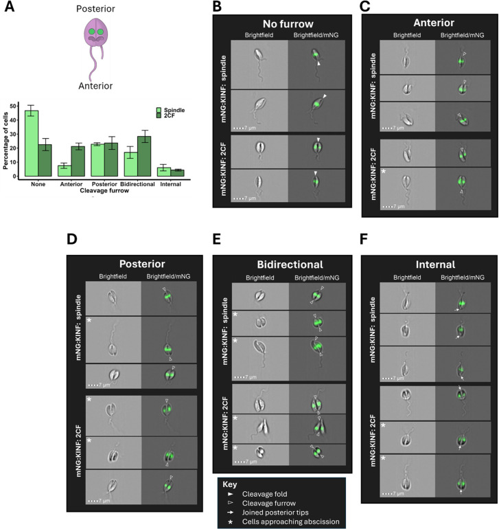
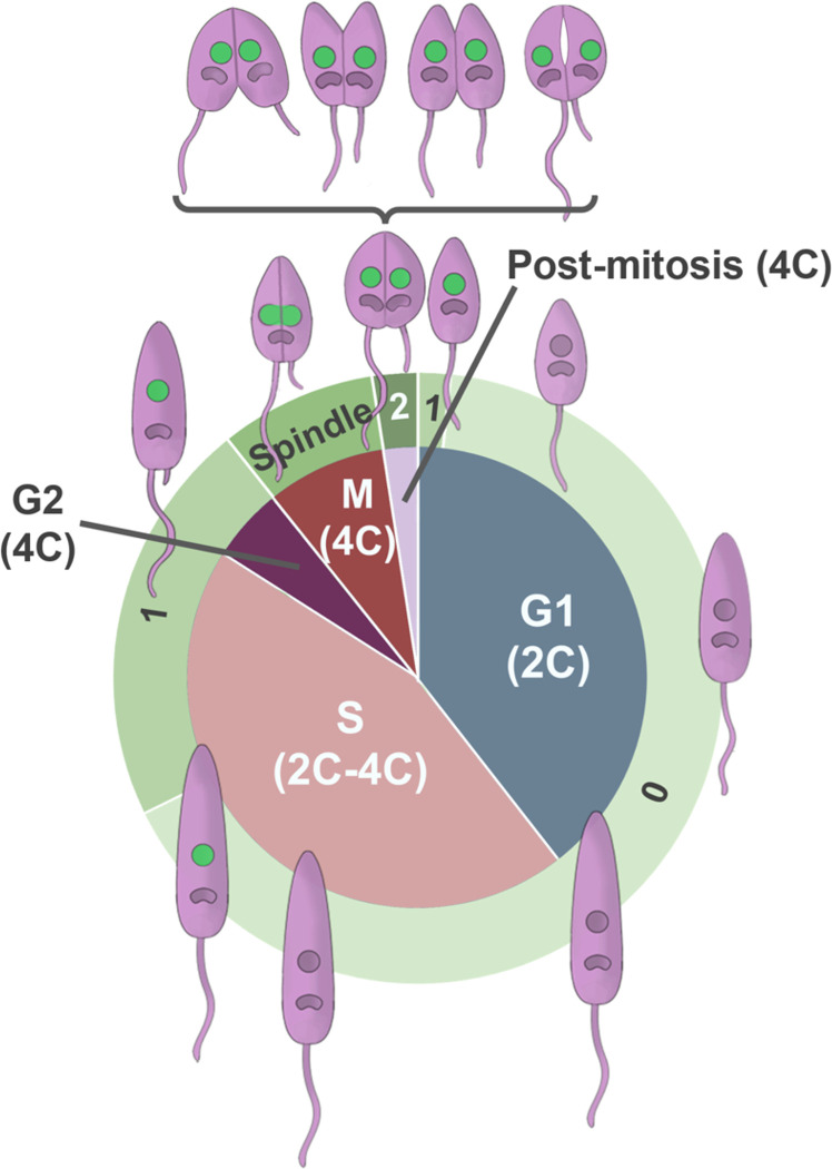
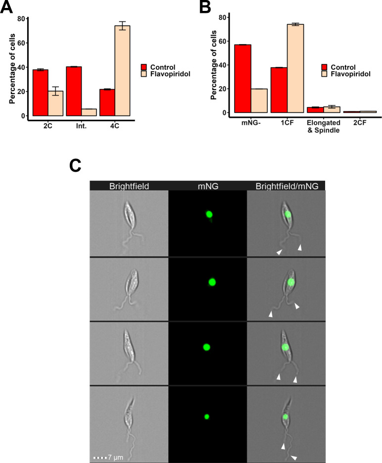
Promastigote Leishmania mexicana have a complex cell division cycle characterised by the ordered replication of several single-copy organelles, a prolonged S phase and rapid G2 and cytokinesis phases, accompanied by cell cycle stage-associated morphological changes. Here we exploit these morphological changes to develop a high-throughput and semi-automated imaging flow cytometry (IFC) pipeline to analyse the cell cycle in live L. mexicana. Firstly, we demonstrate that, unlike several other DNA stains, Vybrant™ DyeCycle™ Orange (DCO) is non-toxic and enables quantitative DNA imaging in live promastigotes. Secondly, by tagging the orphan spindle kinesin, KINF, with mNeonGreen, we describe KINF’s cell cycle-dependent expression and localisation. Then, by combining manual gating of DCO DNA intensity profiles with automated masking and morphological measurements of parasite images, visual determination of the number of flagella per cell, and automated masking and analysis of mNG:KINF fluorescence, we provide a newly detailed description of L. mexicana promastigote cell cycle events that, for the first time, includes the durations of individual G2, mitosis and post-mitosis phases, and identifies G1 cells within the first 12 minutes of the new cell cycle. Our custom-developed masking and gating scheme allowed us to identify elusive G2 cells and to demonstrate that the CDK-inhibitor, flavopiridol, arrests cells in G2 phase, rather than mitosis, providing proof-of-principle of the utility of IFC for drug mechanism-of-action studies. Further, the high-throughput nature of IFC allowed the close examination of promastigote cytokinesis, revealing considerable flexibility in both the timing of cytokinesis initiation and the direction of furrowing, in contrast to the related kinetoplastid parasite, Trypanosoma brucei and many other cell types. Our new pipeline offers many advantages over traditional methods of cell cycle analysis such as fluorescence microscopy and flow cytometry and paves the way for novel high-throughput analysis of Leishmania cell division.
Overview
The claims and statistics of this paper haven’t been checked yet.
Run the check — free, no account needed. It reads the open-access text and checks the sources it cites.
Integrity signals
Registry
Cites
Statistics
Figures
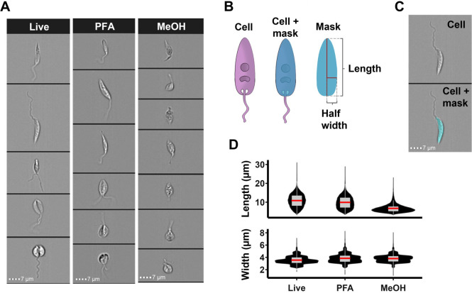
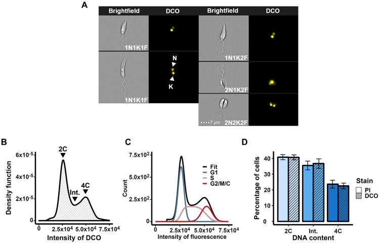
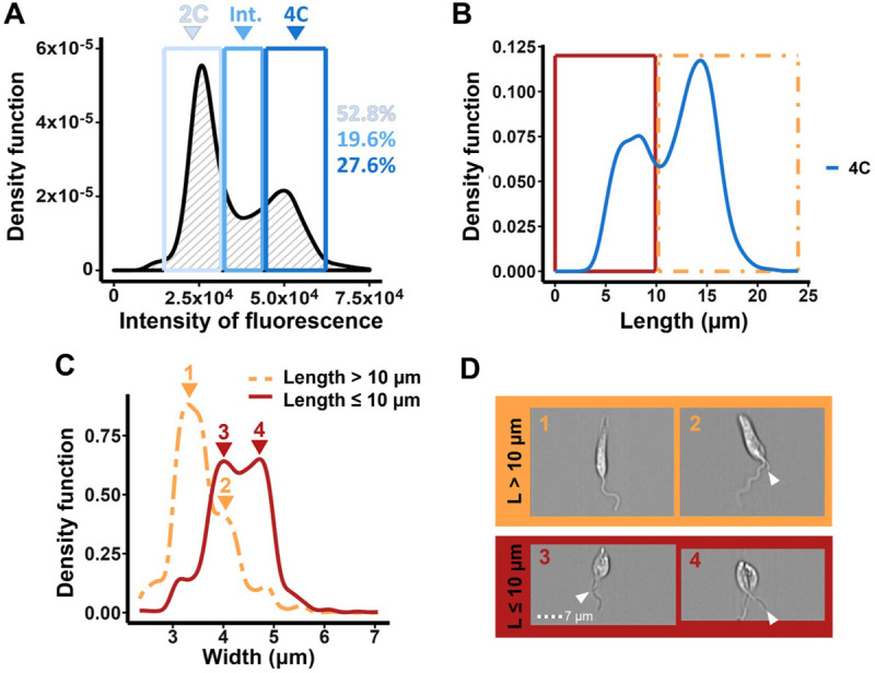
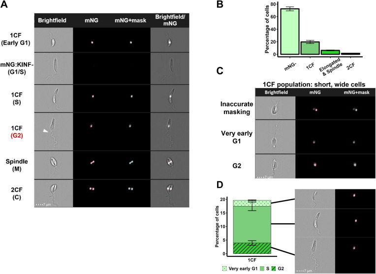
Verification
Claims the paper makes
The claims and statistics of this paper haven’t been checked yet.
Run the check — free, no account needed. It reads the open-access text and checks the sources it cites.
Claims other papers make about this one
Each is checked against this paper’s own text: does it say what the citing paper says it does?
The source backs part of the claim, not all of it — or backs it for a different population, condition or comparison.
In the citing paper
“This method can be used to evaluate the proliferation rates of microalgae depending on the cultivation conditions, e.g., changes in nutrient availability or light intensity [235].”
From Multi-Sensing Monitoring of the Microalgae Biomass Cultivation Systems for Biofuels and Added Value Products Synthesis—Challenges and Opportunities (2025) · cites this paper as [235]
Experimental — accuracy not yet measured
Self-check
Percentages, sums and reported differences re-computed from the paper’s own numbers.
The self-check needs the paper’s full text.
Citations
Cites
NIH Image to ImageJ: 25 years of image analysis
Schneider, C. A., Rasband, W. S., & Eliceiri, K. W. (2012) · Nature Methods · doi:10.1038/nmeth.2089 · Cited by 64,638
A CRISPR Cas9 high-throughput genome editing toolkit for kinetoplastids
Beneke, T., Madden, R., Makin, L., Valli, J., Sunter, J. D., & Gluenz, E. (2017) · Royal Society Open Science · doi:10.1098/rsos.170095 · Cited by 392
Label-free cell cycle analysis for high-throughput imaging flow cytometry
Blasi, T., Hennig, H., Summers, H. D., Theis, F. J., Cerveira, J., Patterson, J. O., Davies, D. C., Filby, A. J., Carpenter, A. E., & Rees, P. C. T. (2016) · Nature Communications · doi:10.1038/ncomms10256 · Cited by 301
Reconstructing cell cycle and disease progression using deep learning
Eulenberg, P., Köhler, N., Blasi, T., Filby, A. J., Carpenter, A. E., Rees, P. C. T., Theis, F. J., & Wolf, F. A. (2017) · Nature Communications · doi:10.1038/s41467-017-00623-3 · Cited by 278
A pragmatic approach to the analysis of DNA histograms with a definable G1 peak
Watson, J. V., Chambers, S. H., & Smith, P. J. (1987) · Cytometry · doi:10.1002/cyto.990080101 · Cited by 258
The cell cycle of Leishmania: morphogenetic events and their implications for parasite biology
Wheeler, R. J., Gluenz, E., & Gull, K. (2010) · Molecular Microbiology · doi:10.1111/j.1365-2958.2010.07479.x · Cited by 222
mScarlet3: a brilliant and fast-maturing red fluorescent protein
Gadella, T. W. J., van Weeren, L., Stouthamer, J., Hink, M. A., Wolters, A. H. G., Giepmans, B. N. G., Aumonier, S., Dupuy, J., & Royant, A. (2023) · Nature Methods · doi:10.1038/s41592-023-01809-y · Cited by 218
Sunter, J. D., & Gull, K. (2017) · Open Biology · doi:10.1098/rsob.170165 · Cited by 192
Rees, P. C. T., Summers, H. D., Filby, A. J., Carpenter, A. E., & Doan, M. (2022) · Nature Reviews Methods Primers · doi:10.1038/s43586-022-00167-x · Cited by 180
A model for the computer analysis of synchronous DNA distributions obtained by flow cytometry
Fox, M. H. (1980) · Cytometry · doi:10.1002/cyto.990010114 · Cited by 146
Wheeler, R. J., Gull, K., & Gluenz, E. (2012) · BMC Biology · doi:10.1186/1741-7007-10-1 · Cited by 139
Dynamics of Microbial Populations
WILLIAMS, F. M. (1971) · Elsevier eBooks · doi:10.1016/b978-0-12-547201-2.50011-x · Cited by 131
Synchronization of Leishmania tarentolae by Hydroxyurea
Simpson, L., & Braly, P. S. (1970) · The Journal of Protozoology · doi:10.1111/j.1550-7408.1970.tb04719.x · Cited by 121
Meiosis and Haploid Gametes in the Pathogen Trypanosoma brucei
Peacock, L., Bailey, M., Carrington, M., & Gibson, W. C. (2014) · Current Biology · doi:10.1016/j.cub.2013.11.044 · Cited by 119
Lippeveld, M., Knill, C., Ladlow, E., Fuller, A., Michaelis, L. J., Saeys, Y., Filby, A. J., & Peralta, D. (2019) · Cytometry Part A · doi:10.1002/cyto.a.23920 · Cited by 117
Hurrell, B. P., Schuster, S., Grün, E., Coutaz, M., Williams, R., Held, W., MALISSEN, B., Malissen, M., Yousefi, S., Simon, H., et al. (2015) · PLoS Pathogens · doi:10.1371/journal.ppat.1004929 · Cited by 115
Morphological Events during the Cell Cycle of Leishmania major
Ambit, A., Woods, K. L., Cull, B., Coombs, G. H., & Mottram, J. C. (2011) · Eukaryotic Cell · doi:10.1128/ec.05118-11 · Cited by 108
Pollen analysis using multispectral imaging flow cytometry and deep learning
Dunker, S., Švara, E. M., Rákosy, D., Boho, D., Mäder, P., Hornick, T., & Knight, T. M. (2020) · New Phytologist · doi:10.1111/nph.16882 · Cited by 107
Hammarton, T. C., Krämer, S., Tetley, L., Boshart, M., & Mottram, J. C. (2007) · Molecular Microbiology · doi:10.1111/j.1365-2958.2007.05866.x · Cited by 106
Self-citationSádlová, J., Price, H. P., Smith, B. A., Votýpka, J., Volf, P., & Smith, D. F. (2010) · Cellular Microbiology · doi:10.1111/j.1462-5822.2010.01507.x · Cited by 105
Chapter 18 DNA Content Histogram and Cell-Cycle Analysis
Rabinovitch, P. S. (1994) · Methods in cell biology · doi:10.1016/s0091-679x(08)61723-9 · Cited by 102
The CRK3 protein kinase is essential for cell cycle progression of Leishmania mexicana
Hassan, P., Fergusson, D., Grant, K. M., & Mottram, J. C. (2001) · Molecular and Biochemical Parasitology · doi:10.1016/s0166-6851(01)00220-1 · Cited by 100
Kanada, M., Nagasaki, A., & Uyeda, T. Q. P. (2005) · Molecular Biology of the Cell · doi:10.1091/mbc.e05-03-0233 · Cited by 95
The Mechanics of Mitotic Cell Rounding
Taubenberger, A., Baum, B., & Matthews, H. K. (2020) · Frontiers in Cell and Developmental Biology · doi:10.3389/fcell.2020.00687 · Cited by 92
γδ T cells suppress Plasmodium falciparum blood-stage infection by direct killing and phagocytosis
Junqueira, C., Polidoro, R. B., de Castro, G. D., Absalon, S., Liang, Z., Santara, S. S., Crespo, Â. C., Pereira, D. B., Gazzinelli, R. T., Dvorin, J. D., et al. (2021) · Nature Immunology · doi:10.1038/s41590-020-00847-4 · Cited by 91
Dunker, S., Boho, D., Wäldchen, J., & Mäder, P. (2018) · BMC Ecology · doi:10.1186/s12898-018-0209-5 · Cited by 88
LeishGEdit: A Method for Rapid Gene Knockout and Tagging Using CRISPR-Cas9
Beneke, T., & Gluenz, E. (2019) · Methods in molecular biology · doi:10.1007/978-1-4939-9210-2_9 · Cited by 87
Coordination of the Cell Cycle in Trypanosomes
Wheeler, R. J., Keith Gull, B., & Sunter, J. D. (2019) · Annual Review of Microbiology · doi:10.1146/annurev-micro-020518-115617 · Cited by 78
Rodrigues, M. A., Beaton-Green, L. A., Wilkins, R. C., & Fenech, M. F. (2018) · Mutation Research/Genetic Toxicology and Environmental Mutagenesis · doi:10.1016/j.mrgentox.2018.05.003 · Cited by 75
Filby, A. J., Perucha, E., Summers, H. D., Rees, P. C. T., Chana, P. S., Heck, S., Lord, G. M., & Davies, D. C. (2011) · Cytometry Part A · doi:10.1002/cyto.a.21091 · Cited by 74
Wójcik, K., & Dobrucki, J. W. (2008) · Cytometry Part A · doi:10.1002/cyto.a.20573 · Cited by 74
Acetylation of histone H4K4 is cell cycle regulated and mediated by HAT3 in Trypanosoma brucei
Siegel, T. N., Kawahara, T., DeGrasse, J. A., Janzen, C. J., Horn, D., & Cross, G. (2007) · Molecular Microbiology · doi:10.1111/j.1365-2958.2007.06079.x · Cited by 69
Infection-adapted emergency hematopoiesis promotes visceral leishmaniasis
Abidin, B. M., Hammami, A., Stäger, S., & Heinonen, K. M. (2017) · PLoS Pathogens · doi:10.1371/journal.ppat.1006422 · Cited by 67
Cellular landmarks of Trypanosoma brucei and Leishmania mexicana
Halliday, C., Billington, K., Wang, Z., Madden, R., Dean, S., Sunter, J. D., & Wheeler, R. J. (2018) · Molecular and Biochemical Parasitology · doi:10.1016/j.molbiopara.2018.12.003 · Cited by 67
Genome-wide mapping reveals single-origin chromosome replication in Leishmania, a eukaryotic microbe
Marques, C. A., Dickens, N. J., Paape, D., Campbell, S. J., & McCulloch, R. (2015) · Genome biology · doi:10.1186/s13059-015-0788-9 · Cited by 65
Calvert, M., Lannigan, J. A., & Pemberton, L. F. (2008) · Cytometry Part A · doi:10.1002/cyto.a.20609 · Cited by 64
Imaging flow cytometry analysis of intracellular pathogens
Haridas, V., Ranjbar, S., Vorobjev, I. A., Goldfeld, A. E., & Barteneva, N. S. (2016) · Methods · doi:10.1016/j.ymeth.2016.09.007 · Cited by 64
Miller, H. W., Suleiman, R. L., & Ralston, K. S. (2019) · mBio · doi:10.1128/mbio.00068-19 · Cited by 56
Dekel, E., Rivkin, A., Heidenreich, M., Nadav, Y., Ofir‐Birin, Y., Porat, Z., & Regev‐Rudzki, N. (2016) · Methods · doi:10.1016/j.ymeth.2016.06.021 · Cited by 53
Dent, B., Ogle, L. F., O'Donnell, R. L., Hayes, N., Mallick, U. K., Curtin, N. J., Boddy, A. V., Plummer, E. R., Edmondson, R. J., Reeves, H. L., et al. (2015) · International Journal of Cancer · doi:10.1002/ijc.29680 · Cited by 49
Minocha, N., Kumar, D., Rajanala, K., & Saha, S. (2011) · Journal of Eukaryotic Microbiology · doi:10.1111/j.1550-7408.2011.00539.x · Cited by 46
Protein diversity in discrete structures at the distal tip of the trypanosome flagellum
Varga, V., Moreira-Leite, F. F., Portman, N., & Gull, K. (2017) · Proceedings of the National Academy of Sciences · doi:10.1073/pnas.1703553114 · Cited by 45
Christian, D. A., Koshy, A. A., Reuter, M. A., Betts, M. R., Boothroyd, J. C., & Hunter, C. A. (2014) · Infection and Immunity · doi:10.1128/iai.01643-14 · Cited by 43
Carlsen, E. D., Jie, Z., Liang, Y., Henard, C. A., Hay, C., Sun, J., Guedes, H. L. D. M., & Soong, L. (2015) · Journal of Innate Immunity · doi:10.1159/000373923 · Cited by 42
Muchlińska, A., Smentoch, J., Zaczek, A. J., & Bednarz‐Knoll, N. (2022) · Cancers · doi:10.3390/cancers14174178 · Cited by 41
TLR7 Sensing by Neutrophils Is Critical for the Control of Cutaneous Leishmaniasis
Regli, I. B., Passelli, K., Martínez-Salazar, B., Amore, J., Hurrell, B. P., Müller, A. J., & Tacchini‐Cottier, F. (2020) · Cell Reports · doi:10.1016/j.celrep.2020.107746 · Cited by 37
Wills, J. W., Verma, J. R., Rees, B. J., Harte, D. S. G., Haxhiraj, Q., Barnes, C. M., Barnes, R., Rodrigues, M. A., Doan, M., Filby, A. J., et al. (2021) · Archives of Toxicology · doi:10.1007/s00204-021-03113-0 · Cited by 37
Niswander, L. M., McGrath, K. E., Kennedy, J. C., & Palis, J. (2014) · Cytometry Part A · doi:10.1002/cyto.a.22438 · Cited by 36
Power, A. L., Barber, D., Groenhof, S. R. M., Wagley, S., Liu, P., Parker, D. A., & Love, J. (2021) · Frontiers in Cellular and Infection Microbiology · doi:10.3389/fcimb.2021.716592 · Cited by 36
Deep learning‐enabled imaging flow cytometry for high‐speed Cryptosporidium and Giardia detection
Luo, S., Nguyen, K. T., Nguyễn, B. T. T., Feng, S., Shi, Y., Elsayed, A. A., Zhang, Y., Zhou, X., Wen, B., Chierchia, G., et al. (2021) · Cytometry Part A · doi:10.1002/cyto.a.24321 · Cited by 35
DNA histogram debris theory and compensation
Bagwell, C. B., Mayo, S. W., Whetstone, S. D., Hitchcox, S. A., Baker, D. R., Herbert, D. J., Weaver, D. L., Jones, M. A., & Lovett, E. J. (1991) · Cytometry · doi:10.1002/cyto.990120203 · Cited by 33
Chandra, U., Yadav, A., Kumar, D., & Saha, S. (2017) · PLoS Pathogens · doi:10.1371/journal.ppat.1006615 · Cited by 31
Genome duplication in Leishmania major relies on persistent subtelomeric DNA replication
Damasceno, J. D., Marques, C. A., Beraldi, D., Crouch, K., Lapsley, C. T., Obonaga, R., Tosi, L. R. O., & McCulloch, R. (2020) · eLife · doi:10.7554/elife.58030 · Cited by 30
Applications of Imaging Flow Cytometry for Microalgae
Hildebrand, M., Davis, A. K., Abbriano, R. M., Pugsley, H. R., Traller, J. C., Smith, S. R., Shrestha, R. P., Cook, O., Sánchez-Alvarez, E. L., Manandhar-Shrestha, K., et al. (2015) · Methods in molecular biology · doi:10.1007/978-1-4939-3302-0_4 · Cited by 29
Uncovering Leishmania–macrophage interplay using imaging flow cytometry
Terrazas, C. A., Oghumu, S., Varikuti, S., Martínez‐Saucedo, D., Beverley, S. M., & Satoskar, A. R. (2015) · Journal of Immunological Methods · doi:10.1016/j.jim.2015.04.022 · Cited by 28
Lipid Polarization during Cytokinesis
Govind, K., Acharya, U. R., & Acharya, J. (2022) · Cells · doi:10.3390/cells11243977 · Cited by 27
Hammarton, T. C. (2019) · Frontiers in Cellular and Infection Microbiology · doi:10.3389/fcimb.2019.00397 · Cited by 26
Self-citationCarnielli, J. B. T., Dave, A., Romano, A., Forrester, S. J., de Faria, P. R., Monti-Rocha, R., Costa, C. H. N., Dietze, R., Graham, I. A., & Mottram, J. C. (2022) · EBioMedicine · doi:10.1016/j.ebiom.2022.104378 · Cited by 25
An Imaging Flow Cytometry-based approach to analyse the fission yeast cell cycle in fixed cells
Patterson, J. O., Swaffer, M. P., & Filby, A. J. (2015) · Methods · doi:10.1016/j.ymeth.2015.04.026 · Cited by 24
Formation and three-dimensional architecture of Leishmania adhesion in the sand fly vector
Yanase, R., Moreira-Leite, F. F., Rea, E., Wilburn, L., Sádlová, J., Vojtková, B., Pružinová, K., Taniguchi, A., Nonaka, S., Volf, P., et al. (2023) · eLife · doi:10.7554/elife.84552 · Cited by 22
Corrales, R. M., Vaselek, S., Neish, R., Berry, L., Brunet, C. D., Crobu, L., Kuk, N., Langerak, J. M., Robinson, D. R., Volf, P., et al. (2021) · PLoS Pathogens · doi:10.1371/journal.ppat.1009666 · Cited by 21
Kabani, S., Waterfall, M., & Matthews, K. R. (2009) · Molecular and Biochemical Parasitology · doi:10.1016/j.molbiopara.2009.08.008 · Cited by 20
The Analysis of Cell Cycle, Proliferation, and Asymmetric Cell Division by Imaging Flow Cytometry
Filby, A. J., Day, W. P., Purewal, S., & Martínez, N. (2015) · Methods in molecular biology · doi:10.1007/978-1-4939-3302-0_5 · Cited by 18
A novel mode of cytokinesis without cell-substratum adhesion
Taira, R., & Yumura, S. (2017) · Scientific Reports · doi:10.1038/s41598-017-17477-w · Cited by 17
Zakharova, A. A., Albanaz, A. T. S., Opperdoes, F. R., Škodová‐Sveráková, I., Zagirova, D., Saura, A., Chmelová, Ľ., Gerasimov, E. S., Leštinová, T., Bečvář, T., et al. (2022) · PLoS neglected tropical diseases · doi:10.1371/journal.pntd.0010510 · Cited by 17
Sickle Cell Imaging Flow Cytometry Assay (SIFCA)
Fertrin, K. Y., Samsel, L., van Beers, E. J., Mendelsohn, L. G., Kato, G. J., & McCoy, J. P. (2015) · Methods in molecular biology · doi:10.1007/978-1-4939-3302-0_20 · Cited by 16
Chiboub, O., Sifaoui, I., Abderrabba, M., Mejri, M., Fernández, J. J., Díaz‐Marrero, A. R., Lorenzo‐Morales, J., & Piñero, J. E. (2021) · Parasites & Vectors · doi:10.1186/s13071-021-04693-7 · Cited by 15
Whole cell reconstructions of Leishmania mexicana through the cell cycle
Hair, M., Yanase, R., Moreira-Leite, F. F., Wheeler, R. J., Sádlová, J., Volf, P., Vaughan, S., & Sunter, J. D. (2024) · PLoS Pathogens · doi:10.1371/journal.ppat.1012054 · Cited by 15
In Flow Cytometry, Image Is Everything
Goda, K., Filby, A. J., & Nitta, N. (2019) · Cytometry Part A · doi:10.1002/cyto.a.23778 · Cited by 14
Morphogenesis Dynamics in Leishmania Differentiation
Dandugudumula, R., Fischer-Weinberger, R., & Zilberstein, D. (2022) · Pathogens · doi:10.3390/pathogens11090952 · Cited by 14
Włodkowic, D., & Darżynkiewicz, Z. (2008) · Cytometry Part A · doi:10.1002/cyto.a.20622 · Cited by 13
Centrosome function is critical during terminal erythroid differentiation
Tátrai, P., & Gergely, F. V. (2022) · The EMBO Journal · doi:10.15252/embj.2021108739 · Cited by 13
ImageJ for Partially and Fully Automated Analysis of Trypanosome Micrographs
Wheeler, R. J. (2020) · Methods in molecular biology · doi:10.1007/978-1-0716-0294-2_24 · Cited by 12
Chia, W. N., Lee, Y. Q., & Tan, K. S. W. (2016) · Methods · doi:10.1016/j.ymeth.2016.07.002 · Cited by 11
Magalhães, T. B. D. S., Silva, D. K. C., Teixeira, J. D. S., Lima, J. D. T. D., Barbosa‐Filho, J. M., Moreira, D. R. M., Guimarães, E. T., & Soares, M. B. P. (2022) · Frontiers in Pharmacology · doi:10.3389/fphar.2022.846123 · Cited by 11
A fixable probe for visualizing flagella and plasma membranes of the African trypanosome
Wiedeman, J., & Mensa‐Wilmot, K. (2018) · PLoS ONE · doi:10.1371/journal.pone.0197541 · Cited by 10
Baron, N., Tupperwar, N., Dahan, I., Hadad, U., Davidov, G., Zarivach, R., & Shapira, M. (2021) · PLoS neglected tropical diseases · doi:10.1371/journal.pntd.0008352 · Cited by 10
In and out: Leishmania metastasis by hijacking lymphatic system and migrating immune cells
Jha, B., Reverte, M., Ronet, C., Prével, F., Morgenthaler, F. D., Desponds, C., Lye, L., Owens, K. L., Scarpellino, L., Dubey, L. K., et al. (2022) · Frontiers in Cellular and Infection Microbiology · doi:10.3389/fcimb.2022.941860 · Cited by 10
Dynamics of a family of cyan fluorescent proteins probed by incoherent neutron scattering
Golub, M., Guillon, V., Gotthard, G., Zeller, D., Martínez, N., Seydel, T., Koza, M. M., Lafaye, C., Clavel, D., von Stetten, D., et al. (2019) · Journal of The Royal Society Interface · doi:10.1098/rsif.2018.0848 · Cited by 9
Ambaru, B., Muthu, G., Subramanya, H. S., & Gupta, C. M. (2022) · PLoS ONE · doi:10.1371/journal.pone.0265692 · Cited by 9
Synchronization of Leishmania amazonensis Cell Cycle Using Hydroxyurea
de Oliveira, B. C. D., Assis, L. H. C., Shiburah, M. E., de Paiva, S. C., Fontes, V. S., de Oliveira, L. S., da Silva, V. L., da Silva, M. S., & Cano, M. I. N. (2022) · Methods in molecular biology · doi:10.1007/978-1-0716-2736-5_10 · Cited by 9
Wortzel, I., Porat, Z., Seger, R., & Maik-Rachline, G. (2022) · STAR Protocols · doi:10.1016/j.xpro.2022.101278 · Cited by 8
Imaging Flow Cytometry of Multi-Nuclearity
Vorobjev, I. A., Bekbayev, S., Temirgaliyev, A., Tlegenova, M., & Barteneva, N. S. (2023) · Methods in molecular biology · doi:10.1007/978-1-0716-3020-4_5 · Cited by 8
Detection and Characterization of Rare Circulating Endothelial Cells by Imaging Flow Cytometry
Samsel, L., & McCoy, J. P. (2015) · Methods in molecular biology · doi:10.1007/978-1-4939-3302-0_18 · Cited by 6
Analysis of Nucleocytoplasmic Protein Shuttling by Imaging Flow Cytometry
Fasler‐Kan, E., Baiken, Y., Vorobjev, I. A., & Barteneva, N. S. (2015) · Methods in molecular biology · doi:10.1007/978-1-4939-3302-0_8 · Cited by 6
Cited by
Dębowski, M., Kazimierowicz, J., & Zieliński, M. (2025) · Applied Sciences · doi:10.3390/app15137324 · Cited by 30
Kumari, D., Kour, J., Kumar, A., Sangral, M., Singh, S. K., Sawant, S. D., & Singh, K. (2025) · Bioorganic Chemistry · doi:10.1016/j.bioorg.2025.108804 · Cited by 14
Dual immunomodulatory and antileishmanial potential of TLR7/8 agonists against Leishmania donovani
Thakur, S., Kaushik, D., Kumar, K., Kaur, S., Kumar, R., Salunke, D. B., & Kaur, S. (2025) · Infection and Immunity · doi:10.1128/iai.00323-25 · Cited by 6
Howell, J., Hall, N. M., Omwenga, S., Hammarton, T. C., & Jimenez, M. (2025) · The Analyst · doi:10.1039/d4an01288g · Cited by 2
Self-citationGupta, A. R., Beg, M. A., Badhwar, S., Srivastava, S., Srivastava, R., Puri, N., Saxena, A. K., Abdin, M. Z., & Selvapandiyan, A. (2025) · Microbial Pathogenesis · doi:10.1016/j.micpath.2025.108192 · Cited by 1
Metadata
- Authors
- Howell, J., Omwenga, S., Jimenez, M., & Hammarton, T. C.
- Affiliations
- University of Glasgow (GB)
- Corresponding author
- Hammarton, Tansy C.
- Venue
- PLOS ONE
- Publisher
- Public Library of Science (PLoS)
- Published
- 2024-10-03
- Volume
- 19
- Issue
- 10
- Pages
- e0311367
- ISSN
- 1932-6203 (ISSN-L)
- PMC
- PMC11449296
- OpenAlex
- W4403092611
- Cited by
- 10
- References
- 85
- Citations by year
- 2026: 5 · 2025: 5
- FWCI
- 4.14
- Type
- journal article
- Language
- en
- Open access
- gold
- doi.org
- Licence
- Locations
- PLoS ONE (publishedVersion, cc-by)
- PubMed Central (publishedVersion, cc-by)
- PubMed (publishedVersion)
- ENLIGHTEN (Jurnal Bimbingan dan Konseling Islam) (acceptedVersion, cc-by-sa)
- Strathprints: The University of Strathclyde institutional repository (University of Strathclyde) (acceptedVersion)
- DOAJ (DOAJ: Directory of Open Access Journals) (submittedVersion, cc-by-sa)
- Funders
- Engineering and Physical Sciences Research Council (EP/R513222/1)
- Royal Academy of Engineering (RF/201718/1741)
- The Cunningham Trust (PhD-CT-19-14)
- Topics
- Health Sciences › Medicine › Public Health, Environmental and Occupational Health › Research on Leishmaniasis Studies
- Life Sciences › Biochemistry, Genetics and Molecular Biology › Biophysics › Cell Image Analysis Techniques
- Physical Sciences › Computer Science › Computer Vision and Pattern Recognition › Digital Imaging for Blood Diseases
- Keywords
- Leishmania mexicana, promastigotes, cell cycle, imaging flow cytometry, mNeonGreen tagging, G2 phase, cytokinesis, mitosis, flavopiridol, cyclin-dependent kinase inhibitors, cell morphology, mechanism of action
- MeSH terms
- Cell Cycle, Flow Cytometry, Leishmania mexicana, Mitosis, Protozoan Proteins, Kinesins
About these checks
Registry, retraction, Cites and statistics results are computed, not judged, with one exception: a printed reference that no index lists is read once by a model before it counts as a citation error. Claims are extracted by a model and kept only when their quote appears verbatim in the text; each cited source is then checked, with a model’s help, for whether it says what the claim relies on. Those verdicts can be wrong — read the claim’s quote and the cited source before relying on one.
Statistics and arithmetic are recomputed, not judged.
Confidence bands are shown only for fields calibrated on our annotated test set. None is calibrated yet, so no band is shown.
What counts as a claim ›How we measure confidence ›How we check this ›
Are you an author, or do you see a problem with this page? Request a review
