Cited by 2Malaria Research and ControlMicrotubule and mitosis dynamicsBird parasitology and diseases

The DOI matches its Crossref record; 4 of the 4 references it cites were found in a registry or an index.

Plasmodium actin-like proteins are essential for DNA segregation during male gametogenesis and malaria transmission

Open access
Varshney, Aastha, Pandey, Eisha, Nirdosh, Mishra, Satish·2025·PLOS Pathogens

Published version of a preprint

Abstract

Protozoan parasites of the genus Plasmodium cause malaria and involve infection of multiple hosts and cell types during the life cycle. Producing sexually fit gametocytes is essential for transmitting the Plasmodium parasite into an anopheline mosquito vector. After the uptake of malaria parasites, male gametocytes undergo three rounds of DNA replication to produce eight nucleated flagellar gametes. Here, we report that the actin-like proteins Alp5a and Alp5b are involved in DNA segregation during male gametogenesis. The Plasmodium -specific Alp5a and Alp5b can be superimposed on human Arp2 and Arp3, localize to the nucleus, and interact with each other. Alp5a and Alp5b are individually dispensable for the development of P. berghei blood stages, but are simultaneously indispensable for parasite viability. Consistent with genetic studies, the inhibitory activity of the Arp2/3 complex inhibitor in Plasmodium supports an essential role for this complex during the blood stage. Deletion of Alp5a or Alp5b had no impact on actin nucleation, parasite growth, or gametocytemia during the blood stage. The knockout parasites were able to invade the mosquito midgut and form oocysts; however, these oocysts were significantly smaller in size and failed to mature, ultimately leading to their death. Genetic crosses revealed defects in male gamete integrity. We found that the reduced oocyst development was due to impaired DNA segregation during male gametogenesis. Our study provides molecular insights into the fundamental requirements of the Alps in Plasmodium , which are essential for malaria transmission.

Overview

The claims and statistics of this paper haven’t been checked yet.

Run the check — free, no account needed. It reads the open-access text and checks the sources it cites.

A few free checks a day · papers already verified open instantly · results are public

Integrity signals

Registry

Crossref · 10.1371/journal.ppat.1013687

Cites

4 of 4 references verified0 citation errors · 0 not checkable

Statistics

Figures

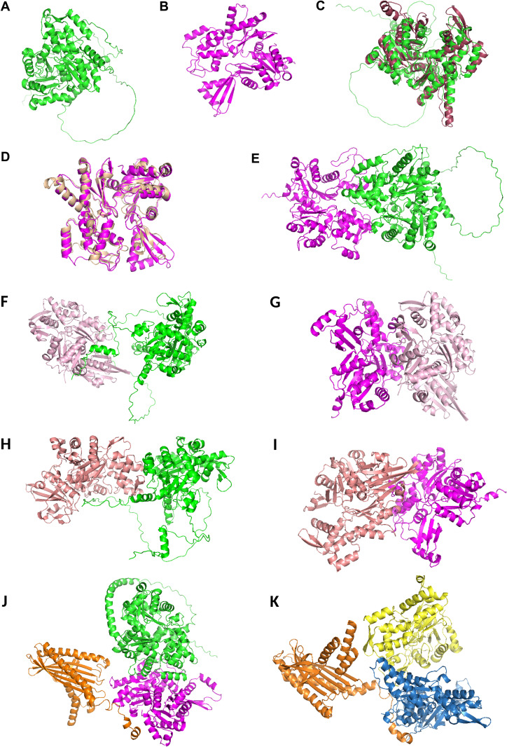
Fig 1 Plasmodium Alp5s are structurally conserved actin-related proteins (Arps). (A) The cartoon structure of P. berghei Alp5a was visualized using PyMOL, with α-helices, β-sheets, and loop regions depicted to highlight the protein’s overall fold and key secondary structural features. (B) Cartoon structure of Alp5b. (C) The structural superposition of P. berghei Alp5a (green) with human Arp3 (chocolate) reveals a RMSD of 2.961 Å. This RMSD value quantifies the degree of structural similarity between the two proteins, indicating both conserved regions and conformational differences. (D) Structural superposition of P. berghei Alp5b (magenta) and human Arp2 (brown) reveals a RMSD of 1.328 Å, indicating a high degree of structural conservation. This similarity suggests that key functional domains are maintained across species, despite evolutionary divergence. (E) The interaction between P. berghei Alp5a and 5b, as predicted by ClusPro. The docking model illustrates the predicted binding interface and interaction sites between the two proteins, highlighting their potential functional relationships. (F) Predicted interaction between P. falciparum Actin I (PfActin I; PDB ID: 6I4H, pink) and P. berghei Alp5a (green). (G) Interaction between PfActin I (6I4H; pink) and PbAlp5b (magenta). (H) Modeled complex of PbActin II (PbActin II; PDB ID: 6I4M, salmon red) with PbAlp5a (green). (I) Predicted interaction between PbActin II (6I4M, salmon red) and PbAlp5b (magenta). (J) Predicted ternary complex of PbAlp5a (green) and PbAlp5b (magenta) interacting with F-actin (orange), illustrating their potential binding interfaces and relative orientations. (K) Predicted ternary complex of Arp2 (yellow) and Arp3 (sky blue) bound to F-actin (orange), representing a canonical actin-branching complex.
Fig 1 Plasmodium Alp5s are structurally conserved actin-related proteins (Arps). (A) The cartoon structure of P. berghei Alp5a was visualized using PyMOL, with α-helices, β-sheets, and loop regions depicted to highlight the protein’s overall fold and key secondary structural features. (B) Cartoon structure of Alp5b. (C) The structural superposition of P. berghei Alp5a (green) with human Arp3 (chocolate) reveals a RMSD of 2.961 Å. This RMSD value quantifies the degree of structural similarity between the two proteins, indicating both conserved regions and conformational differences. (D) Structural superposition of P. berghei Alp5b (magenta) and human Arp2 (brown) reveals a RMSD of 1.328 Å, indicating a high degree of structural conservation. This similarity suggests that key functional domains are maintained across species, despite evolutionary divergence. (E) The interaction between P. berghei Alp5a and 5b, as predicted by ClusPro. The docking model illustrates the predicted binding interface and interaction sites between the two proteins, highlighting their potential functional relationships. (F) Predicted interaction between P. falciparum Actin I (PfActin I; PDB ID: 6I4H, pink) and P. berghei Alp5a (green). (G) Interaction between PfActin I (6I4H; pink) and PbAlp5b (magenta). (H) Modeled complex of PbActin II (PbActin II; PDB ID: 6I4M, salmon red) with PbAlp5a (green). (I) Predicted interaction between PbActin II (6I4M, salmon red) and PbAlp5b (magenta). (J) Predicted ternary complex of PbAlp5a (green) and PbAlp5b (magenta) interacting with F-actin (orange), illustrating their potential binding interfaces and relative orientations. (K) Predicted ternary complex of Arp2 (yellow) and Arp3 (sky blue) bound to F-actin (orange), representing a canonical actin-branching complex.CC BY · PubMed Central
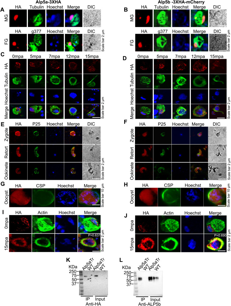
Fig 2 Expression and localization of Alp5s and their interaction. (A and B) Representative confocal immunofluorescence images of male and female gametocytes immunostained with anti-HA, anti-tubulin and anti-g377 antibodies. The Alp5a-3XHA and Alp5b-3XHA-mCherry parasites show their localization. Nuclei were stained with Hoechst. DIC: differential interference contrast. (C and D) Confocal immunofluorescence analyses of microgametes after gametocyte activation. The cells were fixed and immunostained with anti-HA and anti-tubulin antibodies. Alp5a and Alp5b localize from the nucleus to a microtubule-rich structure, likely the spindle or axoneme. (E and F) Immunofluorescence analyses of Alp5a-3XHA and Alp5b-3XHA-mCherry revealed their localization at the zygote, retort, and ookinete stages. Parasites were immunostained with anti-HA and anti-P25 antibodies. (G and H) Expression of Alp5a-3XHA and Alp5b-3XHA-mCherry parasites in oocysts. The midgut oocysts were labeled with anti-HA and anti-CSP antibodies. (I and J) Male gametocytes of Alp5 transgenic parasites were immunostained with anti-HA and anti-actin antibodies at 0- and 15-min post-activation, and subsequently imaged using a confocal microscope. At 0 minutes post-activation (mpa), Alp5 was predominantly localized to the nucleus, whereas actin was distributed in both the cytosol and the nucleus. Following activation at 15 mpa, Alp5 and actin are redistributed around the nucleus and exhibit colocalization. (K and L) Interaction of Alp5s in the parasite. The protein complex was immunoprecipitated from gametocytes of Alp5a-3XHA transgenic parasites using anti-HA magnetic beads. The Alp5a and Alp5b bands were detected by immunoblotting with anti-HA and anti-Alp5b antibodies.
Fig 2 Expression and localization of Alp5s and their interaction. (A and B) Representative confocal immunofluorescence images of male and female gametocytes immunostained with anti-HA, anti-tubulin and anti-g377 antibodies. The Alp5a-3XHA and Alp5b-3XHA-mCherry parasites show their localization. Nuclei were stained with Hoechst. DIC: differential interference contrast. (C and D) Confocal immunofluorescence analyses of microgametes after gametocyte activation. The cells were fixed and immunostained with anti-HA and anti-tubulin antibodies. Alp5a and Alp5b localize from the nucleus to a microtubule-rich structure, likely the spindle or axoneme. (E and F) Immunofluorescence analyses of Alp5a-3XHA and Alp5b-3XHA-mCherry revealed their localization at the zygote, retort, and ookinete stages. Parasites were immunostained with anti-HA and anti-P25 antibodies. (G and H) Expression of Alp5a-3XHA and Alp5b-3XHA-mCherry parasites in oocysts. The midgut oocysts were labeled with anti-HA and anti-CSP antibodies. (I and J) Male gametocytes of Alp5 transgenic parasites were immunostained with anti-HA and anti-actin antibodies at 0- and 15-min post-activation, and subsequently imaged using a confocal microscope. At 0 minutes post-activation (mpa), Alp5 was predominantly localized to the nucleus, whereas actin was distributed in both the cytosol and the nucleus. Following activation at 15 mpa, Alp5 and actin are redistributed around the nucleus and exhibit colocalization. (K and L) Interaction of Alp5s in the parasite. The protein complex was immunoprecipitated from gametocytes of Alp5a-3XHA transgenic parasites using anti-HA magnetic beads. The Alp5a and Alp5b bands were detected by immunoblotting with anti-HA and anti-Alp5b antibodies.CC BY · PubMed Central
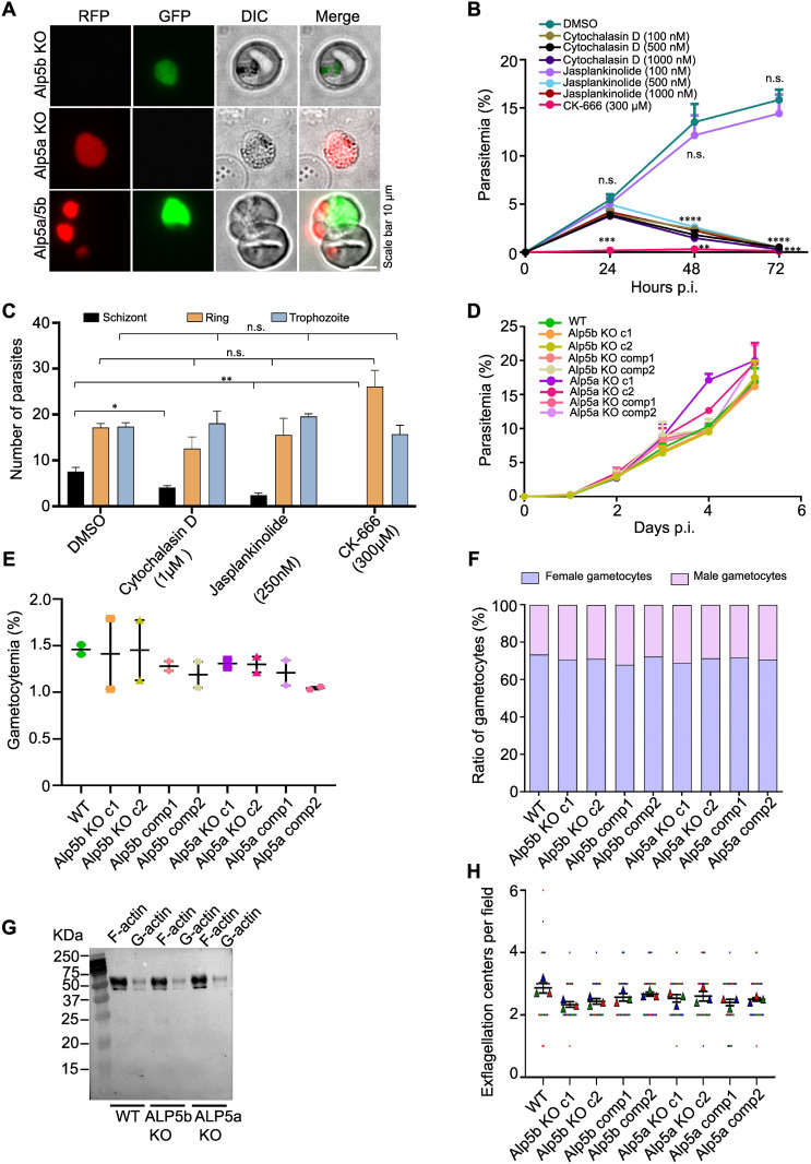
Fig 3 Alp5a and Alp5b are not individually required for asexual blood stage propagation, but simultaneously, they are indispensable for parasite viability. (A) Parasites transfected with constructs targeting Alp5a and Alp5b, tagged with mCherry and GFP respectively, predominantly gave rise to single knockout populations expressing only one of the two fluorophores. Rare instances of parasites expressing both mCherry and GFP were observed within the same red blood cell; however, the lack of fluorescence colocalization suggests that these are not true double knockouts. Representative images were acquired using a widefield fluorescence microscope. (B) Effect of inhibitors on the growth of P. falciparum. Parasitemia was measured at 24, 48, and 72 h after treatment with the indicated concentrations of cytochalasin D, jasplakinolide, and CK-666. At 24 h post-treatment, no significant difference in parasitemia was observed between DMSO-treated controls and cultures treated with either cytochalasin D or jasplakinolide (P = 0.2631; one-way ANOVA). Inhibition was observed in the CK-666-treated cultures, DMSO vs CK-666, ***P = 0.0008; unpaired Student’s t test. A reduction in growth was observed at 48 h, DMSO vs cytochalasin D or jasplankinolide (500 nm-1 µM) ****P < 0.0001; one-way ANOVA, DMSO vs CK-666, **P = 0.0021; unpaired Student’s t test. The growth further decreased at 72 h. DMSO vs cytochalasin D or jasplankinolide (500 nm-1 µM), ****P < 0.0001 and DMSO vs CK-666 ***P = 0.0001; unpaired Student’s t test. Compared with DMSO, 100 nM jasplankinolide did not affect parasite growth at 48 h (P = 0.6447) or 72 h (P = 0.5590). The data are presented as the mean ± SEM and were analyzed via one-way ANOVA or unpaired Student’s t tests; n = 3 biological replicates. (C) P. berghei blood cultures were treated with the inhibitors cytochalasin D (1 µM), jasplankinolide (250 nM) and CK-666 (300 µM). Counting of ring, trophozoite, and schizont stages in P. berghei cultures after treatment. There was no effect on the ring (cytochalasin D, P = 0.1605; jasplankinolide, P = 0.6896; and CK-666, P = 0.0693) or trophozoite stage (cytochalasin D, P = 0.8053; jasplankinolide, P = 0.1031; and CK-666 (P = 0.4832). The number of schizonts decreased significantly in all the treated groups (cytochalasin D, *P = 0.0257; jasplankinolide, **P = 0.0082; and CK-666, **P = 0.0012). The data are presented as the mean ± SEM and were analyzed via unpaired Student’s t tests; n = 3 biological replicates. (D) Asexual blood-stage propagation of control and KO parasites. The data are presented as the mean ± SEM, with no significant differences (P = 0.9998; Brown-Forsythe ANOVA). (E) Estimation of gametocytemia. The data are presented as the mean ± SEM with no significant differences (P = 0.7776; Brown-Forsythe ANOVA). (F) The male and female gametocyte ratios were comparable in WT and KO parasites. The data are presented as the mean ± SEM, with no significant differences (P = 0.9864; one-way ANOVA). Data are presented from two biological replicates, each with 5 mice per group in D, E, and F. (G) Western blot analysis of F-actin and G-actin in blood-stage parasites. Parasites were homogenized in F-actin stabilization buffer, followed by centrifugation to separate F-actin from the G-actin pool. Immunoblotting was performed with an anti-actin antibody to detect actin. Western blot quantitation of F-actin and G-actin in blood-stage parasites and data are presented as the F/G actin ratio. There was no difference between WT and Alp5 KO parasites (P = 0.5903; one-way ANOVA). (H) Comparison of exflagellation centers in WT and Alp5 KO parasites revealed no difference (P = 0.1274, Brown Forsythe and Welch ANOVA). Counts were checked from 10 random fields from three biological replicates. The data are presented as the mean ± SEM.
Fig 3 Alp5a and Alp5b are not individually required for asexual blood stage propagation, but simultaneously, they are indispensable for parasite viability. (A) Parasites transfected with constructs targeting Alp5a and Alp5b, tagged with mCherry and GFP respectively, predominantly gave rise to single knockout populations expressing only one of the two fluorophores. Rare instances of parasites expressing both mCherry and GFP were observed within the same red blood cell; however, the lack of fluorescence colocalization suggests that these are not true double knockouts. Representative images were acquired using a widefield fluorescence microscope. (B) Effect of inhibitors on the growth of P. falciparum. Parasitemia was measured at 24, 48, and 72 h after treatment with the indicated concentrations of cytochalasin D, jasplakinolide, and CK-666. At 24 h post-treatment, no significant difference in parasitemia was observed between DMSO-treated controls and cultures treated with either cytochalasin D or jasplakinolide (P = 0.2631; one-way ANOVA). Inhibition was observed in the CK-666-treated cultures, DMSO vs CK-666, ***P = 0.0008; unpaired Student’s t test. A reduction in growth was observed at 48 h, DMSO vs cytochalasin D or jasplankinolide (500 nm-1 µM) ****P < 0.0001; one-way ANOVA, DMSO vs CK-666, **P = 0.0021; unpaired Student’s t test. The growth further decreased at 72 h. DMSO vs cytochalasin D or jasplankinolide (500 nm-1 µM), ****P < 0.0001 and DMSO vs CK-666 ***P = 0.0001; unpaired Student’s t test. Compared with DMSO, 100 nM jasplankinolide did not affect parasite growth at 48 h (P = 0.6447) or 72 h (P = 0.5590). The data are presented as the mean ± SEM and were analyzed via one-way ANOVA or unpaired Student’s t tests; n = 3 biological replicates. (C) P. berghei blood cultures were treated with the inhibitors cytochalasin D (1 µM), jasplankinolide (250 nM) and CK-666 (300 µM). Counting of ring, trophozoite, and schizont stages in P. berghei cultures after treatment. There was no effect on the ring (cytochalasin D, P = 0.1605; jasplankinolide, P = 0.6896; and CK-666, P = 0.0693) or trophozoite stage (cytochalasin D, P = 0.8053; jasplankinolide, P = 0.1031; and CK-666 (P = 0.4832). The number of schizonts decreased significantly in all the treated groups (cytochalasin D, *P = 0.0257; jasplankinolide, **P = 0.0082; and CK-666, **P = 0.0012). The data are presented as the mean ± SEM and were analyzed via unpaired Student’s t tests; n = 3 biological replicates. (D) Asexual blood-stage propagation of control and KO parasites. The data are presented as the mean ± SEM, with no significant differences (P = 0.9998; Brown-Forsythe ANOVA). (E) Estimation of gametocytemia. The data are presented as the mean ± SEM with no significant differences (P = 0.7776; Brown-Forsythe ANOVA). (F) The male and female gametocyte ratios were comparable in WT and KO parasites. The data are presented as the mean ± SEM, with no significant differences (P = 0.9864; one-way ANOVA). Data are presented from two biological replicates, each with 5 mice per group in D, E, and F. (G) Western blot analysis of F-actin and G-actin in blood-stage parasites. Parasites were homogenized in F-actin stabilization buffer, followed by centrifugation to separate F-actin from the G-actin pool. Immunoblotting was performed with an anti-actin antibody to detect actin. Western blot quantitation of F-actin and G-actin in blood-stage parasites and data are presented as the F/G actin ratio. There was no difference between WT and Alp5 KO parasites (P = 0.5903; one-way ANOVA). (H) Comparison of exflagellation centers in WT and Alp5 KO parasites revealed no difference (P = 0.1274, Brown Forsythe and Welch ANOVA). Counts were checked from 10 random fields from three biological replicates. The data are presented as the mean ± SEM.CC BY · PubMed Central
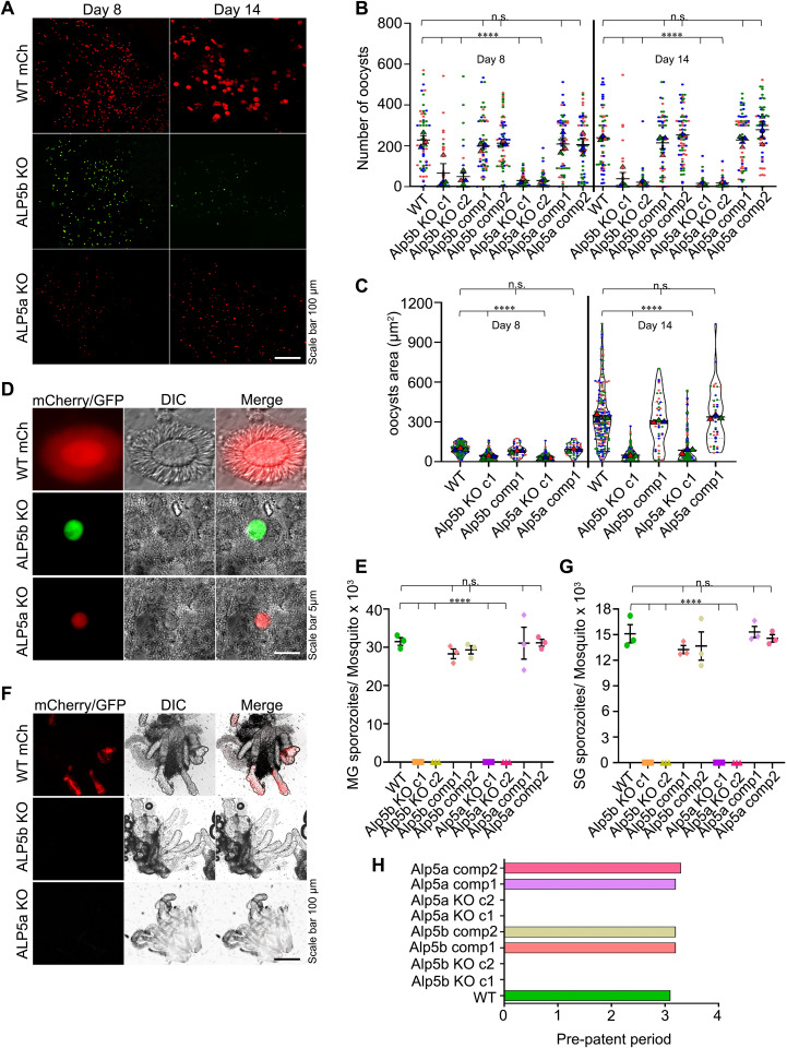
Fig 4 Alp5s are essential for oocyst development and malaria transmission. (A) Representative widefield fluorescence microscopy images of mosquito midguts showing oocysts on the indicated days post-infection. (B) Quantification of oocyst numbers. There was a significant difference in oocyst number between WT and KO parasites on days 8 and 14 (****P < 0.0001). In contrast, no differences were detected between the complement lines and WT on days 8 (P = 0.8721) and 14 (P = 0.1075). Sixty midguts from all the groups were dissected each day. (C) Determination of the oocyst area. The data from independent clones were pooled, and a significant difference was detected between the WT and KO lines on days 8 and 14 (****P < 0.0001), whereas no differences were detected between the complemented lines and the WT line on days 8 (P = 0.1058) and 14 (P = 0.8983). The Kruskal‒Wallis test was used to determine the significance. A total of 200 oocyst areas were measured for each of the WT, Alp5a KO, and Alp5b KO lines, and 40 oocyst areas for each of the corresponding complemented lines, on the indicated days. Data represent three independent biological replicates. The triangle represents the mean individual count in each experiment, and each dot represents an individual count. (D) Widefield fluorescence microscopy of WT oocysts undergoing sporogony. No sporogony was observed in Alp5a or Alp5b KO oocysts. (E) Midgut sporozoite count. The data revealed a significant difference between the WT and knockout lines (****P < 0.0001; one-way ANOVA). No difference was observed between the WT and complemented lines (P = 0.7737). The total number of mosquitoes dissected in each group was 95 (WT GFP), 235 (Alp5b c1), 235 (Alp5b c2), 95 (Alp5b comp1), 70 (Alp5b comp2), 260 (Alp5a c1), 245 (Alp5a c2), 80 (Alp5a comp1), and 85 (Alp5a comp2). Data are presented as the mean ± SEM from three independent biological replicates. (F) Widefield fluorescence microscopy reveals sporozoite-specific fluorescence in WT but not KO-infected salivary glands. (G) Salivary gland sporozoite count. The total number of mosquitoes dissected in each group was 85 (WT GFP), 118 (Alp5b c1), 170 (Alp5b c2), 70 (Alp5b comp1), 100 (Alp5b c2), 135 (Alp5a c1), 235 (Alp5a c2), 70 (Alp5a comp1), and 70 (Alp5a comp2). The data revealed a significant difference between the WT and knockout lines (****P < 0.0001; one-way ANOVA). No difference was detected between the WT and complemented lines (P = 0.5317; one-way ANOVA). Data are presented as the mean ± SEM from three independent biological replicates. (H) Transmission of sporozoites to mice via mosquito bites (n = 5 mice per group) was assessed. Infection was not detected in mice bitten by mosquitoes infected with the KO parasites. The data represent pooled results from two independent biological replicates.
Fig 4 Alp5s are essential for oocyst development and malaria transmission. (A) Representative widefield fluorescence microscopy images of mosquito midguts showing oocysts on the indicated days post-infection. (B) Quantification of oocyst numbers. There was a significant difference in oocyst number between WT and KO parasites on days 8 and 14 (****P < 0.0001). In contrast, no differences were detected between the complement lines and WT on days 8 (P = 0.8721) and 14 (P = 0.1075). Sixty midguts from all the groups were dissected each day. (C) Determination of the oocyst area. The data from independent clones were pooled, and a significant difference was detected between the WT and KO lines on days 8 and 14 (****P < 0.0001), whereas no differences were detected between the complemented lines and the WT line on days 8 (P = 0.1058) and 14 (P = 0.8983). The Kruskal‒Wallis test was used to determine the significance. A total of 200 oocyst areas were measured for each of the WT, Alp5a KO, and Alp5b KO lines, and 40 oocyst areas for each of the corresponding complemented lines, on the indicated days. Data represent three independent biological replicates. The triangle represents the mean individual count in each experiment, and each dot represents an individual count. (D) Widefield fluorescence microscopy of WT oocysts undergoing sporogony. No sporogony was observed in Alp5a or Alp5b KO oocysts. (E) Midgut sporozoite count. The data revealed a significant difference between the WT and knockout lines (****P < 0.0001; one-way ANOVA). No difference was observed between the WT and complemented lines (P = 0.7737). The total number of mosquitoes dissected in each group was 95 (WT GFP), 235 (Alp5b c1), 235 (Alp5b c2), 95 (Alp5b comp1), 70 (Alp5b comp2), 260 (Alp5a c1), 245 (Alp5a c2), 80 (Alp5a comp1), and 85 (Alp5a comp2). Data are presented as the mean ± SEM from three independent biological replicates. (F) Widefield fluorescence microscopy reveals sporozoite-specific fluorescence in WT but not KO-infected salivary glands. (G) Salivary gland sporozoite count. The total number of mosquitoes dissected in each group was 85 (WT GFP), 118 (Alp5b c1), 170 (Alp5b c2), 70 (Alp5b comp1), 100 (Alp5b c2), 135 (Alp5a c1), 235 (Alp5a c2), 70 (Alp5a comp1), and 70 (Alp5a comp2). The data revealed a significant difference between the WT and knockout lines (****P < 0.0001; one-way ANOVA). No difference was detected between the WT and complemented lines (P = 0.5317; one-way ANOVA). Data are presented as the mean ± SEM from three independent biological replicates. (H) Transmission of sporozoites to mice via mosquito bites (n = 5 mice per group) was assessed. Infection was not detected in mice bitten by mosquitoes infected with the KO parasites. The data represent pooled results from two independent biological replicates.CC BY · PubMed Central
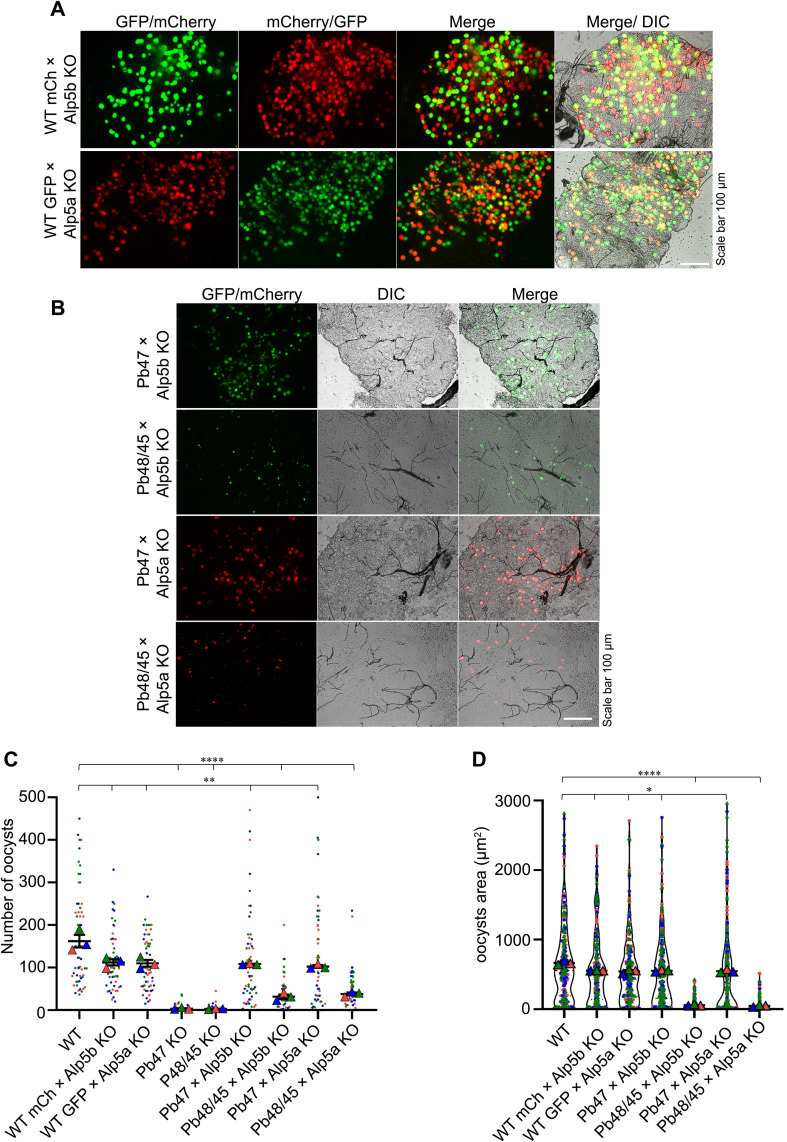
Fig 5 Genetic cross revealed a defect in the male gamete in Alp5 KO parasites. (A) Widefield fluorescence microscopy of oocysts at day 15 post-feeding following genetic crosses between WT-mCherry×Alp5b KO or WT-GFP × Alp5a KO parasites. (B) Genetic crosses of Alp5 KO parasites with male or female gamete-defective lines. The phenotype was restored in the KO lines crossed with the female gamete-defective lines. Genetic crosses with the male gamete-defective lines did not restore the KO phenotype. (C) Quantification of oocysts on day 15. Sixty-two midguts from each group were dissected and observed. Significant differences were observed in WT vs WT-mCherry×Alp5b KO (**P = 0.0038), WT vs WT-GFP × Alp5a KO (**P = 0.0019), WT vs Pb47 KO (****P < 0.0001), WT vs Pb48/45 KO (****P < 0.0001), WT vs Pb47 KO × Alp5b KO (**P = 0.0071), WT vs Pb48/45 KO × Alp5b KO (****P < 0.0001), WT vs Pb47 KO × Alp5a KO (**P = 0.0050), and WT vs Pb48/45 KO × Alp5a KO (****P < 0.0001). (D) Oocyst area on day 15 post-infection. A total of 210 oocyst areas were quantified in each group. Significant differences were observed in WT vs WT-mCherry×Alp5b KO (*P = 0.0377), WT vs WT-GFP × Alp5a KO (*P = 0.0203), WT vs Pb47 KO × Alp5b KO (*P = 0.0334), WT vs Pb48/45 KO × Alp5b KO (****P < 0.0001), WT vs Pb47 KO × Alp5a KO (*P = 0.0435), and WT vs Pb48/45 KO × Alp5a KO (****P < 0.0001). Unpaired Student’s t test was used to determine the statistical significance. The data were pooled from three independent biological replicates and are presented as the mean ± SEM in panels C and D.
Fig 5 Genetic cross revealed a defect in the male gamete in Alp5 KO parasites. (A) Widefield fluorescence microscopy of oocysts at day 15 post-feeding following genetic crosses between WT-mCherry×Alp5b KO or WT-GFP × Alp5a KO parasites. (B) Genetic crosses of Alp5 KO parasites with male or female gamete-defective lines. The phenotype was restored in the KO lines crossed with the female gamete-defective lines. Genetic crosses with the male gamete-defective lines did not restore the KO phenotype. (C) Quantification of oocysts on day 15. Sixty-two midguts from each group were dissected and observed. Significant differences were observed in WT vs WT-mCherry×Alp5b KO (**P = 0.0038), WT vs WT-GFP × Alp5a KO (**P = 0.0019), WT vs Pb47 KO (****P < 0.0001), WT vs Pb48/45 KO (****P < 0.0001), WT vs Pb47 KO × Alp5b KO (**P = 0.0071), WT vs Pb48/45 KO × Alp5b KO (****P < 0.0001), WT vs Pb47 KO × Alp5a KO (**P = 0.0050), and WT vs Pb48/45 KO × Alp5a KO (****P < 0.0001). (D) Oocyst area on day 15 post-infection. A total of 210 oocyst areas were quantified in each group. Significant differences were observed in WT vs WT-mCherry×Alp5b KO (*P = 0.0377), WT vs WT-GFP × Alp5a KO (*P = 0.0203), WT vs Pb47 KO × Alp5b KO (*P = 0.0334), WT vs Pb48/45 KO × Alp5b KO (****P < 0.0001), WT vs Pb47 KO × Alp5a KO (*P = 0.0435), and WT vs Pb48/45 KO × Alp5a KO (****P < 0.0001). Unpaired Student’s t test was used to determine the statistical significance. The data were pooled from three independent biological replicates and are presented as the mean ± SEM in panels C and D.CC BY · PubMed Central
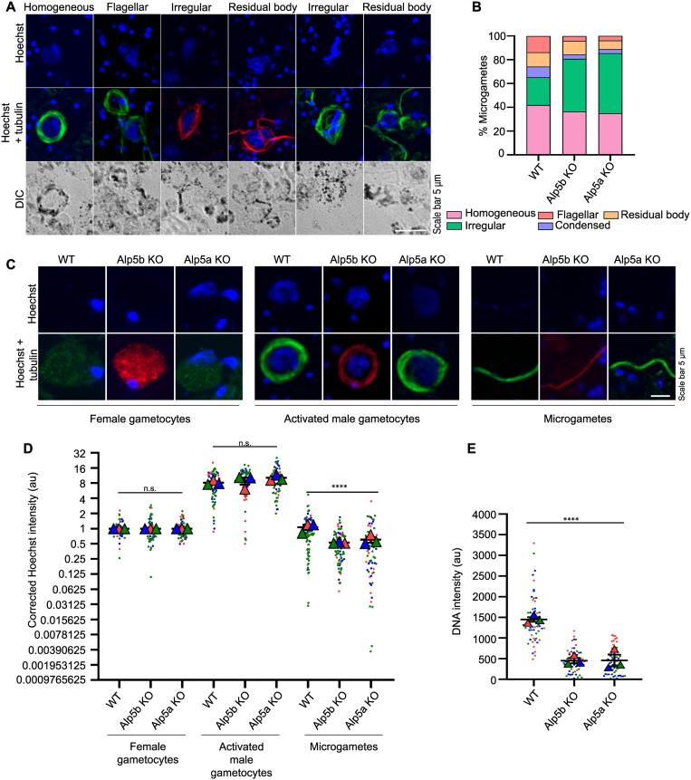
Fig 6 Impaired DNA segregation in Alp5 KO parasites. (A) Gametocytes were activated and fixed at 15 mpa (minutes post-activation). DNA was stained with Hoechst, and an anti-tubulin antibody was used to identify activated male gametocytes. Images were acquired using a confocal microscope. (B) Different forms of microgametes were identified based on quantification of DNA localization patterns in activated male gametocytes. A total of 167 (WT), 192 (Alp5b KO), and 295 (Alp5a KO) male gametocytes were analyzed from three biological replicates. (C) Confocal IFA images of female gametocytes, activated male gametocytes and microgametes. Activated male gametocytes and microgametes were identified by anti-tubulin antibody staining. The female gametocytes were identified via diffuse tubulin staining. DNA was stained with Hoechst. (D) We quantified the DNA content of activated male and female gametocytes as well as microgametes, normalizing all measurements to the mean DNA content of female gametocytes from the same slide. Analysis included 70 female gametocytes, alongside male gametocytes from WT (68), Alp5b KO (69), and Alp5a KO (65) lines. Microgametes were similarly assessed with sample sizes of 92 (WT), 89 (Alp5b KO), and 64 (Alp5a KO). The data were comparable between WT and Alp5 KO lines in terms of the number of female gametocytes (P = 0.3126) and activated male gametocytes (P = 0.148). The DNA content significantly differed between WT and Alp5 KO microgametes (****P < 0.0001). Data from three independent biological replicates were analyzed using the Kruskal–Wallis test to assess statistical differences among groups. (E) Quantification of the zygote DNA content (a.u., arbitrary unit). We detected significant differences among the WT, Alp5a KO and Alp5b KO parasites (****P < 0.0001). The DNA content of 60 zygotes per group was analyzed. Data from three independent biological replicates were analyzed using Student’s t test to assess statistical differences.
Fig 6 Impaired DNA segregation in Alp5 KO parasites. (A) Gametocytes were activated and fixed at 15 mpa (minutes post-activation). DNA was stained with Hoechst, and an anti-tubulin antibody was used to identify activated male gametocytes. Images were acquired using a confocal microscope. (B) Different forms of microgametes were identified based on quantification of DNA localization patterns in activated male gametocytes. A total of 167 (WT), 192 (Alp5b KO), and 295 (Alp5a KO) male gametocytes were analyzed from three biological replicates. (C) Confocal IFA images of female gametocytes, activated male gametocytes and microgametes. Activated male gametocytes and microgametes were identified by anti-tubulin antibody staining. The female gametocytes were identified via diffuse tubulin staining. DNA was stained with Hoechst. (D) We quantified the DNA content of activated male and female gametocytes as well as microgametes, normalizing all measurements to the mean DNA content of female gametocytes from the same slide. Analysis included 70 female gametocytes, alongside male gametocytes from WT (68), Alp5b KO (69), and Alp5a KO (65) lines. Microgametes were similarly assessed with sample sizes of 92 (WT), 89 (Alp5b KO), and 64 (Alp5a KO). The data were comparable between WT and Alp5 KO lines in terms of the number of female gametocytes (P = 0.3126) and activated male gametocytes (P = 0.148). The DNA content significantly differed between WT and Alp5 KO microgametes (****P < 0.0001). Data from three independent biological replicates were analyzed using the Kruskal–Wallis test to assess statistical differences among groups. (E) Quantification of the zygote DNA content (a.u., arbitrary unit). We detected significant differences among the WT, Alp5a KO and Alp5b KO parasites (****P < 0.0001). The DNA content of 60 zygotes per group was analyzed. Data from three independent biological replicates were analyzed using Student’s t test to assess statistical differences.CC BY · PubMed Central
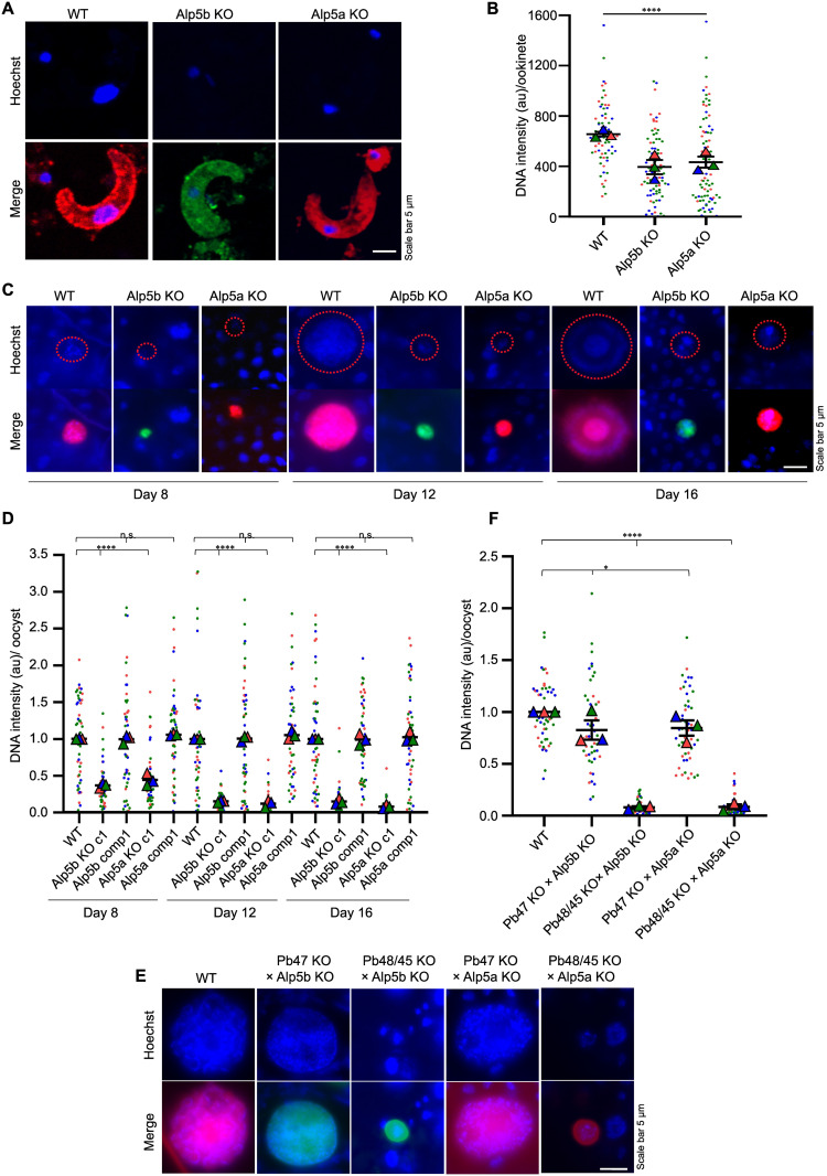
Fig 7 DNA content is reduced in the ookinete and oocyst of Alp5 KO parasites. (A) Representative confocal immunofluorescence images of ookinetes. Compared with WT ookinetes, Alp5 KO ookinetes presented a lower DNA content. (B) Quantification of ookinete DNA content (a.u., arbitrary unit). We detected significant differences between WT and Alp5 KO parasites (****P < 0.0001). The DNA contents of 70 (WT), 83 (Alp5b), and 83 (Alp5a) ookinetes were quantified. Data from three independent biological replicates were analyzed using the Kruskal–Wallis test to assess statistical differences among groups. (C) Widefield fluorescence microscopy images of Hoechst-stained oocysts showing the DNA content on different days. (D) The average DNA intensity of 50 oocysts was significantly different between WT and Alp5 KO parasites on days 8 (****P < 0.0001), 12 (****P < 0.0001) and 16 (****P < 0.0001). The DNA content was comparable between WT, ALP5b comp and ALP5a comp on day 8 (P = 0.7347), day 12 (P = 0.6874), and day 16 (P = 0. 8970). Data from three independent biological replicates were analyzed using the Kruskal–Wallis test to assess statistical differences among groups. (E) Widefield fluorescence microscopy images of Hoechst-stained oocysts illustrate the DNA content after the genetic crosses. The DNA content was restored in Alp5 KO parasites crossed with female gamete-defective lines. In contrast, genetic crosses with male gamete-defective lines did not rescue the DNA content. (F) The DNA content of 50 oocysts in Alp5 KO parasites was quantified and normalized to the mean DNA intensity of WT oocysts. A significant difference was observed in WT vs Pb47 KO × Alp5b KO (*P = 0.0330), WT vs Pb48/45 KO × Alp5b KO (****P < 0.0001), WT vs Pb47 KO × Alp5a KO (*P = 0.0172), and WT vs Pb48/45 KO × Alp5a KO (****P < 0.0001). Data from three independent biological replicates were analyzed using Student’s t test to assess statistical differences.
Fig 7 DNA content is reduced in the ookinete and oocyst of Alp5 KO parasites. (A) Representative confocal immunofluorescence images of ookinetes. Compared with WT ookinetes, Alp5 KO ookinetes presented a lower DNA content. (B) Quantification of ookinete DNA content (a.u., arbitrary unit). We detected significant differences between WT and Alp5 KO parasites (****P < 0.0001). The DNA contents of 70 (WT), 83 (Alp5b), and 83 (Alp5a) ookinetes were quantified. Data from three independent biological replicates were analyzed using the Kruskal–Wallis test to assess statistical differences among groups. (C) Widefield fluorescence microscopy images of Hoechst-stained oocysts showing the DNA content on different days. (D) The average DNA intensity of 50 oocysts was significantly different between WT and Alp5 KO parasites on days 8 (****P < 0.0001), 12 (****P < 0.0001) and 16 (****P < 0.0001). The DNA content was comparable between WT, ALP5b comp and ALP5a comp on day 8 (P = 0.7347), day 12 (P = 0.6874), and day 16 (P = 0. 8970). Data from three independent biological replicates were analyzed using the Kruskal–Wallis test to assess statistical differences among groups. (E) Widefield fluorescence microscopy images of Hoechst-stained oocysts illustrate the DNA content after the genetic crosses. The DNA content was restored in Alp5 KO parasites crossed with female gamete-defective lines. In contrast, genetic crosses with male gamete-defective lines did not rescue the DNA content. (F) The DNA content of 50 oocysts in Alp5 KO parasites was quantified and normalized to the mean DNA intensity of WT oocysts. A significant difference was observed in WT vs Pb47 KO × Alp5b KO (*P = 0.0330), WT vs Pb48/45 KO × Alp5b KO (****P < 0.0001), WT vs Pb47 KO × Alp5a KO (*P = 0.0172), and WT vs Pb48/45 KO × Alp5a KO (****P < 0.0001). Data from three independent biological replicates were analyzed using Student’s t test to assess statistical differences.CC BY · PubMed Central
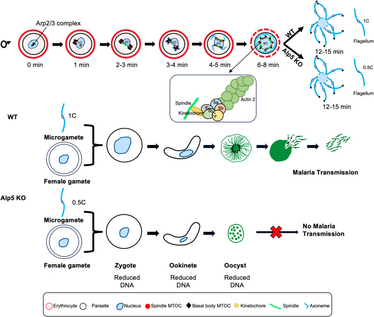
Fig 8 Model of non-canonical Arp2/3 complex-mediated actin nucleation during male gametogony and its impact on parasite development. During male gametogony, actin stabilizes the endomitotic spindle assembly, ensuring proper chromosomal segregation. Parasites deficient in Alp5 exhibit reduced DNA content transitioning from microgametes to oocysts. This compromised genomic integrity in the male lineage likely impairs DNA replication, leading to defective oocyst development and maturation.
Fig 8 Model of non-canonical Arp2/3 complex-mediated actin nucleation during male gametogony and its impact on parasite development. During male gametogony, actin stabilizes the endomitotic spindle assembly, ensuring proper chromosomal segregation. Parasites deficient in Alp5 exhibit reduced DNA content transitioning from microgametes to oocysts. This compromised genomic integrity in the male lineage likely impairs DNA replication, leading to defective oocyst development and maturation.CC BY · PubMed Central

Verification

Claims the paper makes

The claims and statistics of this paper haven’t been checked yet.

Run the check — free, no account needed. It reads the open-access text and checks the sources it cites.

A few free checks a day · papers already verified open instantly · results are public

Self-check

Percentages, sums and reported differences re-computed from the paper’s own numbers.

The self-check needs the paper’s full text.

Citations

Cites

Research works printed but not linked: 74

  1. H Matthews, 2018, Checks and balances? DNA replication and the cell cycle in Plasmodium, Parasit Vectors, 11, 216, 10.1186/s13071-018-2800-1 · DOI
  2. DS Guttery, 2023, Meiosis in Plasmodium: how does it work?, Trends Parasitol, 39, 812, 10.1016/j.pt.2023.07.002 · DOI
  3. A Gandarillas, 2018, Mammalian endoreplication emerges to reveal a potential developmental timer, Cell Death Differ, 25, 471, 10.1038/s41418-017-0040-0 · DOI
  4. M Roques, 2023, Same, same but different: Exploring Plasmodium cell division during liver stage development, PLoS Pathog, 19, 10.1371/journal.ppat.1011210 · DOI
  5. Y Voß, 2023, Plasmodium schizogony, a chronology of the parasite’s cell cycle in the blood stage, PLoS Pathog, 19, 10.1371/journal.ppat.1011157 · DOI
  6. N Gerald, 2011, Mitosis in the human malaria parasite Plasmodium falciparum, Eukaryot Cell, 10, 474, 10.1128/EC.00314-10 · DOI
  7. CS Simon, 2021, An extended DNA-free intranuclear compartment organizes centrosome microtubules in malaria parasites, Life Sci Alliance, 4, 10.26508/lsa.202101199 · DOI
  8. J Schrével, 1977, Ultrastructural study of multiple mitoses during sporogony of Plasmodium b. berghei, J Ultrastruct Res, 59, 332, 10.1016/S0022-5320(77)90043-0 · DOI
  9. C Arquint, 2014, Centrosomes as signalling centres, Philos Trans R Soc Lond B Biol Sci, 369, 20130464, 10.1098/rstb.2013.0464 · DOI
  10. M Zeeshan, 2024, Plasmodium NEK1 coordinates MTOC organisation and kinetochore attachment during rapid mitosis in male gamete formation, PLoS Biol, 22, 10.1371/journal.pbio.3002802 · DOI
  11. DS Guttery, 2022, Division and Transmission: Malaria Parasite Development in the Mosquito, Annu Rev Microbiol, 76, 113, 10.1146/annurev-micro-041320-010046 · DOI
  12. K Venugopal, 2020, Plasmodium asexual growth and sexual development in the haematopoietic niche of the host, Nat Rev Microbiol, 18, 177, 10.1038/s41579-019-0306-2 · DOI
  13. RE Sinden, 2010, The flagellum in malarial parasites, Curr Opin Microbiol, 13, 491, 10.1016/j.mib.2010.05.016 · DOI
  14. R Tewari, 2010, The systematic functional analysis of Plasmodium protein kinases identifies essential regulators of mosquito transmission, Cell Host Microbe, 8, 377, 10.1016/j.chom.2010.09.006 · DOI
  15. BM Invergo, 2017, Sub-minute Phosphoregulation of Cell Cycle Systems during Plasmodium Gamete Formation, Cell Rep, 21, 2017, 10.1016/j.celrep.2017.10.071 · DOI
  16. DS Guttery, 2014, Genome-wide functional analysis of Plasmodium protein phosphatases reveals key regulators of parasite development and differentiation, Cell Host Microbe, 16, 128, 10.1016/j.chom.2014.05.020 · DOI
  17. F Liu, 2021, A conserved malaria parasite antigen Pb22 plays a critical role in male gametogenesis in Plasmodium berghei, Cell Microbiol, 23
  18. C Ramakrishnan, 2023, Radial spoke protein 9 is necessary for axoneme assembly in Plasmodium but not in trypanosomatid parasites, J Cell Sci, 136, 10.1242/jcs.260655 · DOI
  19. C Zhang, 2022, Pyp25α is required for male gametocyte exflagellation, Pathog Dis, 80, 10.1093/femspd/ftac043 · DOI
  20. P Singh, 2019, Role of a patatin-like phospholipase in Plasmodium falciparum gametogenesis and malaria transmission, Proc Natl Acad Sci U S A, 116, 17498, 10.1073/pnas.1900266116 · DOI
  21. DS Guttery, 2024, The molecular mechanisms driving Plasmodium cell division, Biochem Soc Trans, 52, 593, 10.1042/BST20230403 · DOI
  22. KM Skillman, 2011, Evolutionarily divergent, unstable filamentous actin is essential for gliding motility in apicomplexan parasites, PLoS Pathog, 7, 10.1371/journal.ppat.1002280 · DOI
  23. AJ Lopez, 2023, Structure and function of Plasmodium actin II in the parasite mosquito stages, PLoS Pathog, 19, 10.1371/journal.ppat.1011174 · DOI
  24. JG Wesseling, 1988, Extremely diverged actin proteins in Plasmodium falciparum, Mol Biochem Parasitol, 30, 143, 10.1016/0166-6851(88)90107-7 · DOI
  25. M Andreadaki, 2014, Genetic crosses and complementation reveal essential functions for the Plasmodium stage-specific actin2 in sporogonic development, Cell Microbiol, 16, 751, 10.1111/cmi.12274 · DOI
  26. S Das, 2017, Multiple essential functions of Plasmodium falciparum actin-1 during malaria blood-stage development, BMC Biol, 15, 70, 10.1186/s12915-017-0406-2 · DOI
  27. E Deligianni, 2011, Critical role for a stage-specific actin in male exflagellation of the malaria parasite, Cell Microbiol, 13, 1714, 10.1111/j.1462-5822.2011.01652.x · DOI
  28. I Rupp, 2011, Malaria parasites form filamentous cell-to-cell connections during reproduction in the mosquito midgut, Cell Res, 21, 683, 10.1038/cr.2010.176 · DOI
  29. KM Skillman, 2013, The unusual dynamics of parasite actin result from isodesmic polymerization, Nat Commun, 4, 2285, 10.1038/ncomms3285 · DOI
  30. KL Hvorecny, 2024, Toxoplasma gondii actin filaments are tuned for rapid disassembly and turnover, Nat Commun, 15, 1840, 10.1038/s41467-024-46111-3 · DOI
  31. E-P Kumpula, 2017, Apicomplexan actin polymerization depends on nucleation, Sci Rep, 7, 12137, 10.1038/s41598-017-11330-w · DOI
  32. J Vahokoski, 2014, Structural differences explain diverse functions of Plasmodium actins, PLoS Pathog, 10, 10.1371/journal.ppat.1004091 · DOI
  33. H Schüler, 2005, A Plasmodium actin-depolymerizing factor that binds exclusively to actin monomers, Mol Biol Cell, 16, 4013, 10.1091/mbc.e05-02-0086 · DOI
  34. BK Singh, 2011, Crystal structures explain functional differences in the two actin depolymerization factors of the malaria parasite, J Biol Chem, 286, 28256, 10.1074/jbc.M110.211730 · DOI
  35. D Lasiwa, 2025, Functional insights into Plasmodium actin-depolymerizing factor interactions with phosphoinositides, J Biol Chem, 301, 110399, 10.1016/j.jbc.2025.110399 · DOI
  36. S Collier, 2023, Plasmodium falciparum formins are essential for invasion and sexual stage development, Commun Biol, 6, 861, 10.1038/s42003-023-05233-y · DOI
  37. ÁÁ Bendes, 2022, Structure and function of an atypical homodimeric actin capping protein from the malaria parasite, Cell Mol Life Sci, 79, 125, 10.1007/s00018-021-04032-0 · DOI
  38. M Ganter, 2009, Vital role for the Plasmodium actin capping protein (CP) beta-subunit in motility of malaria sporozoites, Mol Microbiol, 74, 1356, 10.1111/j.1365-2958.2009.06828.x · DOI
  39. I Kursula, 2008, Structural basis for parasite-specific functions of the divergent profilin of Plasmodium falciparum, Structure, 16, 1638, 10.1016/j.str.2008.09.008 · DOI
  40. F Hentzschel, 2025, An atypical Arp2/3 complex is required for Plasmodium DNA segregation and malaria transmission, Nat Microbiol, 10, 1775, 10.1038/s41564-025-02023-6 · DOI · Paper page
  41. HM Behrens, 2024, Identification of domains in Plasmodium falciparum proteins of unknown function using DALI search on AlphaFold predictions, Sci Rep, 14, 10527, 10.1038/s41598-024-60058-x · DOI
  42. P Bieling, 2023, From WRC to Arp2/3: Collective molecular mechanisms of branched actin network assembly, Curr Opin Cell Biol, 80, 102156, 10.1016/j.ceb.2023.102156 · DOI
  43. J Pizarro-Cerdá, 2017, The Diverse Family of Arp2/3 Complexes, Trends Cell Biol, 27, 93, 10.1016/j.tcb.2016.08.001 · DOI
  44. JL Gordon, 2005, Comparative genome analysis reveals a conserved family of actin-like proteins in apicomplexan parasites, BMC Genomics, 6, 179, 10.1186/1471-2164-6-179 · DOI
  45. J Baum, 2006, Regulation of apicomplexan actin-based motility, Nat Rev Microbiol, 4, 621, 10.1038/nrmicro1465 · DOI
  46. AM Gautreau, 2022, Nucleation, stabilization, and disassembly of branched actin networks, Trends Cell Biol, 32, 421, 10.1016/j.tcb.2021.10.006 · DOI
  47. LK Fritz-Laylin, 2023, The evolution and diversity of actin-dependent cell migration, Mol Biol Cell, 34, 7
  48. ED Goley, 2006, The ARP2/3 complex: an actin nucleator comes of age, Nat Rev Mol Cell Biol, 7, 713, 10.1038/nrm2026 · DOI
  49. M Plessner, 2019, Centrosomal Actin Assembly Is Required for Proper Mitotic Spindle Formation and Chromosome Congression, iScience, 15, 274, 10.1016/j.isci.2019.04.022 · DOI
  50. D Inoue, 2019, Actin filaments regulate microtubule growth at the centrosome, EMBO J, 38
  51. F Farina, 2019, Local actin nucleation tunes centrosomal microtubule nucleation during passage through mitosis. EMBO J, 38
  52. KG Campellone, 2023, Branching out in different directions: Emerging cellular functions for the Arp2/3 complex and WASP-family actin nucleation factors, Eur J Cell Biol, 102, 151301, 10.1016/j.ejcb.2023.151301 · DOI · Paper page
  53. MD Welch, 1997, The human Arp2/3 complex is composed of evolutionarily conserved subunits and is localized to cellular regions of dynamic actin filament assembly, J Cell Biol, 138, 375, 10.1083/jcb.138.2.375 · DOI
  54. MR van Dijk, 2001, A central role for P48/45 in malaria parasite male gamete fertility, Cell, 104, 153, 10.1016/S0092-8674(01)00199-4 · DOI
  55. MR van Dijk, 2010, Three members of the 6-cys protein family of Plasmodium play a role in gamete fertility, PLoS Pathog, 6, 10.1371/journal.ppat.1000853 · DOI
  56. PM Davidson, 2021, Actin on and around the Nucleus, Trends Cell Biol, 31, 211, 10.1016/j.tcb.2020.11.009 · DOI
  57. E Deligianni, Critical role for a stage‐specific actin in male exflagellation of the malaria
  58. M Zeeshan, 2023, Plasmodium ARK2 and EB1 drive unconventional spindle dynamics, during chromosome segregation in sexual transmission stages, Nat Commun, 14, 5652, 10.1038/s41467-023-41395-3 · DOI
  59. ESC Bushell, 2009, Paternal effect of the nuclear formin-like protein MISFIT on Plasmodium development in the mosquito vector, PLoS Pathog, 5
  60. M Zeeshan, 2019, Plasmodium kinesin-8X associates with mitotic spindles and is essential for oocyst development during parasite proliferation and transmission, PLoS Pathog, 15, 10.1371/journal.ppat.1008048 · DOI
  61. M Roques, 2015, Plasmodium P-Type Cyclin CYC3 Modulates Endomitotic Growth during Oocyst Development in Mosquitoes, PLoS Pathog, 11, 10.1371/journal.ppat.1005273 · DOI
  62. M Mirdita, 2022, ColabFold: making protein folding accessible to all, Nat Methods, 19, 679, 10.1038/s41592-022-01488-1 · DOI · Paper page
  63. D Szklarczyk, 2023, The STRING database in 2023: protein-protein association networks and functional enrichment analyses for any sequenced genome of interest, Nucleic Acids Res, 51, 10.1093/nar/gkac1000 · DOI · Paper page
  64. D Kozakov, 2017, The ClusPro web server for protein-protein docking, Nat Protoc, 12, 255, 10.1038/nprot.2016.169 · DOI
  65. 2015, The PyMOL Molecular Graphics System version 2.0
  66. R Gupta, 2020, Secreted protein with altered thrombospondin repeat (SPATR) is essential for asexual blood stages but not required for hepatocyte invasion by the malaria parasite Plasmodium berghei, Mol Microbiol, 113, 478, 10.1111/mmi.14432 · DOI
  67. C Lambros, 1979, Synchronization of Plasmodium falciparum erythrocytic stages in culture, J Parasitol, 65, 418, 10.2307/3280287 · DOI
  68. CJ Janse, 2006, High-efficiency transfection and drug selection of genetically transformed blood stages of the rodent malaria parasite Plasmodium berghei, Nat Protoc, 1, 346, 10.1038/nprot.2006.53 · DOI
  69. B Nayak, 2024, Neddylation is essential for malaria transmission in Plasmodium berghei, mBio, 15, 10.1128/mbio.00232-24 · DOI
  70. A Mishra, 2023, Regulation of Atg8 membrane deconjugation by cysteine proteases in the malaria parasite Plasmodium berghei, Cell Mol Life Sci, 80, 344, 10.1007/s00018-023-05004-2 · DOI
  71. AR Gomes, 2015, A genome-scale vector resource enables high-throughput reverse genetic screening in a malaria parasite, Cell Host Microbe, 17, 404, 10.1016/j.chom.2015.01.014 · DOI
  72. Nirdosh, 2024, The novel Plasmodium berghei protein S14 is essential for sporozoite gliding motility and infectivity, J Cell Sci, 137
  73. SK Narwal, 2024, Stearoyl-CoA desaturase regulates organelle biogenesis and hepatic merozoite formation in Plasmodium berghei, Mol Microbiol, 121, 940, 10.1111/mmi.15246 · DOI
  74. SK Narwal, 2022, Protein kinase 9 is not required for completion of the Plasmodium berghei life cycle, Microbiol Res, 260, 127051, 10.1016/j.micres.2022.127051 · DOI

Metadata

Authors
Varshney, A., Pandey, E., Nirdosh, & Mishra, S.
Affiliations
Central Drug Research Institute (IN)
Corresponding author
Mishra, Satish
Venue
PLOS Pathogens
Published
2025-11-11
Volume
21
Issue
11
Pages
e1013687
ISSN
  • 1553-7366 (ISSN-L)
  • 1553-7374
OpenAlex
W4416113613
Cited by
2
References
4
Citations by year
2026: 2
FWCI
1.81
Type
journal article
Language
en
Open access
Locations
  • PLoS Pathogens (publishedVersion, cc-by)
  • bioRxiv (Cold Spring Harbor Laboratory) (acceptedVersion, public-domain)
  • Europe PMC (PubMed Central) (publishedVersion, cc-by)
  • PubMed (publishedVersion)
  • DOAJ (DOAJ: Directory of Open Access Journals) (submittedVersion, cc-by-sa)
Funders
  • Indian Council of Medical Research (6/9-7(306)/2022/ECD-II)
  • Anusandhan National Research Foundation (CRG/2022/003848)
Topics
  • Health Sciences › Medicine › Public Health, Environmental and Occupational Health › Malaria Research and Control
  • Life Sciences › Biochemistry, Genetics and Molecular Biology › Cell Biology › Microtubule and mitosis dynamics
  • Life Sciences › Immunology and Microbiology › Parasitology › Bird parasitology and diseases
Keywords
Plasmodium, actin-like protein, Arp2/3 complex, DNA segregation, male gametogenesis, malaria transmission, gametocytes, blood stage, Plasmodium berghei, nuclear actin
MeSH terms
Actins, Animals, Gametogenesis, Humans, Malaria, Male, Plasmodium berghei, Protozoan Proteins, DNA, Protozoan, Mice

About these checks

Registry, retraction, Cites and statistics results are computed, not judged, with one exception: a printed reference that no index lists is read once by a model before it counts as a citation error. Claims are extracted by a model and kept only when their quote appears verbatim in the text; each cited source is then checked, with a model’s help, for whether it says what the claim relies on. Those verdicts can be wrong — read the claim’s quote and the cited source before relying on one.

Statistics and arithmetic are recomputed, not judged.

Confidence bands are shown only for fields calibrated on our annotated test set. None is calibrated yet, so no band is shown.

What counts as a claim ›How we measure confidence ›How we check this ›

Are you an author, or do you see a problem with this page? Request a review