

The DOI matches its Crossref record; 28 of the 29 references it cites were found in a registry or an index.
Cholinergic projections from the basal forebrain of rat to the amygdala
Open accessAbstract
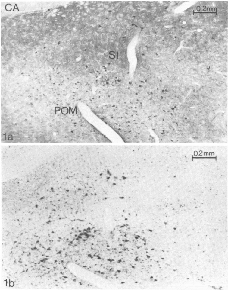
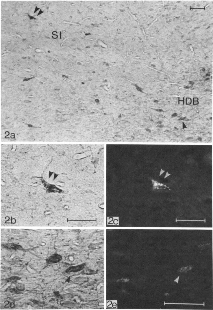
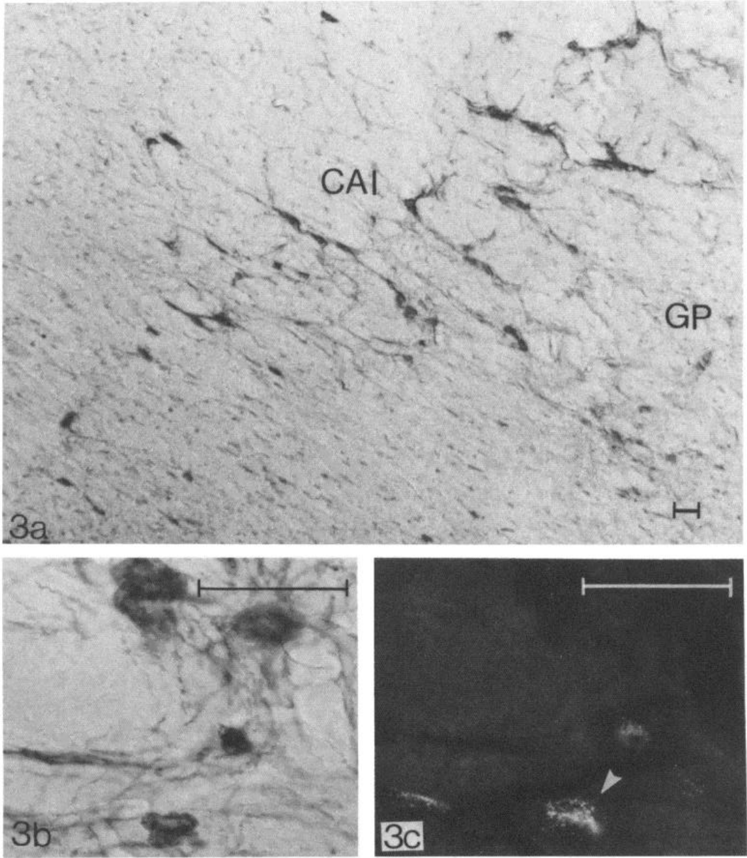
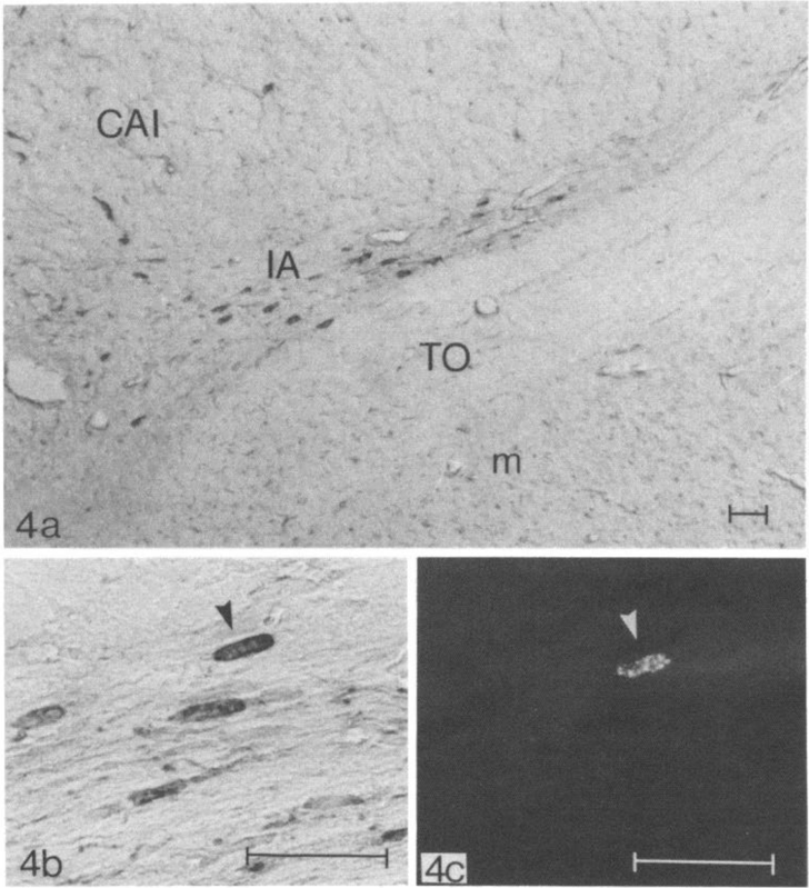
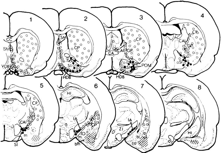
Cholinergic afferents to the amygdala from the basal forebrain were studied using di-isopropyl fluorophosphate-AChE histochemistry in combination with retrograde tracing using various fluorescent dyes. Cells sending their axons to the amygdala and staining intensely for AChE were located mainly in the nucleus of the substantia innominata. They also were found in the ventral part of the globus pallidus, the horizontal limb of the nucleus tractus diagonalis Broca, and the nucleus interstitialis ansae lenticularis. A correspondence was established between these cells and cells staining for choline acetyltransferase by immunohistochemistry in both distribution and morphology. Non-cholinergic neurons which send their axons to the amygdala also were found in the substantia innominata complex.
Overview
The claims and statistics of this paper haven’t been checked yet.
Run the check — free, no account needed. It reads the open-access text and checks the sources it cites.
Integrity signals
Registry
Cites
Statistics
Figures
Verification
Claims the paper makes
The claims and statistics of this paper haven’t been checked yet.
Run the check — free, no account needed. It reads the open-access text and checks the sources it cites.
Claims other papers make about this one
Each is checked against this paper’s own text: does it say what the citing paper says it does?
In the citing paper
“Furthermore, NBM projects to the amygdala and is the primary source of cholinergic input to the basolateral amygdala (44,46,56,57).”
From Comparative Perspectives on Oxytocin and Vasopressin Receptor Research in Rodents and Primates: Translational Implications (2016) · cites this paper as [57]
Self-check
Percentages, sums and reported differences re-computed from the paper’s own numbers.
The self-check needs the paper’s full text.
Citations
Cites
Graham, R. C., & Karnovsky, M. J. (1966) · Journal of Histochemistry & Cytochemistry · doi:10.1177/14.4.291 · Cited by 6,502
Sternberger, L. A., Hardy, P. H., Cuculis, J. J., & Meyer, H. G. (1970) · Journal of Histochemistry & Cytochemistry · doi:10.1177/18.5.315 · Cited by 5,042
A "DIRECT-COLORING" THIOCHOLINE METHOD FOR CHOLINESTERASES
Karnovsky, M. J., & ROOTS, L. (1964) · Journal of Histochemistry & Cytochemistry · doi:10.1177/12.3.219 · Cited by 3,368
The central cholinergic system studied by choline acetyltransferase immunohistochemistry in the cat
Kimura, H., McGeer, P. L., Peng, J. H., & McGeer, E. G. (1981) · The Journal of Comparative Neurology · doi:10.1002/cne.902000202 · Cited by 966
Self-citationCortical and subcortical afferents to the amygdala of the rhesus monkey (Macaca mulatta)
Aggleton, J. P., Burton, M. J., & Passingham, R. E. (1980) · Brain Research · doi:10.1016/0006-8993(80)90279-6 · Cited by 610
An autoradiographic study of the efferent connections of the preoptic region in the rat
Swanson, L. W. (1976) · Journal of Comparative Neurology · doi:10.1002/cne.901670207 · Cited by 588
Further contributions to the study of the evolution of the forebrain
Johnston, J. B. (1923) · Journal of Comparative Neurology · doi:10.1002/cne.900350502 · Cited by 575
Acetylcholinesterase-rich projections from the basal forebrain of the rhesus monkey to neocortex
Mesulam, M.-M., & Hoesen, G. W. V. (1976) · Brain Research · doi:10.1016/0006-8993(76)90385-1 · Cited by 426
Basal Forebrain and Hypothalamic Connection to Frontal and Parietal Cortex in the Rhesus Monkey
Kievit, J., & Kuypers, H. G. J. M. (1975) · Science · doi:10.1126/science.1114317 · Cited by 385
Cholinesterase-Containing Systems of the Brain of the Rat
Shute, C. C. D., & Lewis, P. R. (1963) · Nature · doi:10.1038/1991160a0 · Cited by 359
The projection field of the stria terminalis in the rat brain. An experimental study
de Olmos, J. S., & Ingram, W. R. (1972) · The Journal of Comparative Neurology · doi:10.1002/cne.901460303 · Cited by 355
Choline Acetyltransferase-Containing Neurons in Rodent Brain Demonstrated by Immunohistochemistry
Kimura, H., McGeer, P. L., Peng, F., & McGeer, E. G. (1980) · Science · doi:10.1126/science.6990490 · Cited by 333
Self-citationRetrograde anoxal transport of fluorescent substances in the rat's forebrain
Kuypers, H. G. J. M., Catsman‐Berrevoets, C. E., & Padt, R. E. (1977) · Neuroscience Letters · doi:10.1016/0304-3940(77)90007-6 · Cited by 284
Subcortical afferents of the amygdaloid complex in the rat: an HRP study
Veening, J. G. (1978) · Neuroscience Letters · doi:10.1016/0304-3940(78)90121-0 · Cited by 237
The forebrain of the opossum, didelphis virginiana. Part II. Histology
Loo, Y. T. (1931) · The Journal of Comparative Neurology · doi:10.1002/cne.900520102 · Cited by 232
The hypothalamic distribution of the stria terminalis in the rat
Heimer, L., & Nauta, W. J. H. (1969) · Brain Research · doi:10.1016/0006-8993(69)90288-1 · Cited by 231
Ottersen, O. P. (1980) · The Journal of Comparative Neurology · doi:10.1002/cne.901940113 · Cited by 217
Acetylcholinesterase and the cholinergic neuron
Lehmann, J. C., & Fibiger, H. C. (1979) · Life Sciences · doi:10.1016/0024-3205(79)90596-4 · Cited by 187
Choline acetyltransferase content of limbic nuclei of the rat
Palkovits, M., Saavedra, J. M., Kobayashi, R. M., & Brownstein, M. (1974) · Brain Research · doi:10.1016/0006-8993(74)90442-9 · Cited by 185
Ben‐Ari, Y., Zigmond, R. E., Shute, C. C. D., & Lewis, P. R. (1977) · Brain Research · doi:10.1016/0006-8993(77)90397-3 · Cited by 133
Emson, P. C., Paxinos, G., Salle, G. L. G. L., Ben‐Ari, Y., & Silver, A. (1979) · Brain Research · doi:10.1016/0006-8993(79)90559-6 · Cited by 110
Björklund, A., & Skagerberg, G. (1979) · Journal of Neuroscience Methods · doi:10.1016/0165-0270(79)90038-4 · Cited by 104
Nature and mechanisms of cholinergic-monoaminergic interactions in the brain
Butcher, L. L. (1977) · Life Sciences · doi:10.1016/0024-3205(77)90001-7 · Cited by 96
Subcortical afferents to the monkey amygdala: an HRP study
Norita, M., & Kawamura, K. (1980) · Brain Research · doi:10.1016/0006-8993(80)91171-3 · Cited by 89
Anti-choline acetyltransferase fragments antigen binding (Fab) for immunohistochemistry
Peng, J. H., Kimura, H., McGeer, P. L., & McGeer, E. G. (1981) · Neuroscience Letters · doi:10.1016/0304-3940(81)90217-2 · Cited by 20
Self-citationHarujiro, I. M. A. I., Nakai, K., Kamei, I., Itakura, T., Komai, N., Nagai, T., Kimura, H., Imamoto, K., & Maeda, T. (1980) · PubMed · Cited by 13
Self-citationNagai, T., Satoh, K., Imamoto, K., & Maeda, T. (1981) · PubMed · Cited by 6
Self-citationThe Stria Terminalis, The Ventral Amygdaloid Pathways, and Related Amygdaloid Connections
Hamilton, L. W. (1976) · Springer eBooks · doi:10.1007/978-1-4684-2247-4_7 · Cited by 1
Research works printed but not linked: 1
- Heimer, L., and R. D. WiIIson (1975) The subcortical projec- tions of the allocortex: Similarities in the neural associations of the hippocampus, the piriform cortex, and the neocortex. In Golgi Centennial Symposium, M. Santini, ed., pp. 177- 193, Raven Press, New York.
Cited by
Sorted by citations
Alheid, G. F., & Heimer, L. (1988) · Neuroscience · doi:10.1016/0306-4522(88)90217-5 · Cited by 1,217
Turski, W. A., Cavalheiro, É. A., Schwarz, M., Czuczwar, S. J., Kleinrok, Z., & Turski, L. (1983) · Behavioural Brain Research · doi:10.1016/0166-4328(83)90136-5 · Cited by 1,114
Multiple Parallel Memory Systems in the Brain of the Rat
White, N. M., & McDonald, R. J. (2002) · Neurobiology of Learning and Memory · doi:10.1006/nlme.2001.4008 · Cited by 933
Rye, D. B., Wainer, B. H., Mesulam, M.-M., Mufson, E. J., & Saper, C. B. (1984) · Neuroscience · doi:10.1016/0306-4522(84)90083-6 · Cited by 743
Bigl, V., Woolf, N. J., & Butcher, L. L. (1982) · Brain Research Bulletin · doi:10.1016/0361-9230(82)90101-0 · Cited by 691
Metadata
- Authors
- Nagai, T., Kimura, H., Maeda, T., McGeer, P., Peng, F., & McGeer, E.
- Affiliations
- University of British Columbia (CA)
- ORCID
- Kimura, H
- Venue
- The Journal of Neuroscience
- Publisher
- Society for Neuroscience
- Published
- 1982-04-01
- Volume
- 2
- Issue
- 4
- Pages
- 513-520
- ISSN
- 0270-6474 (ISSN-L)
- 1529-2401
- PMC
- PMC6564247
- OpenAlex
- W1949870697
- Cited by
- 140
- References
- 29
- Citations by year
- 2025: 1 · 2024: 3 · 2022: 2 · 2021: 2 · 2020: 1 · 2019: 1 · 2018: 2 · 2016: 4 · 2015: 1 · 2013: 2
- FWCI
- 7.50
- Type
- journal article
- Language
- en
- Open access
- hybrid
- doi.org
- Licence
- Locations
- Journal of Neuroscience (publishedVersion, cc-by-nc-sa)
- PubMed Central (publishedVersion)
- PubMed (publishedVersion)
- OUKA (Osaka University Knowledge Archive) (Osaka University) (submittedVersion)
- Funders
- Medical Research Council Canada
- Medical Research Council
- Topics
- Life Sciences › Neuroscience › Cognitive Neuroscience › Memory and Neural Mechanisms
- Life Sciences › Neuroscience › Cellular and Molecular Neuroscience › Neuroscience and Neuropharmacology Research
- Health Sciences › Medicine › Pharmacology (medical) › Cholinesterase and Neurodegenerative Diseases
- Keywords
- cholinergic projections, amygdala, basal forebrain, substantia innominata, globus pallidus, choline acetyltransferase, acetylcholinesterase histochemistry, retrograde tracing, cholinergic afferents
- MeSH terms
- Acetylcholinesterase, Amygdala, Animals, Brain, Choline O-Acetyltransferase, Cholinergic Fibers, Globus Pallidus, Isoflurophate, Rats
About these checks
Registry, retraction, Cites and statistics results are computed, not judged, with one exception: a printed reference that no index lists is read once by a model before it counts as a citation error. Claims are extracted by a model and kept only when their quote appears verbatim in the text; each cited source is then checked, with a model’s help, for whether it says what the claim relies on. Those verdicts can be wrong — read the claim’s quote and the cited source before relying on one.
Statistics and arithmetic are recomputed, not judged.
Confidence bands are shown only for fields calibrated on our annotated test set. None is calibrated yet, so no band is shown.
What counts as a claim ›How we measure confidence ›How we check this ›
Are you an author, or do you see a problem with this page? Request a review
