

The DOI matches its Crossref record; 65 of the 65 references it cites were found in a registry or an index.
Selective Activation of Microglia Facilitates Synaptic Strength
Open accessAbstract
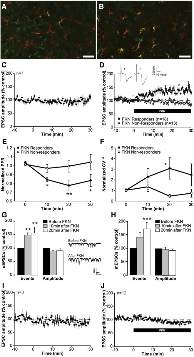
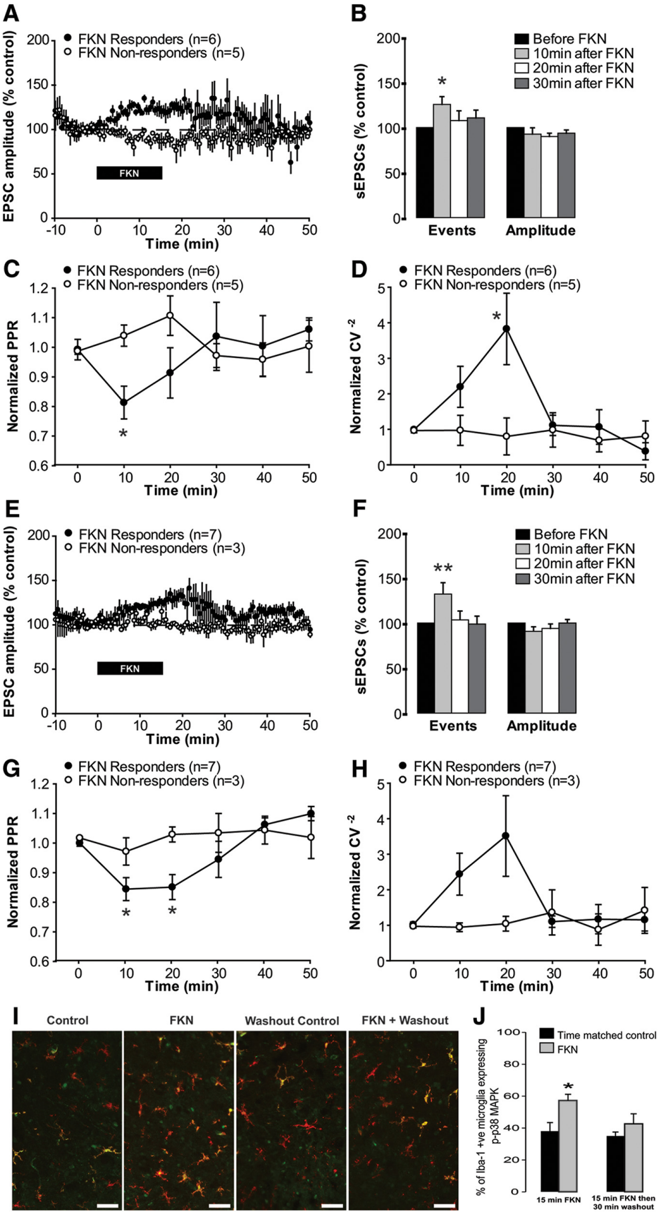
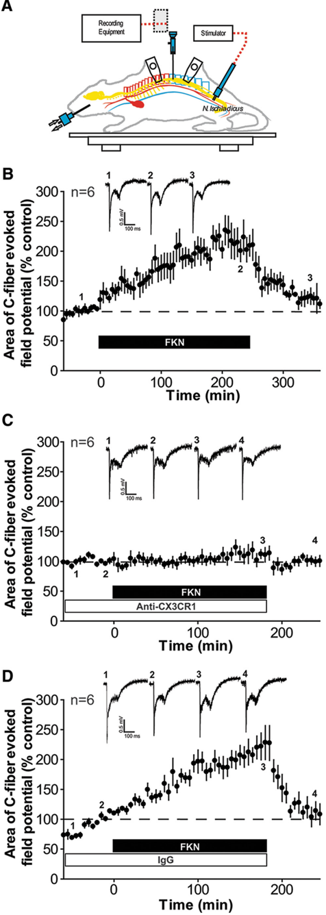
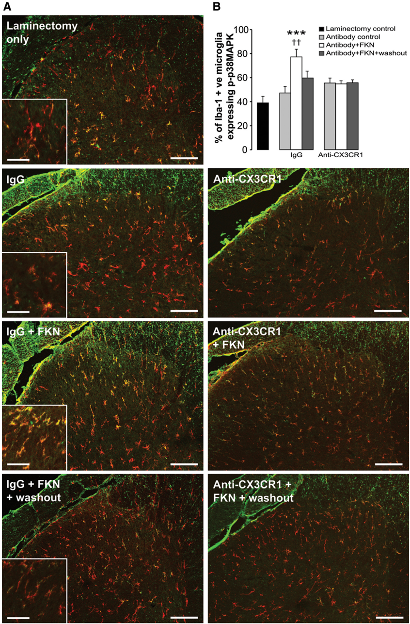
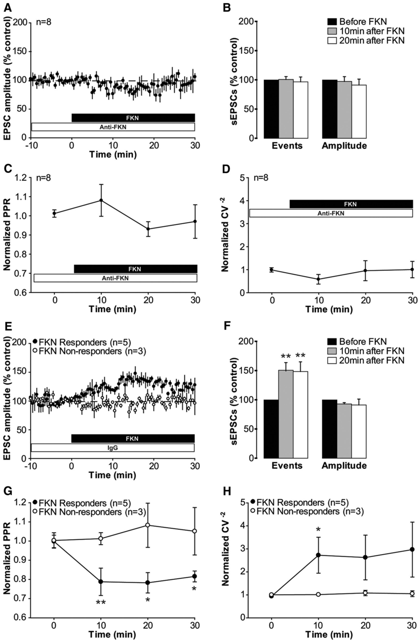
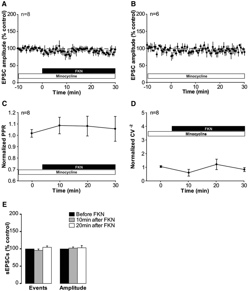
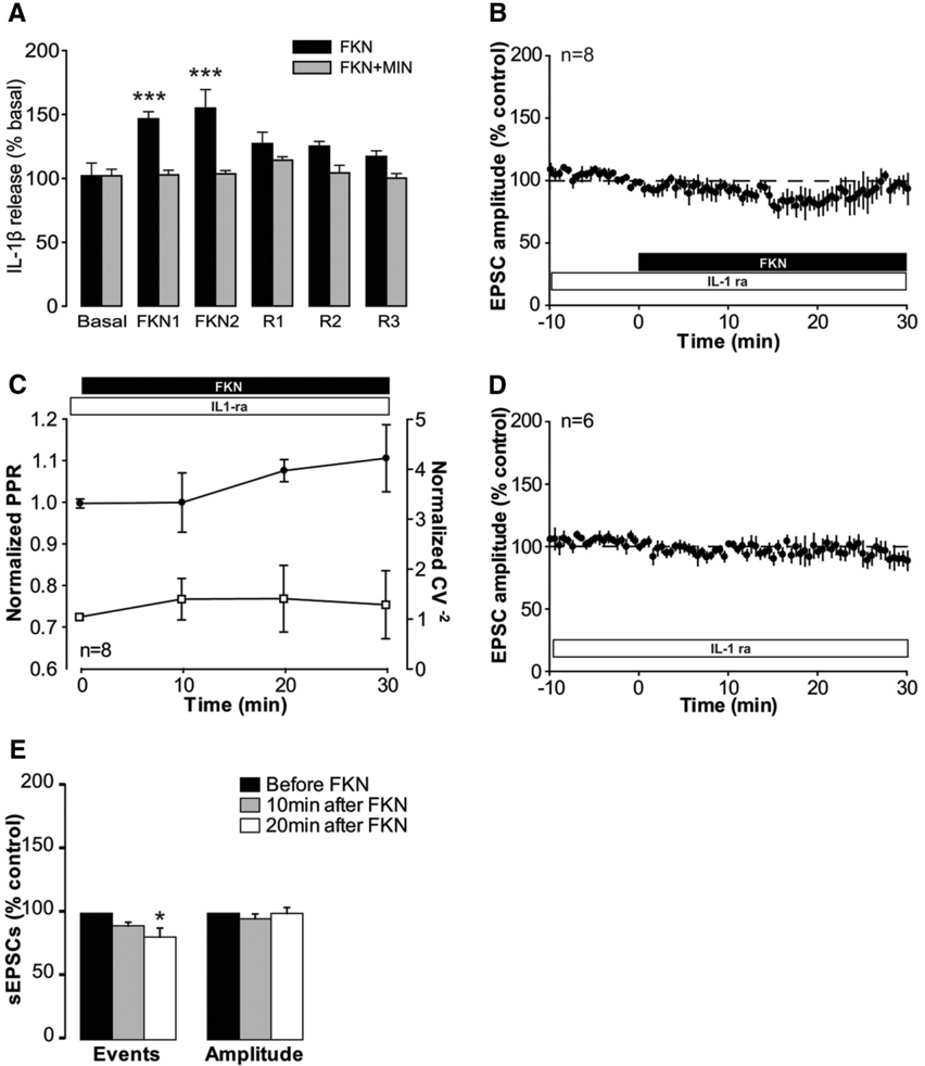
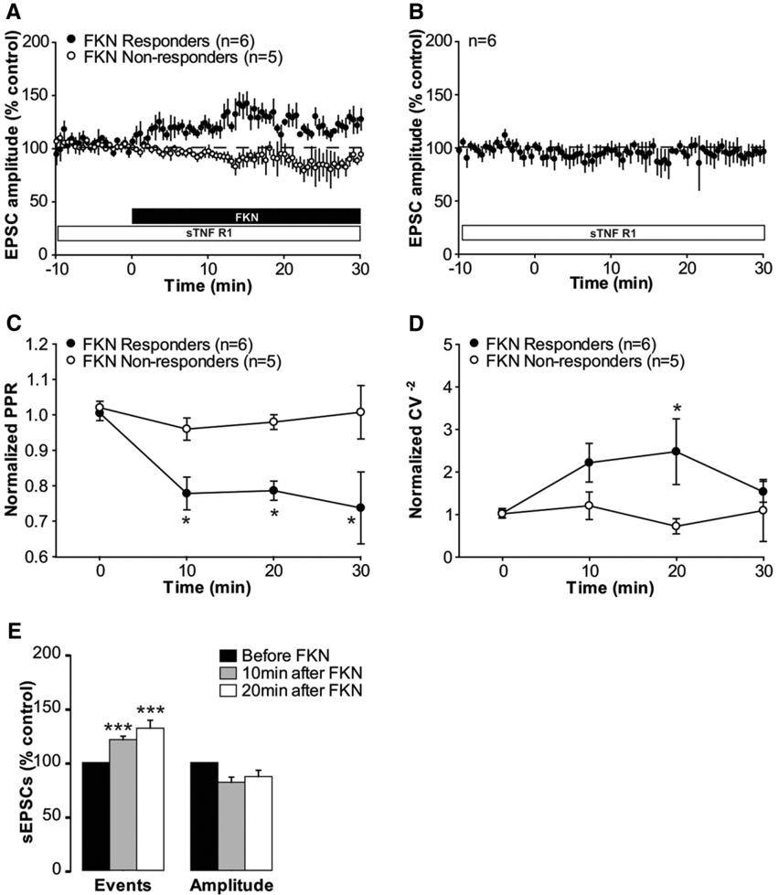
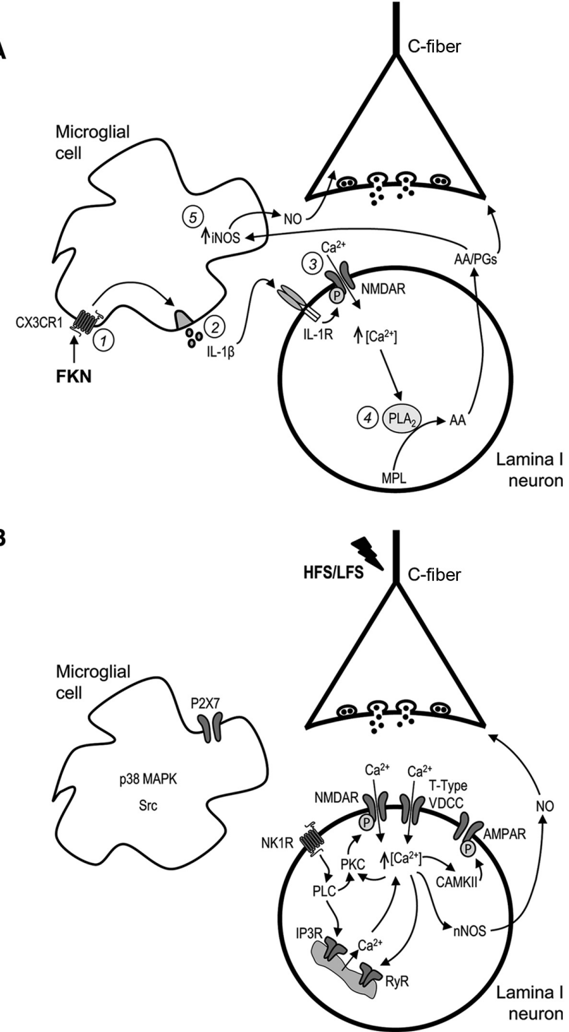
Synaptic plasticity is thought to be initiated by neurons only, with the prevailing view assigning glial cells mere specify supportive functions for synaptic transmission and plasticity. We now demonstrate that glial cells can control synaptic strength independent of neuronal activity. Here we show that selective activation of microglia in the rat is sufficient to rapidly facilitate synaptic strength between primary afferent C-fibers and lamina I neurons, the first synaptic relay in the nociceptive pathway. Specifically, the activation of the CX3CR1 receptor by fractalkine induces the release of interleukin-1β from microglia, which modulates NMDA signaling in postsynaptic neurons, leading to the release of an eicosanoid messenger, which ultimately enhances presynaptic neurotransmitter release. In contrast to the conventional view, this form of plasticity does not require enhanced neuronal activity to trigger the events leading to synaptic facilitation. Augmentation of synaptic strength in nociceptive pathways represents a cellular model of pain amplification. The present data thus suggest that, under chronic pain states, CX3CR1-mediated activation of microglia drives the facilitation of excitatory synaptic transmission in the dorsal horn, which contributes to pain hypersensitivity in chronic pain states.
Overview
The claims and statistics of this paper haven’t been checked yet.
Run the check — free, no account needed. It reads the open-access text and checks the sources it cites.
Integrity signals
Registry
Cites
Statistics
Figures
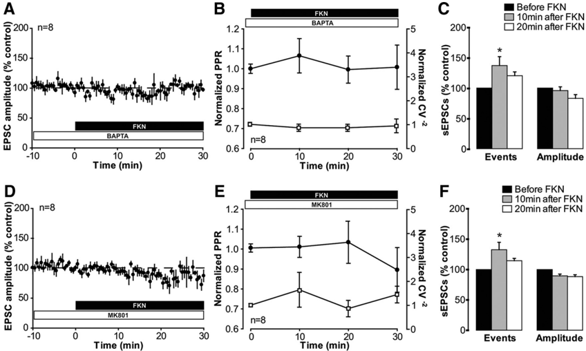
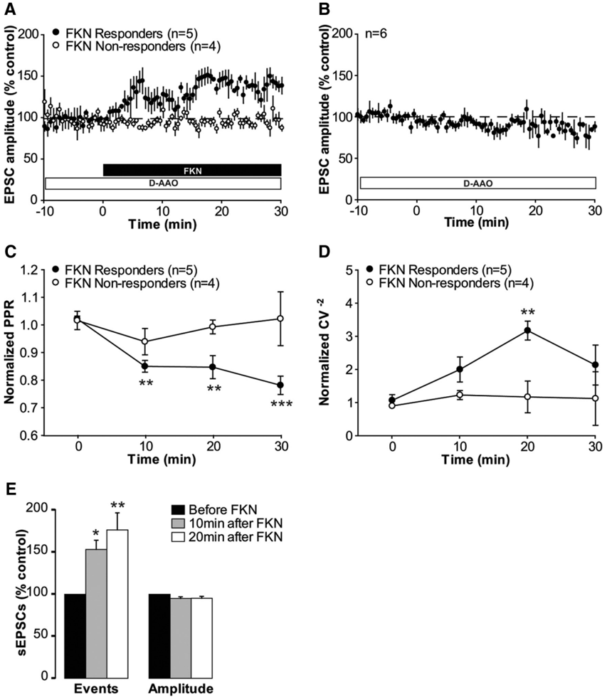
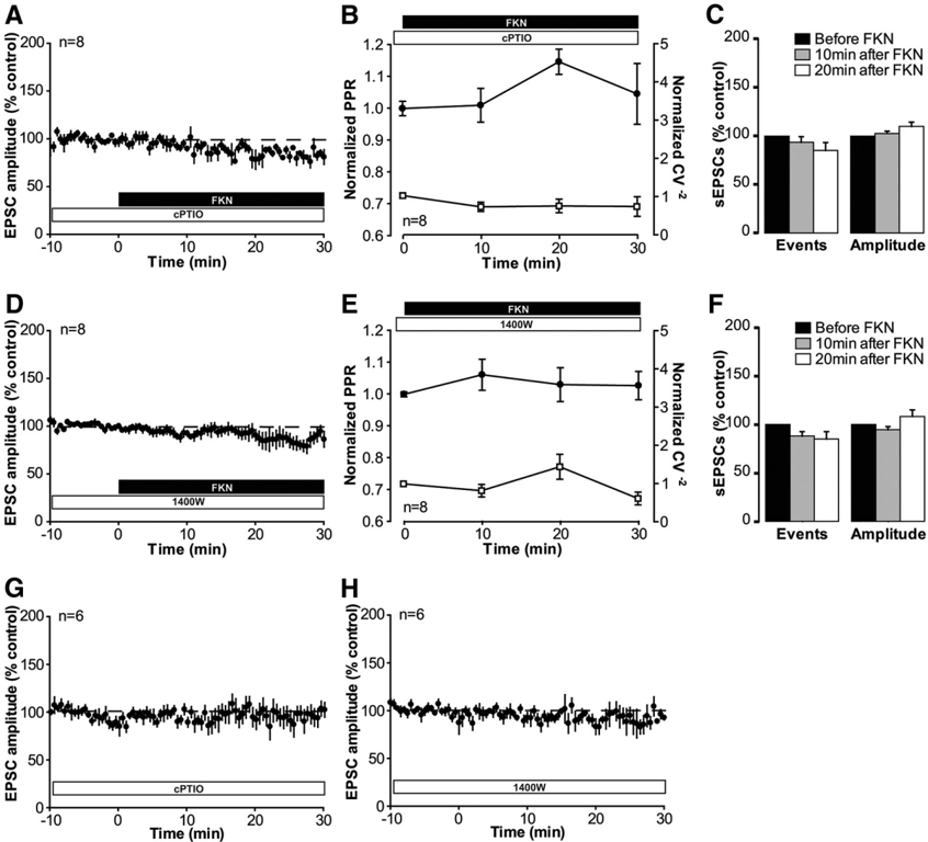
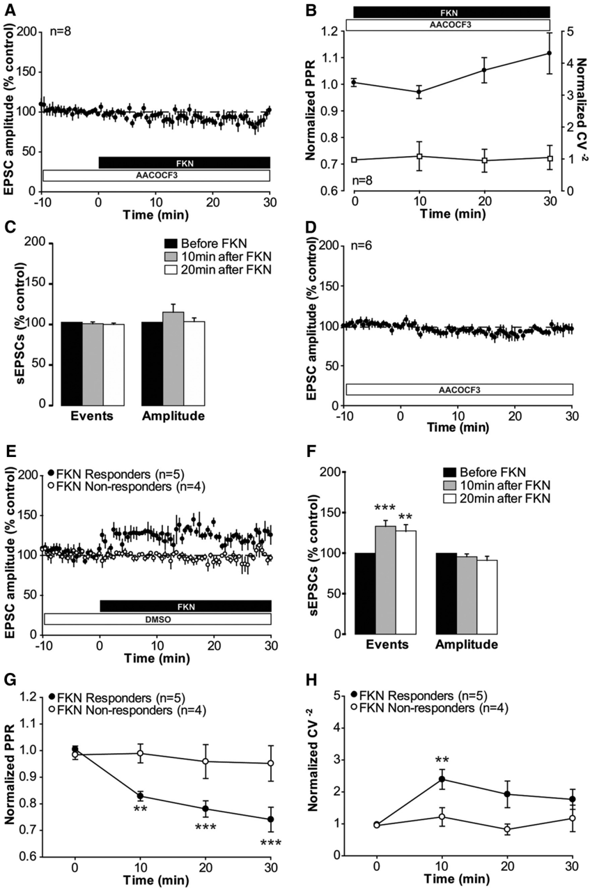
Verification
Claims the paper makes
The claims and statistics of this paper haven’t been checked yet.
Run the check — free, no account needed. It reads the open-access text and checks the sources it cites.
Claims other papers make about this one
Each is checked against this paper’s own text: does it say what the citing paper says it does?
In the citing paper
“The activation of the fractalkine receptor (CX3CR1) in microglial cells, increases synaptic strength. However, deficiency in this receptor results in a reduced hippocampal synaptic plasticity (Rogers et al., 2011; Clark et al., 2015).”
From The role of microglia in retinal neurodegeneration: Alzheimer’s disease, parkinson, and glaucoma. (2017) · cites this paper as Clark et al., 2015
Self-check
Percentages, sums and reported differences re-computed from the paper’s own numbers.
The self-check needs the paper’s full text.
Citations
Cites
The Rat Brain in Stereotaxic Coordinates
Paxinos, G., & Watson, C. (1983) · Elsevier eBooks · doi:10.1016/c2009-0-63235-9 · Cited by 28,585
A synaptic model of memory: long-term potentiation in the hippocampus
Bliss, T. V. P., & Collingridge, G. L. (1993) · Nature · doi:10.1038/361031a0 · Cited by 11,566
Polazzi, E., Mengoni, I., Caprini, M., Peña‐Altamira, E., Kurtys, E., & Monti, B. (2012) · Neurosignals · doi:10.1159/000337115 · Cited by 9,023
AChE-stained horizontal sections of the rat brain in stereotaxic coordinates
Paxinos, G., WATSON, C. R. R., & Emson, P. C. (1980) · Journal of Neuroscience Methods · doi:10.1016/0165-0270(80)90021-7 · Cited by 4,501
Malenka, R. C., & Bear, M. F. (2004) · Neuron · doi:10.1016/j.neuron.2004.09.012 · Cited by 3,968
Microglia: Active sensor and versatile effector cells in the normal and pathologic brain
Hanisch, U.-K., & Kettenmann, H. (2007) · Nature Neuroscience · doi:10.1038/nn1997 · Cited by 3,676
BDNF from microglia causes the shift in neuronal anion gradient underlying neuropathic pain
Coull, J. A. M., Beggs, S., Boudreau, D., Boivin, D., Tsuda, M., Inoue, K., Gravel, C., Salter, M. W., & De Koninck, Y. (2005) · Nature · doi:10.1038/nature04223 · Cited by 1,965
Control of microglial neurotoxicity by the fractalkine receptor
Cardona, A. E., Pioro, E. P., Sasse, M. E., Kostenko, V., Cardona, S. M., Dijkstra, I. M., Huang, D., Kidd, G., Dombrowski, S., Dutta, R., et al. (2006) · Nature Neuroscience · doi:10.1038/nn1715 · Cited by 1,531
Samad, T. A., Moore, K. A., Sapirstein, A., Billet, S. E., Allchorne, A. J., Poole, S., Bonventre, J. V., & Woolf, C. J. (2001) · Nature · doi:10.1038/35068566 · Cited by 1,282
Glia and pain: Is chronic pain a gliopathy?
Ji, R., Berta, T., & Nedergaard, M. (2013) · Pain · doi:10.1016/j.pain.2013.06.022 · Cited by 1,193
Cited by
Sorted by citations
Neuroinflammation and Central Sensitization in Chronic and Widespread Pain
Ji, R.-R., Nackley, A., Huh, Y., Terrando, N., & Maixner, W. (2018) · Anesthesiology · doi:10.1097/aln.0000000000002130 · Cited by 1,630
Microglia in neurodegenerative diseases: mechanism and potential therapeutic targets
Gao, C., Jiang, J., Tan, Y., & Chen, S. (2023) · Signal Transduction and Targeted Therapy · doi:10.1038/s41392-023-01588-0 · Cited by 1,616
Pain regulation by non-neuronal cells and inflammation
Ji, R.-R., Chamessian, A., & Zhang, Y.-Q. (2016) · Science · doi:10.1126/science.aaf8924 · Cited by 1,314
Microglia in neuropathic pain: cellular and molecular mechanisms and therapeutic potential
Inoue, K., & Tsuda, M. (2018) · Nature Reviews Neuroscience · doi:10.1038/nrn.2018.2 · Cited by 908
Microglia in Pain: Detrimental and Protective Roles in Pathogenesis and Resolution of Pain
Chen, G., Zhang, Y., Qadri, Y. J., Serhan, C. N., & Ji, R. (2018) · Neuron · doi:10.1016/j.neuron.2018.11.009 · Cited by 801
Metadata
- Authors
- Clark, A. K., Gruber-Schoffnegger, D., Drdla-Schutting, R., Gerhold, K. J., Malcangio, M., & Sandkühler, J.
- Affiliations
- King's College London (GB)
- Medical University of Vienna (AT)
- ORCID
- Venue
- The Journal of Neuroscience
- Publisher
- Society for Neuroscience
- Published
- 2015-03-18
- Volume
- 35
- Issue
- 11
- Pages
- 4552-4570
- ISSN
- 0270-6474 (ISSN-L)
- 1529-2401
- PMC
- PMC4363384
- OpenAlex
- W2063244116
- Cited by
- 171
- References
- 65
- Citations by year
- 2026: 9 · 2025: 8 · 2024: 12 · 2023: 15 · 2022: 18 · 2021: 23 · 2020: 14 · 2019: 15 · 2018: 18 · 2017: 12 · 2016: 18 · 2015: 9
- FWCI
- 14.00
- Type
- journal article
- Language
- en
- Open access
- hybrid
- doi.org
- Licence
- Locations
- Journal of Neuroscience (publishedVersion, cc-by-nc-sa)
- Europe PMC (PubMed Central) (publishedVersion)
- PubMed Central (publishedVersion)
- PubMed (publishedVersion)
- Research Portal (King's College London) (submittedVersion, cc-by)
- Funders
- Wellcome Trust (093173/Z/10/Z)
- Austrian Science Fund
- Topics
- Health Sciences › Medicine › Physiology (medical) › Pain Mechanisms and Treatments
- Life Sciences › Neuroscience › Neurology › Neuroinflammation and Neurodegeneration Mechanisms
- Life Sciences › Neuroscience › Cellular and Molecular Neuroscience › Neuroscience and Neuropharmacology Research
- Keywords
- microglia, synaptic plasticity, synaptic facilitation, CX3CR1, fractalkine, IL-1β, eicosanoids, presynaptic neurotransmitter release, nociceptive pathways, dorsal horn, C-fibers, pain amplification, chronic pain, glial cells
- MeSH terms
- Animals, Male, Synaptic Transmission, Neuronal Plasticity, Organ Culture Techniques, Spinal Cord, Rats, Sprague-Dawley, Microglia, Excitatory Postsynaptic Potentials, Rats
About these checks
Registry, retraction, Cites and statistics results are computed, not judged, with one exception: a printed reference that no index lists is read once by a model before it counts as a citation error. Claims are extracted by a model and kept only when their quote appears verbatim in the text; each cited source is then checked, with a model’s help, for whether it says what the claim relies on. Those verdicts can be wrong — read the claim’s quote and the cited source before relying on one.
Statistics and arithmetic are recomputed, not judged.
Confidence bands are shown only for fields calibrated on our annotated test set. None is calibrated yet, so no band is shown.
What counts as a claim ›How we measure confidence ›How we check this ›
Are you an author, or do you see a problem with this page? Request a review
