

The DOI matches its Crossref record; 65 of the 65 references it cites were found in a registry or an index.
Selective Activation of Microglia Facilitates Synaptic Strength
Open accessAbstract
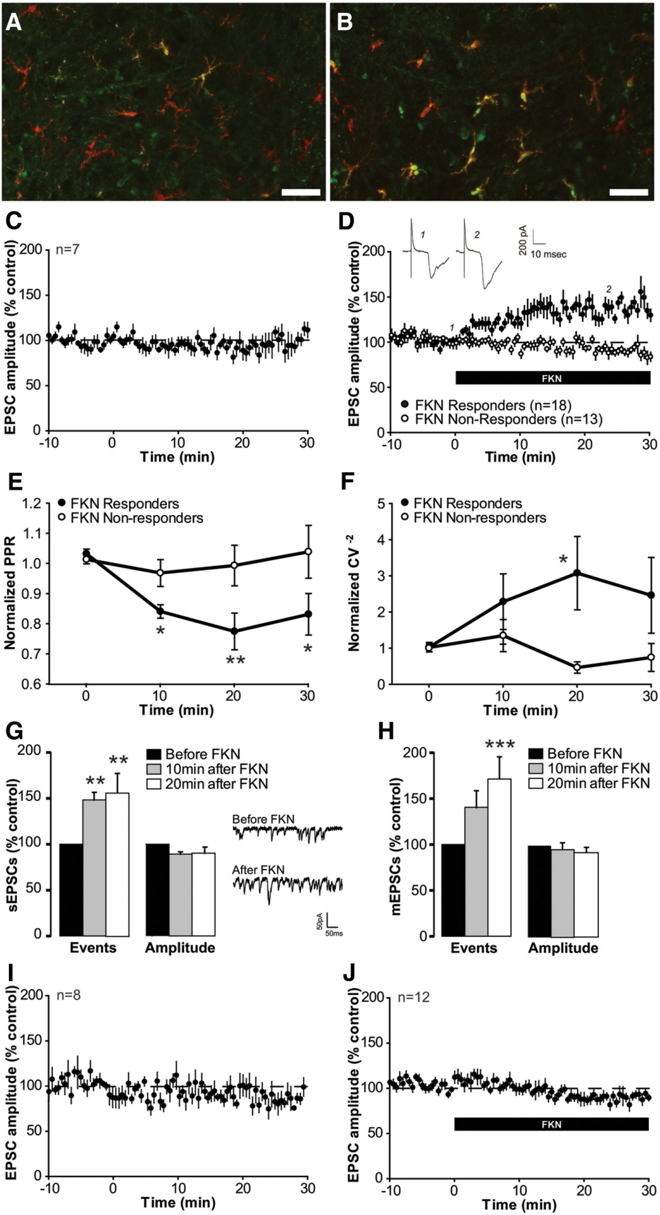
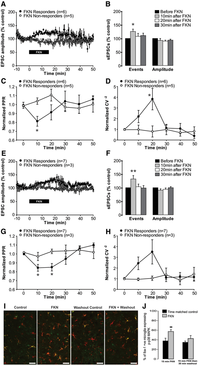
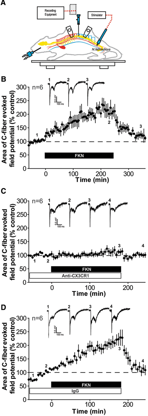
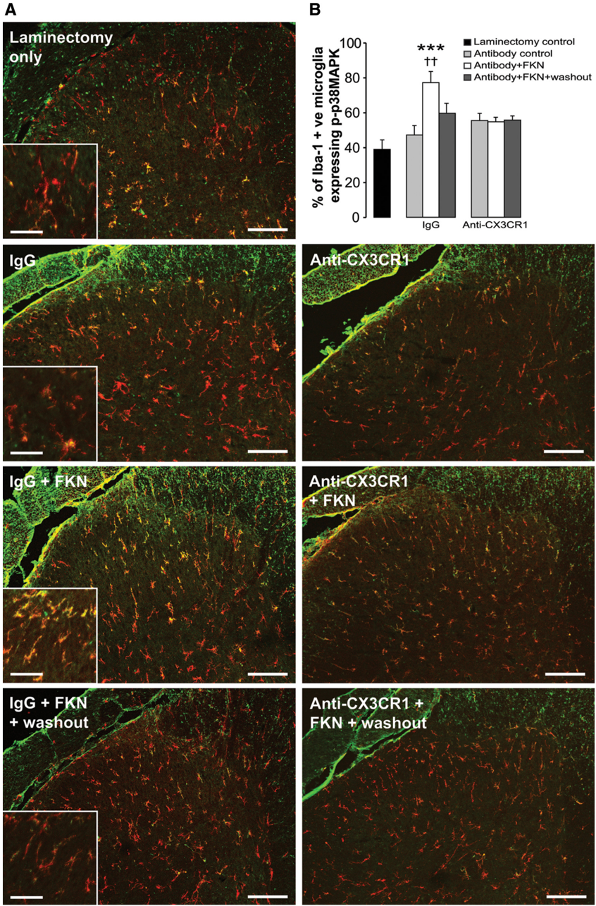
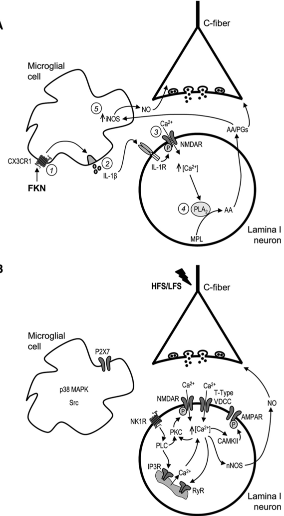
Synaptic plasticity is thought to be initiated by neurons only, with the prevailing view assigning glial cells mere specify supportive functions for synaptic transmission and plasticity. We now demonstrate that glial cells can control synaptic strength independent of neuronal activity. Here we show that selective activation of microglia in the rat is sufficient to rapidly facilitate synaptic strength between primary afferent C-fibers and lamina I neurons, the first synaptic relay in the nociceptive pathway. Specifically, the activation of the CX3CR1 receptor by fractalkine induces the release of interleukin-1β from microglia, which modulates NMDA signaling in postsynaptic neurons, leading to the release of an eicosanoid messenger, which ultimately enhances presynaptic neurotransmitter release. In contrast to the conventional view, this form of plasticity does not require enhanced neuronal activity to trigger the events leading to synaptic facilitation. Augmentation of synaptic strength in nociceptive pathways represents a cellular model of pain amplification. The present data thus suggest that, under chronic pain states, CX3CR1-mediated activation of microglia drives the facilitation of excitatory synaptic transmission in the dorsal horn, which contributes to pain hypersensitivity in chronic pain states.
Overview
The claims and statistics of this paper haven’t been checked yet.
Run the check — free, no account needed. It reads the open-access text and checks the sources it cites.
Integrity signals
Registry
Cites
Statistics
Figures
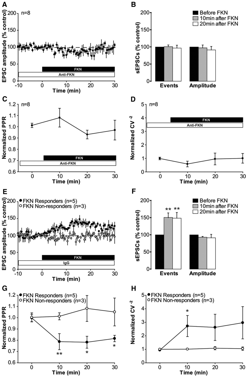
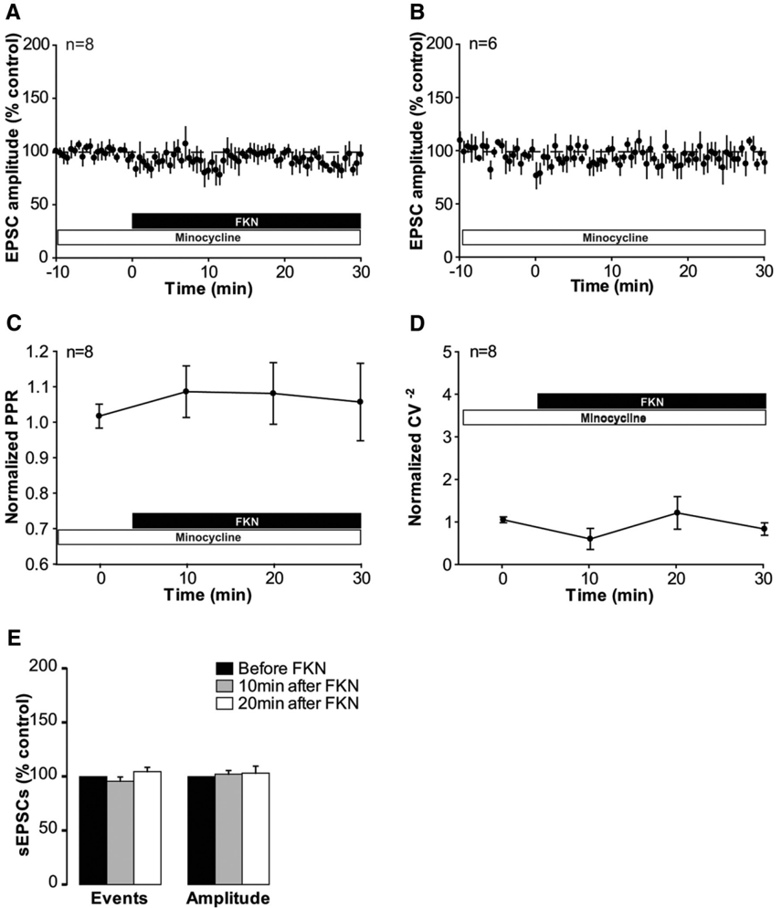
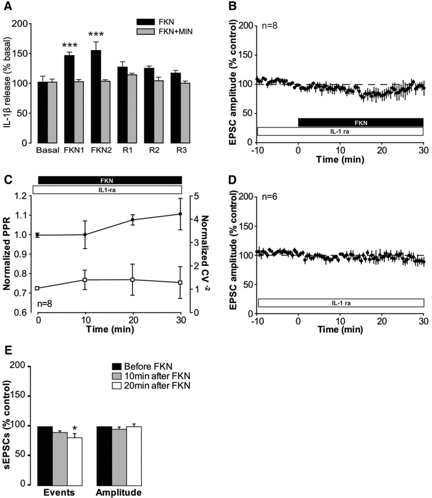
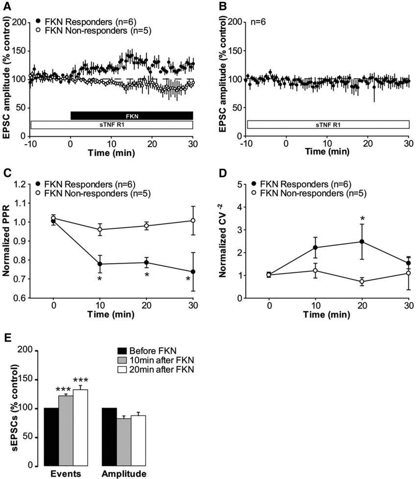
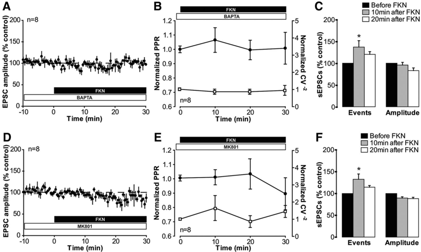
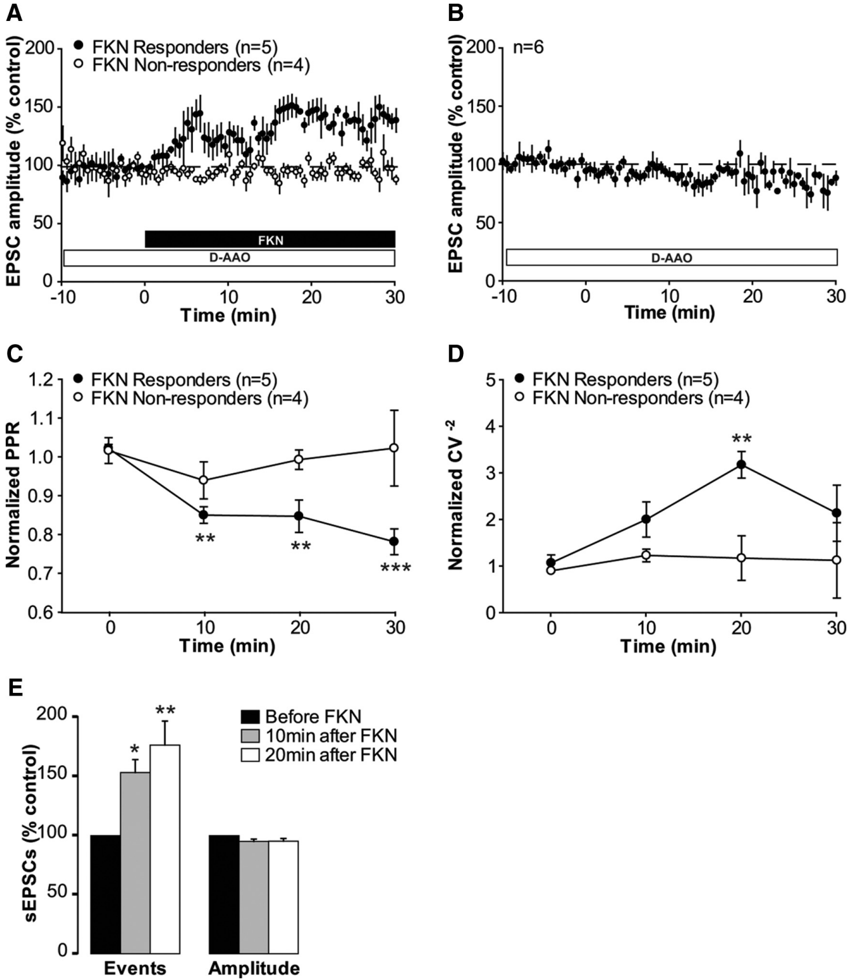
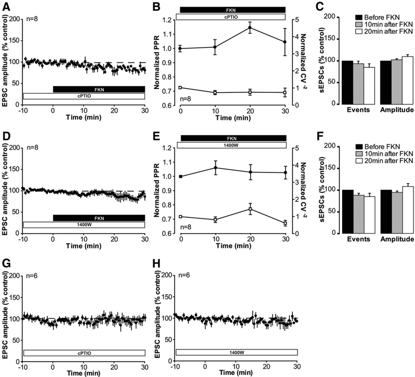
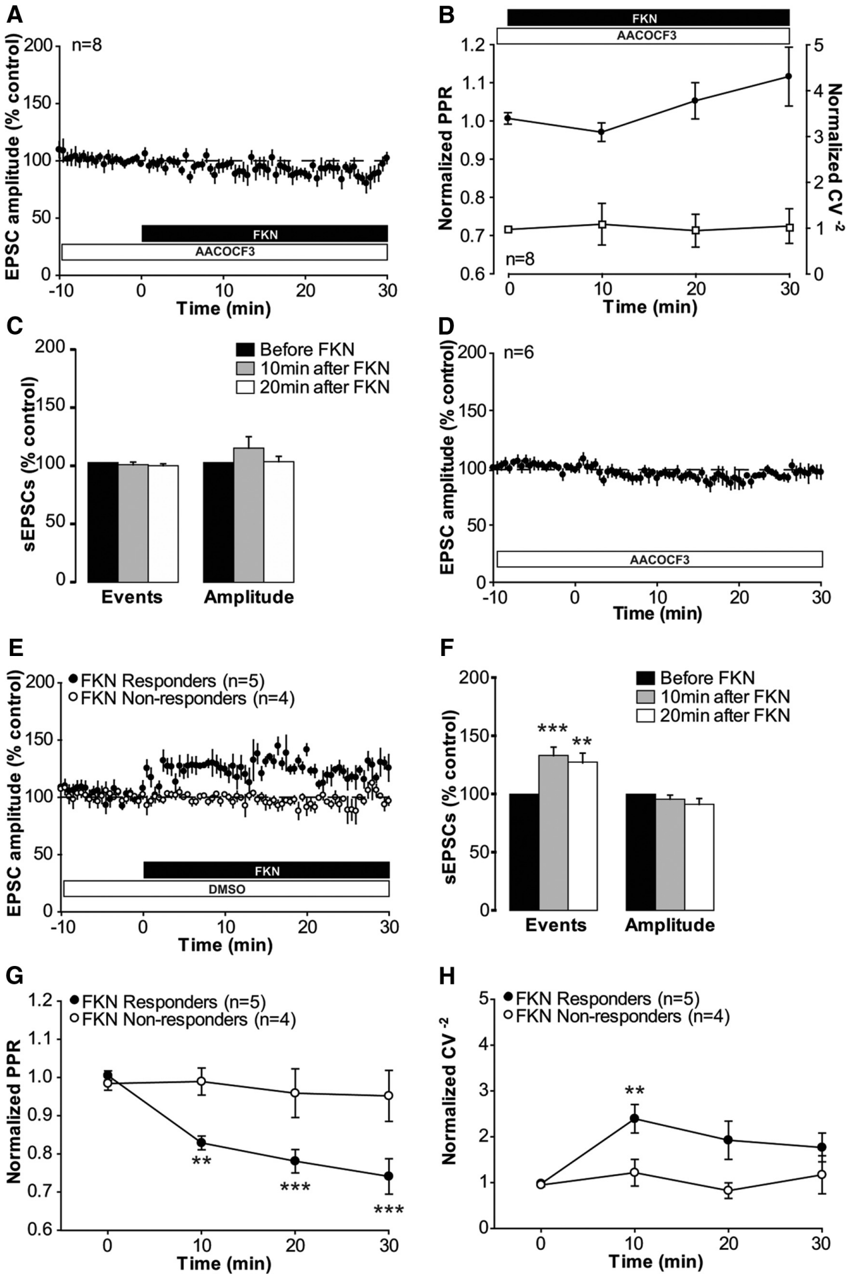
Verification
Claims the paper makes
The claims and statistics of this paper haven’t been checked yet.
Run the check — free, no account needed. It reads the open-access text and checks the sources it cites.
Claims other papers make about this one
Each is checked against this paper’s own text: does it say what the citing paper says it does?
In the citing paper
“The activation of the fractalkine receptor (CX3CR1) in microglial cells, increases synaptic strength. However, deficiency in this receptor results in a reduced hippocampal synaptic plasticity (Rogers et al., 2011; Clark et al., 2015).”
From The role of microglia in retinal neurodegeneration: Alzheimer’s disease, parkinson, and glaucoma. (2017) · cites this paper as Clark et al., 2015
Self-check
Percentages, sums and reported differences re-computed from the paper’s own numbers.
The self-check needs the paper’s full text.
Citations
Cites
The Rat Brain in Stereotaxic Coordinates
Paxinos, G., & Watson, C. (1983) · Elsevier eBooks · doi:10.1016/c2009-0-63235-9 · Cited by 28,585
A synaptic model of memory: long-term potentiation in the hippocampus
Bliss, T. V. P., & Collingridge, G. L. (1993) · Nature · doi:10.1038/361031a0 · Cited by 11,566
Polazzi, E., Mengoni, I., Caprini, M., Peña‐Altamira, E., Kurtys, E., & Monti, B. (2012) · Neurosignals · doi:10.1159/000337115 · Cited by 9,023
AChE-stained horizontal sections of the rat brain in stereotaxic coordinates
Paxinos, G., WATSON, C. R. R., & Emson, P. C. (1980) · Journal of Neuroscience Methods · doi:10.1016/0165-0270(80)90021-7 · Cited by 4,501
Malenka, R. C., & Bear, M. F. (2004) · Neuron · doi:10.1016/j.neuron.2004.09.012 · Cited by 3,968
Microglia: Active sensor and versatile effector cells in the normal and pathologic brain
Hanisch, U.-K., & Kettenmann, H. (2007) · Nature Neuroscience · doi:10.1038/nn1997 · Cited by 3,676
BDNF from microglia causes the shift in neuronal anion gradient underlying neuropathic pain
Coull, J. A. M., Beggs, S., Boudreau, D., Boivin, D., Tsuda, M., Inoue, K., Gravel, C., Salter, M. W., & De Koninck, Y. (2005) · Nature · doi:10.1038/nature04223 · Cited by 1,965
Control of microglial neurotoxicity by the fractalkine receptor
Cardona, A. E., Pioro, E. P., Sasse, M. E., Kostenko, V., Cardona, S. M., Dijkstra, I. M., Huang, D., Kidd, G., Dombrowski, S., Dutta, R., et al. (2006) · Nature Neuroscience · doi:10.1038/nn1715 · Cited by 1,531
Samad, T. A., Moore, K. A., Sapirstein, A., Billet, S. E., Allchorne, A. J., Poole, S., Bonventre, J. V., & Woolf, C. J. (2001) · Nature · doi:10.1038/35068566 · Cited by 1,282
Glia and pain: Is chronic pain a gliopathy?
Ji, R., Berta, T., & Nedergaard, M. (2013) · Pain · doi:10.1016/j.pain.2013.06.022 · Cited by 1,193
Cited by
Sorted by citations
Neuroinflammation and Central Sensitization in Chronic and Widespread Pain
Ji, R.-R., Nackley, A., Huh, Y., Terrando, N., & Maixner, W. (2018) · Anesthesiology · doi:10.1097/aln.0000000000002130 · Cited by 1,630
Microglia in neurodegenerative diseases: mechanism and potential therapeutic targets
Gao, C., Jiang, J., Tan, Y., & Chen, S. (2023) · Signal Transduction and Targeted Therapy · doi:10.1038/s41392-023-01588-0 · Cited by 1,616
Pain regulation by non-neuronal cells and inflammation
Ji, R.-R., Chamessian, A., & Zhang, Y.-Q. (2016) · Science · doi:10.1126/science.aaf8924 · Cited by 1,314
Microglia in neuropathic pain: cellular and molecular mechanisms and therapeutic potential
Inoue, K., & Tsuda, M. (2018) · Nature Reviews Neuroscience · doi:10.1038/nrn.2018.2 · Cited by 908
Microglia in Pain: Detrimental and Protective Roles in Pathogenesis and Resolution of Pain
Chen, G., Zhang, Y., Qadri, Y. J., Serhan, C. N., & Ji, R. (2018) · Neuron · doi:10.1016/j.neuron.2018.11.009 · Cited by 801
Tay, T. L., Savage, J. C., Hui, C. W., Bisht, K., & Tremblay, M. (2017) · The Journal of Physiology · doi:10.1113/jp272134 · Cited by 562
The role of microglia in retinal neurodegeneration: Alzheimer’s disease, parkinson, and glaucoma.
Ramirez, A. I., de Hoz, R., Salobrar-Garcia, E., Salazar, J. J., Rojas, B., Ajoy, D., López-Cuenca, I., Rojas, P., Triviño, A., & Ramírez, J. M. (2017) · Frontiers in Aging Neuroscience · doi:10.3389/fnagi.2017.00214 · Cited by 466
Microglia in CNS development: Shaping the brain for the future
Mosser, C., Baptista, S., Arnoux, I., & Audinat, É. (2017) · Progress in Neurobiology · doi:10.1016/j.pneurobio.2017.01.002 · Cited by 289
Shaping Neuronal Fate: Functional Heterogeneity of Direct Microglia-Neuron Interactions
Cserép, C., Pósfai, B., & Dénes, Á. (2021) · Neuron · doi:10.1016/j.neuron.2020.11.007 · Cited by 256
Fractalkine receptor deficiency impairs microglial and neuronal responsiveness to chronic stress
Milior, G., Lecours, C., Samson, L., Bisht, K., Poggini, S., Pagani, F., Deflorio, C., Lauro, C., Alboni, S., Limatola, C., et al. (2015) · Brain Behavior and Immunity · doi:10.1016/j.bbi.2015.07.024 · Cited by 259
Chemokines in chronic pain: cellular and molecular mechanisms and therapeutic potential
Jiang, B., Liu, T., & Gao, Y. (2020) · Pharmacology & Therapeutics · doi:10.1016/j.pharmthera.2020.107581 · Cited by 242
Microglia-neuron crosstalk: Signaling mechanism and control of synaptic transmission
Marinelli, S., Basilico, B., Marrone, M. C., & Ragozzino, D. (2019) · Seminars in Cell & Developmental Biology · doi:10.1016/j.semcdb.2019.05.017 · Cited by 233
Role of the immune system in neuropathic pain
Malcangio, M. (2019) · Scandinavian Journal of Pain · doi:10.1515/sjpain-2019-0138 · Cited by 236
Self-citationMicroglia Are Indispensable for Synaptic Plasticity in the Spinal Dorsal Horn and Chronic Pain
Zhou, L.-J., Peng, J., Xu, Y.-N., Zeng, W.-J., Zhang, J., Wei, X., Mai, C.-L., Lin, Z.-J., Liu, Y., Murugan, M., et al. (2019) · Cell Reports · doi:10.1016/j.celrep.2019.05.087 · Cited by 228
Fumagalli, G., Monza, L., Cavaletti, G., Rigolio, R., & Meregalli, C. (2021) · Frontiers in Immunology · doi:10.3389/fimmu.2020.626687 · Cited by 183
Gliogenic LTP spreads widely in nociceptive pathways
Kronschläger, M. T., Drdla-Schutting, R., Gassner, M., Honsek, S. D., Teuchmann, H. L., & Sandkühler, J. (2016) · Science · doi:10.1126/science.aah5715 · Cited by 173
Self-citationNeuron–Microglia Interactions in Mental Health Disorders: “For Better, and For Worse”
Wohleb, E. S. (2016) · Frontiers in Immunology · doi:10.3389/fimmu.2016.00544 · Cited by 170
A pharmacological basis of herbal medicines for epilepsy
Sucher, N. J., & Carles, M. (2015) · Epilepsy & Behavior · doi:10.1016/j.yebeh.2015.05.012 · Cited by 160
Peripheral and central oxidative stress in chemotherapy-induced neuropathic pain
Shim, H. S., Bae, C., Wang, J., Lee, K. H., Hankerd, K., Kim, H. K., Chung, J. M., & La, J. (2019) · Molecular Pain · doi:10.1177/1744806919840098 · Cited by 155
Chemogenetic manipulation of microglia inhibits neuroinflammation and neuropathic pain in mice
Yi, M., Liu, Y., Liu, K., Chen, T., Bosco, D. B., Zheng, J., Xie, M., Zhou, L., Qu, W., & Wu, L. (2020) · Brain Behavior and Immunity · doi:10.1016/j.bbi.2020.11.030 · Cited by 143
Stress induced microglial activation contributes to depression
Li, B., Yang, W., Ge, T., Wang, Y., & Cui, R. (2022) · Pharmacological Research · doi:10.1016/j.phrs.2022.106145 · Cited by 151
Eyo, U. B., Peng, J., Murugan, M., Mo, M., Lalani, A., Xie, P., Xu, P., Margolis, D. J., & Wu, L. (2016) · eNeuro · doi:10.1523/eneuro.0209-16.2016 · Cited by 117
Shao, S., Xu, C.-B., Chen, C.-J., Shi, G.-N., Guo, Q.-L., Zhou, Y., Wei, Y.-Z., Wu, L., Shi, J.-G., & Zhang, T.-T. (2021) · Journal of Neuroinflammation · doi:10.1186/s12974-021-02178-z · Cited by 112
Ion Channels and Receptors as Determinants of Microglial Function
Izquierdo, P., Attwell, D., & Madry, C. (2019) · Trends in Neurosciences · doi:10.1016/j.tins.2018.12.007 · Cited by 102
Boakye, P. A., Tang, S.-J., & Smith, P. A. (2021) · Frontiers in Pain Research · doi:10.3389/fpain.2021.698157 · Cited by 102
Microglial modulation of neuronal activity in the healthy brain
York, E. M., Bernier, L., & MacVicar, B. A. (2018) · Developmental Neurobiology · doi:10.1002/dneu.22571 · Cited by 98
Li, X., Shi, H., Zhang, D., Jing, B., Chen, Z., Zheng, Y., Chang, S., Gao, L., & Zhao, G. (2023) · CNS Neuroscience & Therapeutics · doi:10.1111/cns.14211 · Cited by 96
Dou, B., Li, Y., Ma, J., Xu, Z., Fan, W., Tian, L., Chen, Z., Li, N., Gong, Y., Lyu, Z., et al. (2021) · Frontiers in Neuroscience · doi:10.3389/fnins.2021.695670 · Cited by 92
Freria, C. M., Hall, J. C. E., Wei, P. Y., Guan, Z., McTigue, D. M., & Popovich, P. G. (2017) · Journal of Neuroscience · doi:10.1523/jneurosci.2841-16.2017 · Cited by 93
Rossetti, A. C., Papp, M., Gruca, P., Paladini, M. S., Racagni, G., Riva, M. A., & Molteni, R. (2015) · Pharmacological Research · doi:10.1016/j.phrs.2015.10.022 · Cited by 90
Neuroimmune signatures in chronic low back pain subtypes
Alshelh, Z., Brusaferri, L., Saha, A., Morrissey, E. J., Knight, P. C., Kim, M., Zhang, Y., Hooker, J. M., Albrecht, D., Torrado-Carvajal, Á., et al. (2021) · Brain · doi:10.1093/brain/awab336 · Cited by 86
Rosa, J. M., Farré‐Alins, V., Ortega, M. C., Navarrete, M., López-Rodríguez, A. B., Palomino‐Antolín, A., Fernández‐López, E., Sol, V. V., Decouty‐Pérez, C., Narros‐Fernández, P., et al. (2021) · British Journal of Pharmacology · doi:10.1111/bph.15488 · Cited by 81
Substance P spinal signaling induces glial activation and nociceptive sensitization after fracture
Li, W.-W., Guo, T.-Z., Shi, X., Sun, Y., Wei, T., Clark, D. J., & Kingery, W. S. (2015) · Neuroscience · doi:10.1016/j.neuroscience.2015.09.036 · Cited by 80
Nociceptive signaling mediated by P2X3, P2X4 and P2X7 receptors
Inoue, K., & Tsuda, M. (2021) · Biochemical Pharmacology · doi:10.1016/j.bcp.2020.114309 · Cited by 78
Microglia-Mediated Regulation of Neuropathic Pain: Molecular and Cellular Mechanisms
Tsuda, M. (2019) · Biological and Pharmaceutical Bulletin · doi:10.1248/bpb.b19-00715 · Cited by 79
l ‐DOPA‐induced dyskinesia and neuroinflammation: do microglia and astrocytes play a role?
Carta, A. R., Mulas, G., Bortolanza, M., Duarte, T. T., Pillai, E., Fisone, G., Raisman‐Vozari, R., & Bel, E. A. D. (2016) · European Journal of Neuroscience · doi:10.1111/ejn.13482 · Cited by 80
The neuroinflammatory component of negative affect in patients with chronic pain
Albrecht, D., Kim, M., Akeju, O., Torrado-Carvajal, Á., Edwards, R. R., Zhang, Y., Bergan, C., Protsenko, E., Kucyi, A., Wasan, A. D., et al. (2019) · Molecular Psychiatry · doi:10.1038/s41380-019-0433-1 · Cited by 80
Neuroinflammation in Alzheimer’s disease: Pleiotropic roles for cytokines and neuronal pentraxins
Swanson, A., Wolf, T., Sitzmann, A., & Willette, A. A. (2018) · Behavioural Brain Research · doi:10.1016/j.bbr.2018.02.015 · Cited by 72
Role of microglia and P2X4 receptors in chronic pain
Kohno, K., & Tsuda, M. (2021) · PAIN Reports · doi:10.1097/pr9.0000000000000864 · Cited by 66
How microglia contribute to the induction and maintenance of neuropathic pain
Malcangio, M., & Sideris-Lampretsas, G. (2025) · Nature reviews. Neuroscience · doi:10.1038/s41583-025-00914-5 · Cited by 69
Self-citationMicroglia and Parkinson's disease: footprints to pathology
Lazdon, E., Stolero, N., & Frenkel, D. (2020) · Journal of Neural Transmission · doi:10.1007/s00702-020-02154-6 · Cited by 61
Tay, T. L., Carrier, M., & Tremblay, M. (2019) · Advances in experimental medicine and biology · doi:10.1007/978-981-13-9913-8_6 · Cited by 61
Microglia maintain the normal structure and function of the hippocampal astrocyte network
Du, Y., Brennan, F. H., Popovich, P. G., & Zhou, M. (2022) · Glia · doi:10.1002/glia.24179 · Cited by 61
Dodds, K. N., Beckett, E. A. H., Evans, S., Grace, P. M., Watkins, L. R., & Hutchinson, M. R. (2016) · Translational Psychiatry · doi:10.1038/tp.2016.168 · Cited by 61
Fan, Z., Dou, B., Wang, J., Wu, Y., Du, S., Li, J., Yao, K., Li, Y., Wang, S., Gong, Y., et al. (2024) · Frontiers in Neuroscience · doi:10.3389/fnins.2023.1239839 · Cited by 59
Mironets, E., Osei‐Owusu, P., Bracchi-Ricard, V. C., Fischer, R., Owens, E. A., Ricard, J., Wu, D., Saltos, T. M., Collyer, E., Hou, S., et al. (2018) · Journal of Neuroscience · doi:10.1523/jneurosci.2376-17.2018 · Cited by 60
Mechanism of the JAK2/STAT3-CAV-1-NR2B signaling pathway in painful diabetic neuropathy
Chuan-da, L., Zhao, J., Chen, J., Lu, J., Zhang, M., Huang, Q., Cao, Y., Jia, G., Tao, Y., Li, J., et al. (2019) · Endocrine · doi:10.1007/s12020-019-01880-6 · Cited by 58
The force awakens: insights into the origin and formation of microglia
Tay, T. L., Hagemeyer, N., & Prinz, M. (2016) · Current Opinion in Neurobiology · doi:10.1016/j.conb.2016.04.003 · Cited by 59
Cantaut-Belarif, Y., Antri, M., Pizzarelli, R., Colasse, S., Vaccari, I., Soares, S., Renner, M., Dallel, R., Triller, A., & Bessis, A. (2017) · Journal of Cell Biology · doi:10.1083/jcb.201607048 · Cited by 56
Activated microglia facilitate the transmission of α-synuclein in Parkinson's disease
Zheng, T., & Zhang, Z. (2021) · Neurochemistry International · doi:10.1016/j.neuint.2021.105094 · Cited by 57
Synaptoimmunology - roles in health and disease
Nisticò, R., Salter, E. W., Nicolas, C. S., Feligioni, M., Mango, D., Bortolotto, Z. A., Gressèns, P., Collingridge, G. L., & Peineau, S. (2017) · Molecular Brain · doi:10.1186/s13041-017-0308-9 · Cited by 53
Kambrun, C., Roca‐Lapirot, O., Salio, C., Landry, M., Moqrich, A., & Feuvre, Y. L. (2018) · Cell Reports · doi:10.1016/j.celrep.2018.02.068 · Cited by 59
Microglia exit the CNS in spinal root avulsion
Green, L. A., Nebiolo, J. C., & Smith, C. J. (2019) · PLoS Biology · doi:10.1371/journal.pbio.3000159 · Cited by 52
Microglia Dystrophy Following Binge-Like Alcohol Exposure in Adolescent and Adult Male Rats
Marshall, S. A., McClain, J. A., Wooden, J. I., & Nixon, K. (2020) · Frontiers in Neuroanatomy · doi:10.3389/fnana.2020.00052 · Cited by 51
Chemogenetic and Optogenetic Manipulations of Microglia in Chronic Pain
Parusel, S., Yi, M., Hunt, C., & Wu, L. (2022) · Neuroscience Bulletin · doi:10.1007/s12264-022-00937-3 · Cited by 53
Immune drivers of physiological and pathological pain
Jain, A., Hakim, S., & Woolf, C. J. (2024) · The Journal of Experimental Medicine · doi:10.1084/jem.20221687 · Cited by 58
“Neuroinflammation”: does it have a role in chronic pain? Evidence from human imaging
Loggia, M. L. (2024) · Pain · doi:10.1097/j.pain.0000000000003342 · Cited by 51
Si, W., Li, X., Jing, B., Chang, S., Zheng, Y., Chen, Z., Zhao, G., & Zhang, D. (2023) · Phytotherapy Research · doi:10.1002/ptr.8039 · Cited by 53
Fractalkine/CX3CR1-Dependent Modulation of Synaptic and Network Plasticity in Health and Disease
Camacho-Hernández, N. P., & Peña‐Ortega, F. (2023) · Neural Plasticity · doi:10.1155/2023/4637073 · Cited by 52
Neuropathic pain; what we know and what we should do about it
Smith, P. A. (2023) · Frontiers in Pain Research · doi:10.3389/fpain.2023.1220034 · Cited by 53
Neuroinflammation in l-DOPA-induced dyskinesia: beyond the immune function
Pisanu, A., Boi, L., Mulas, G., Spiga, S., Fenu, S., & Carta, A. R. (2018) · Journal of Neural Transmission · doi:10.1007/s00702-018-1874-4 · Cited by 49
Spinal microglia-neuron interactions in chronic pain
Ho, I. H. T., Chan, M. T. V., Wu, W. K. K., & Liu, X. (2020) · Journal of Leukocyte Biology · doi:10.1002/jlb.3mr0520-695r · Cited by 50
Hu, D., Zhu, S., & Potas, J. R. (2016) · Journal of Neuroinflammation · doi:10.1186/s12974-016-0679-3 · Cited by 47
Montague-Cardoso, K., Pitcher, T., Chisholm, K. I., Salera, G., Lindström, E., Hewitt, E. L., Solito, E., & Malcangio, M. (2019) · Brain Behavior and Immunity · doi:10.1016/j.bbi.2019.10.018 · Cited by 44
Self-citationThe chemokine CXCL16 modulates neurotransmitter release in hippocampal CA1 area
Castro, M. A. D., Trettel, F., Milior, G., Maggi, L., Ragozzino, D., & Limatola, C. (2016) · Scientific Reports · doi:10.1038/srep34633 · Cited by 45
Microglia in health and pain: impact of noxious early life events
Burke, N. N., Fan, C. Y., & Trang, T. (2016) · Experimental Physiology · doi:10.1113/ep085714 · Cited by 45
Zhang, T., Zhang, M., Cui, S., Liang, W., Jia, Z., Guo, F., Ou, W., Wu, Y., & Zhang, S. (2023) · Brain and Behavior · doi:10.1002/brb3.2868 · Cited by 45
Activity-triggered tetrapartite neuron–glial interactions following peripheral injury
Ren, K., & Dubner, R. A. (2015) · Current Opinion in Pharmacology · doi:10.1016/j.coph.2015.09.006 · Cited by 42
The Role of ATP Receptors in Pain Signaling
Inoue, K. (2022) · Neurochemical Research · doi:10.1007/s11064-021-03516-6 · Cited by 42
Regulation of neuropathic pain by microglial Orai1 channels
Tsujikawa, S., DeMeulenaere, K. E., Centeno, M. V., Ghazisaeidi, S., Martin, M. E., Tapies, M. G. R., Maneshi, M. M., Yamashita, M., Stauderman, K. A., Apkarian, A. V., et al. (2023) · Science Advances · doi:10.1126/sciadv.ade7002 · Cited by 45
Fractalkine/CX3CR1 Pathway in Neuropathic Pain: An Update
Silva, R., & Malcangio, M. (2021) · Frontiers in Pain Research · doi:10.3389/fpain.2021.684684 · Cited by 41
Self-citationMicroglia in the CNS and Neuropathic Pain
Tsuda, M. (2018) · Advances in experimental medicine and biology · doi:10.1007/978-981-13-1756-9_7 · Cited by 39
The Role of Neuro-Immune Interactions in Chronic Pain: Implications for Clinical Practice
Su, P. P., Zhang, L., He, L., Zhao, N., & Guan, Z. (2022) · Journal of Pain Research · doi:10.2147/jpr.s246883 · Cited by 44
Barkaway, A., Attwell, D., & Korte, N. (2022) · Neurophotonics · doi:10.1117/1.nph.9.3.031914 · Cited by 38
Presynaptic Inhibition of Primary Nociceptive Signals to Dorsal Horn Lamina I Neurons by Dopamine
Lu, Y., Doroshenko, M., Lauzadis, J., Kanjiya, M. P., Rebecchi, M. J., Kaczocha, M., & Puopolo, M. (2018) · Journal of Neuroscience · doi:10.1523/jneurosci.0323-18.2018 · Cited by 38
Microglial heterogeneity in chronic pain
Sideris-Lampretsas, G., & Malcangio, M. (2021) · Brain Behavior and Immunity · doi:10.1016/j.bbi.2021.06.005 · Cited by 40
Self-citationSpinal microglia contribute to sustained inflammatory pain via amplifying neuronal activity
Gu, N., Yi, M., Murugan, M., Xie, M., Parusel, S., Peng, J., Eyo, U. B., Hunt, C., Dong, H., & Wu, L. (2022) · Molecular Brain · doi:10.1186/s13041-022-00970-3 · Cited by 36
Complement 3a receptor in dorsal horn microglia mediates pronociceptive neuropeptide signaling
Doolen, S., Cook, J., Riedl, M. S., Kitto, K. F., Kohsaka, S., Honda, C. N., Fairbanks, C. A., Taylor, B. K., & Vulchanova, L. (2017) · Glia · doi:10.1002/glia.23208 · Cited by 35
Yang, N. J., Isensee, J., Neel, D. V., de Quadros, A. U., Zhang, H.-X. B., Lauzadis, J., Liu, S. M., Shiers, S. I., Belu, A., Palan, S., et al. (2021) · Nature Neuroscience · doi:10.1038/s41593-021-00973-8 · Cited by 34
Diversity of composition and function of sodium channels in peripheral sensory neurons
Dib‐Hajj, S. D., & Waxman, S. G. (2015) · Pain · doi:10.1097/j.pain.0000000000000353 · Cited by 35
Adult and endemic neurogenesis in the vestibular nuclei after unilateral vestibular neurectomy
Rastoldo, G., Mahmoudi, N. E., Marouane, E., Péricat, D., Watabe, I., Toneto, A., López‐Juárez, A., Chabbert, C., & Tighilet, B. (2020) · Progress in Neurobiology · doi:10.1016/j.pneurobio.2020.101899 · Cited by 34
Spinal pain processing in arthritis: Neuron and glia (inter)actions
Schaible, H., König, C., & Ebersberger, A. (2022) · Journal of Neurochemistry · doi:10.1111/jnc.15742 · Cited by 33
VGF: A prospective biomarker and therapeutic target for neuroendocrine and nervous system disorders
Wang, Y., Qin, X., Han, Y. J., & Li, B. (2022) · Biomedicine & Pharmacotherapy · doi:10.1016/j.biopha.2022.113099 · Cited by 33
Schwann cell-secreted PGE2 promotes sensory neuron excitability during development
Kantarci, H., Elvira, P. D., Thottumkara, A. P., O’Connell, E. M., Iyer, M., Donovan, L. J., Dugan, M. Q., Ambiel, N., Granados, A. A., Zeng, H., et al. (2024) · Cell · doi:10.1016/j.cell.2024.07.033 · Cited by 34
Microglial role in the development of chronic pain
Suter, M. R. (2016) · Current Opinion in Anaesthesiology · doi:10.1097/aco.0000000000000373 · Cited by 30
Sauerstein, K., Liebelt, J., Namer, B., Schmidt, R., Rukwied, R., & Schmelz, M. (2018) · Neuroscience · doi:10.1016/j.neuroscience.2018.03.006 · Cited by 31
The role of glia in Parkinson's disease: Emerging concepts and therapeutic applications
Kuter, K. Z., Cenci, M. A., & Carta, A. R. (2020) · Progress in brain research · doi:10.1016/bs.pbr.2020.02.004 · Cited by 31
Marouane, E., Mahmoudi, N. E., Rastoldo, G., Péricat, D., Watabe, I., Lapôtre, A., Tonetto, A., Xavier, F., Dumas, O., Chabbert, C., et al. (2021) · Cells · doi:10.3390/cells10123377 · Cited by 31
Hippocampal neuroplasticity and inflammation: relevance for multiple sclerosis
Mancini, A., Gaetani, L., Gregorio, M. D., Tozzi, A., Ghiglieri, V., Calabresi, P., & Filippo, M. D. (2017) · Multiple Sclerosis and Demyelinating Disorders · doi:10.1186/s40893-017-0019-1 · Cited by 30
Liang, J.-H., Yu, H., Xia, C., Zheng, Y., Zhang, Z., Chen, Y., Raza, M. A., Wu, L. H., & Yan, H. (2024) · Biomedicine & Pharmacotherapy · doi:10.1016/j.biopha.2024.117006 · Cited by 30
Reischer, G., Heinke, B., & Sandkühler, J. (2020) · Molecular Pain · doi:10.1177/1744806920917249 · Cited by 29
Self-citationSnapshot of microglial physiological functions
Verkhratsky, A., Sun, D., & Tanaka, J. (2021) · Neurochemistry International · doi:10.1016/j.neuint.2021.104960 · Cited by 28
Lecchi, C., Costa, E. D., Lebelt, D., Ferrante, V., Canali, E., Ceciliani, F., Stucke, D., & Minero, M. (2017) · animal · doi:10.1017/s1751731117001525 · Cited by 28
The Effects of Peripheral Inflammation on the Brain—A Neuroimaging Perspective
Millett, C. E., Burdick, K. E., & Kubicki, M. R. (2022) · Harvard Review of Psychiatry · doi:10.1097/hrp.0000000000000323 · Cited by 27
Sessler, K., Blechschmidt, V., Hoheisel, U., Mense, S., Schirmer, L., & Treede, R. (2021) · Journal of Neurophysiology · doi:10.1152/jn.00348.2020 · Cited by 27
Zhang, J., Mense, S., Treede, R., & Hoheisel, U. (2017) · Journal of Neurophysiology · doi:10.1152/jn.00680.2016 · Cited by 28
López-Rodríguez, A. B., Decouty‐Pérez, C., Farré‐Alins, V., Palomino‐Antolín, A., Narros‐Fernández, P., & Egea, J. (2022) · Pharmaceutics · doi:10.3390/pharmaceutics14081550 · Cited by 25
Espinoza, A. P. V., Na, N., Campanati, L., Ngo, P., Baldwin, K. K., & Cline, H. T. (2025) · Proceedings of the National Academy of Sciences · doi:10.1073/pnas.2516103122 · Cited by 23
VGF: a biomarker and potential target for the treatment of neuropathic pain?
Soliman, N., Okuse, K., & Rice, A. S. C. (2019) · PAIN Reports · doi:10.1097/pr9.0000000000000786 · Cited by 22
Dodds, K. N., Beckett, E. A. H., Evans, S., & Hutchinson, M. R. (2018) · Reproductive Sciences · doi:10.1177/1933719118773405 · Cited by 24
Metadata
- Authors
- Clark, A. K., Gruber-Schoffnegger, D., Drdla-Schutting, R., Gerhold, K. J., Malcangio, M., & Sandkühler, J.
- Affiliations
- King's College London (GB)
- Medical University of Vienna (AT)
- ORCID
- Venue
- The Journal of Neuroscience
- Publisher
- Society for Neuroscience
- Published
- 2015-03-18
- Volume
- 35
- Issue
- 11
- Pages
- 4552-4570
- ISSN
- 0270-6474 (ISSN-L)
- 1529-2401
- PMC
- PMC4363384
- OpenAlex
- W2063244116
- Cited by
- 171
- References
- 65
- Citations by year
- 2026: 9 · 2025: 8 · 2024: 12 · 2023: 15 · 2022: 18 · 2021: 23 · 2020: 14 · 2019: 15 · 2018: 18 · 2017: 12 · 2016: 18 · 2015: 9
- FWCI
- 14.00
- Type
- journal article
- Language
- en
- Open access
- hybrid
- doi.org
- Licence
- Locations
- Journal of Neuroscience (publishedVersion, cc-by-nc-sa)
- Europe PMC (PubMed Central) (publishedVersion)
- PubMed Central (publishedVersion)
- PubMed (publishedVersion)
- Research Portal (King's College London) (submittedVersion, cc-by)
- Funders
- Wellcome Trust (093173/Z/10/Z)
- Austrian Science Fund
- Topics
- Health Sciences › Medicine › Physiology (medical) › Pain Mechanisms and Treatments
- Life Sciences › Neuroscience › Neurology › Neuroinflammation and Neurodegeneration Mechanisms
- Life Sciences › Neuroscience › Cellular and Molecular Neuroscience › Neuroscience and Neuropharmacology Research
- Keywords
- microglia, synaptic plasticity, synaptic facilitation, CX3CR1, fractalkine, IL-1β, eicosanoids, presynaptic neurotransmitter release, nociceptive pathways, dorsal horn, C-fibers, pain amplification, chronic pain, glial cells
- MeSH terms
- Animals, Male, Synaptic Transmission, Neuronal Plasticity, Organ Culture Techniques, Spinal Cord, Rats, Sprague-Dawley, Microglia, Excitatory Postsynaptic Potentials, Rats
About these checks
Registry, retraction, Cites and statistics results are computed, not judged, with one exception: a printed reference that no index lists is read once by a model before it counts as a citation error. Claims are extracted by a model and kept only when their quote appears verbatim in the text; each cited source is then checked, with a model’s help, for whether it says what the claim relies on. Those verdicts can be wrong — read the claim’s quote and the cited source before relying on one.
Statistics and arithmetic are recomputed, not judged.
Confidence bands are shown only for fields calibrated on our annotated test set. None is calibrated yet, so no band is shown.
What counts as a claim ›How we measure confidence ›How we check this ›
Are you an author, or do you see a problem with this page? Request a review
