Cited by 103Rabies epidemiology and controlMonoclonal and Polyclonal Antibodies ResearchVirology and Viral Diseases

The DOI matches its Crossref record; 62 of the 63 references it cites were found in a registry or an index.

Development of broad‐spectrum human monoclonal antibodies for rabies post‐exposure prophylaxis

Open access

Abstract

Currently available rabies post-exposure prophylaxis (PEP) for use in humans includes equine or human rabies immunoglobulins (RIG). The replacement of RIG with an equally or more potent and safer product is strongly encouraged due to the high costs and limited availability of existing RIG. In this study, we identified two broadly neutralizing human monoclonal antibodies that represent a valid and affordable alternative to RIG in rabies PEP. Memory B cells from four selected vaccinated donors were immortalized and monoclonal antibodies were tested for neutralizing activity and epitope specificity. Two antibodies, identified as RVC20 and RVC58 (binding to antigenic site I and III, respectively), were selected for their potency and broad-spectrum reactivity. In vitro, RVC20 and RVC58 were able to neutralize all 35 rabies virus (RABV) and 25 non-RABV lyssaviruses. They showed higher potency and breath compared to antibodies under clinical development (namely CR57, CR4098, and RAB1) and commercially available human RIG. In vivo, the RVC20-RVC58 cocktail protected Syrian hamsters from a lethal RABV challenge and did not affect the endogenous hamster post-vaccination antibody response.

Overview

The claims and statistics of this paper haven’t been checked yet.

Run the check — free, no account needed. It reads the open-access text and checks the sources it cites.

A few free checks a day · papers already verified open instantly · results are public

Integrity signals

Registry

Crossref · 10.15252/emmm.201505986

Cites

62 of 62 references verified0 citation errors · 0 not checkable

Statistics

Figures

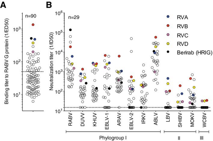
Figure 1 Selection of RABV vaccinees with broadly reactive neutralizing antibodies A panel of 90 sera were tested by ELISA for binding to RABV G protein. Shown are the 1/ED 50 values.Sera samples selected for high binding titers (1/ED 50 > 50) values were tested for the presence of neutralizing antibodies against a panel of 12 pseudotyped viruses. 1/ID 50 values are shown. Black circles indicate HRIG (Berirab®), and colored circles indicate the four donors selected for the memory B‐cell interrogation.
Figure 1 Selection of RABV vaccinees with broadly reactive neutralizing antibodies A panel of 90 sera were tested by ELISA for binding to RABV G protein. Shown are the 1/ED 50 values.Sera samples selected for high binding titers (1/ED 50 > 50) values were tested for the presence of neutralizing antibodies against a panel of 12 pseudotyped viruses. 1/ID 50 values are shown. Black circles indicate HRIG (Berirab®), and colored circles indicate the four donors selected for the memory B‐cell interrogation.CC BY · PubMed Central
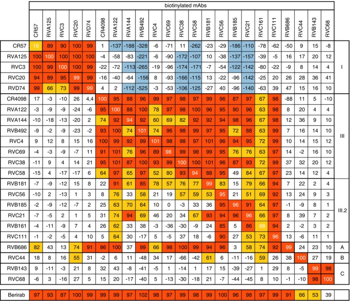
Figure 2 RABV G protein antigenic site mapping using cross‐competition ELISA‐based binding studies Monoclonal antibody cross‐competition matrix performed by ELISA on the 21 isolated antibodies and two reference antibodies of known epitope specificity (CR57 and CR4098). The percentage of binding inhibition of the biotinylated antibodies (upper row) by the unlabeled antibodies listed in the left column is shown. Results are classified using color shading codes with values ≥ 80% in orange, < 80% and ≥ 50% in yellow, < −100% in light blue, and no shading for values < 50%.
Figure 2 RABV G protein antigenic site mapping using cross‐competition ELISA‐based binding studies Monoclonal antibody cross‐competition matrix performed by ELISA on the 21 isolated antibodies and two reference antibodies of known epitope specificity (CR57 and CR4098). The percentage of binding inhibition of the biotinylated antibodies (upper row) by the unlabeled antibodies listed in the left column is shown. Results are classified using color shading codes with values ≥ 80% in orange, < 80% and ≥ 50% in yellow, < −100% in light blue, and no shading for values < 50%.CC BY · PubMed Central
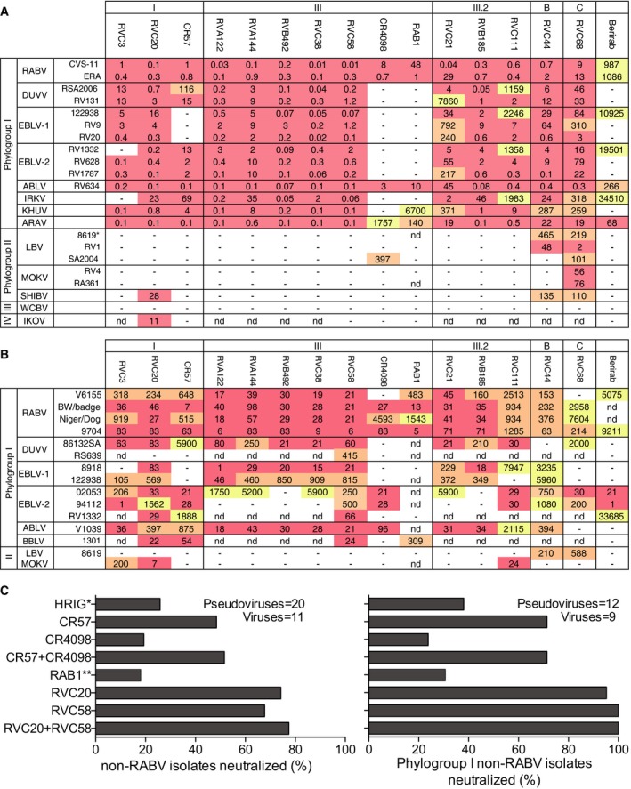
Figure 3 Neutralization of lyssaviruses by human monoclonal antibodies A selection of 12 human monoclonal antibodies, three reference antibodies (CR57, CR4098, and RAB1) and the polyclonal human immunoglobulins (HRIG, Berirab®) were tested for their neutralizing activity against 13 different lyssavirus species using pseudotyped viruses or live viruses. Complete viral strain designations are shown in Appendix Table S4. Results of neutralization assays using 22 pseudotyped viruses expressed as inhibitory concentration 90 (IC 90).Results of neutralization assays using 15 live viruses expressed as inhibitory concentration 50 (IC 50).Summary of the percentage of non‐RABV lyssavirus isolates and phylogroup I non‐RABV lyssavirus isolates neutralized with IC 50 (for viruses) or IC 90 (for pseudotyped viruses) below 10,000 ng/ml for RVC20, RVC58, CR57, CR4098, and RAB1 monoclonal antibodies, HRIG or a combination of RVC20 with RVC58 or CR57 with CR4098. Color coding indicates the potency, with IC 90 (for pseudotyped viruses) or IC 50 (for viruses) < 100 ng/ml in red shading, 100 ng/ml < IC 50 < 1,000 ng/ml in orange shading, and IC 50 ≥ 1,000 ng/ml in yellow shading. IC 50 > 10,000 ng/ml were scored as negative. *HRIG was scored as negative when IC 50 or IC 90 was > 100,000 ng/ml; **RAB1 was tested against 20 pseudotyped viruses and 9 viruses.
Figure 3 Neutralization of lyssaviruses by human monoclonal antibodies A selection of 12 human monoclonal antibodies, three reference antibodies (CR57, CR4098, and RAB1) and the polyclonal human immunoglobulins (HRIG, Berirab®) were tested for their neutralizing activity against 13 different lyssavirus species using pseudotyped viruses or live viruses. Complete viral strain designations are shown in Appendix Table S4. Results of neutralization assays using 22 pseudotyped viruses expressed as inhibitory concentration 90 (IC 90).Results of neutralization assays using 15 live viruses expressed as inhibitory concentration 50 (IC 50).Summary of the percentage of non‐RABV lyssavirus isolates and phylogroup I non‐RABV lyssavirus isolates neutralized with IC 50 (for viruses) or IC 90 (for pseudotyped viruses) below 10,000 ng/ml for RVC20, RVC58, CR57, CR4098, and RAB1 monoclonal antibodies, HRIG or a combination of RVC20 with RVC58 or CR57 with CR4098. Color coding indicates the potency, with IC 90 (for pseudotyped viruses) or IC 50 (for viruses) < 100 ng/ml in red shading, 100 ng/ml < IC 50 < 1,000 ng/ml in orange shading, and IC 50 ≥ 1,000 ng/ml in yellow shading. IC 50 > 10,000 ng/ml were scored as negative. *HRIG was scored as negative when IC 50 or IC 90 was > 100,000 ng/ml; **RAB1 was tested against 20 pseudotyped viruses and 9 viruses.CC BY · PubMed Central
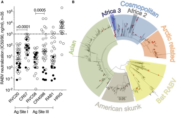
Figure 4 RVC20 and RVC58 potently neutralize multi‐lineage RABV isolates Neutralization of RABV isolates by the selected RVC20 and RVC58 antibodies from our panel, the reference CR57, CR4098, and RAB1 antibodies and HRIG. Tested using pseudotyped viruses (filled circles, n = 9; shown are IC 90 values) or live viruses (empty circles, n = 26; shown are the IC 50 values). The dotted line indicates a threshold for neutralization above 1,000 ng/ml. The geometric mean value for each data set is shown. The P‐values of Wilcoxon matched‐pairs signed‐rank tests are shown.Phylogenetic tree of 2,215G protein sequences retrieved from public databases. Highlighted with red dots are the 40 sequences of the RABV viruses tested in this work (the G protein sequence of CV9.13 strain, was not available and was therefore not included in the tree). Source data are available online for this figure.
Figure 4 RVC20 and RVC58 potently neutralize multi‐lineage RABV isolates Neutralization of RABV isolates by the selected RVC20 and RVC58 antibodies from our panel, the reference CR57, CR4098, and RAB1 antibodies and HRIG. Tested using pseudotyped viruses (filled circles, n = 9; shown are IC 90 values) or live viruses (empty circles, n = 26; shown are the IC 50 values). The dotted line indicates a threshold for neutralization above 1,000 ng/ml. The geometric mean value for each data set is shown. The P‐values of Wilcoxon matched‐pairs signed‐rank tests are shown.Phylogenetic tree of 2,215G protein sequences retrieved from public databases. Highlighted with red dots are the 40 sequences of the RABV viruses tested in this work (the G protein sequence of CV9.13 strain, was not available and was therefore not included in the tree). Source data are available online for this figure.CC BY · PubMed Central
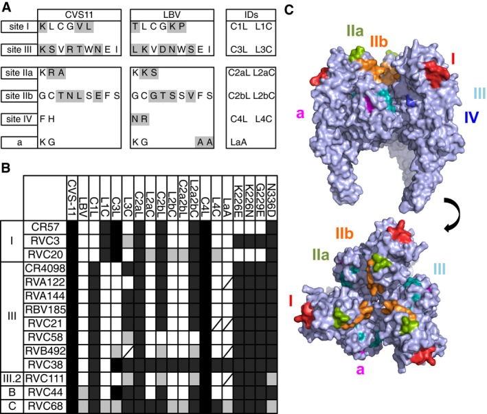
Figure 5 Antibodies epitope mapping using antigenic site swapping in chimeric pseudotyped viruses Sequence of the CVS‐11 and LBV antigenic sites I, III, IIa, IIb, IV, and a. Highlighted in gray are the residues that differ between CVS‐11 and LBV.The scheme shows the results of neutralization of CVS‐11, LBV (strain NIG56‐RV1), different chimeric CVS‐11 and LBV variants and different CVS‐11 mutants by the panel of 12 selected monoclonal antibodies and the reference antibodies CR57 and CR4098. Black cells indicate full neutralization, gray cells partial neutralization, and white cells no neutralization. Strikethrough cells, not tested. Schematic showing generation of epitope swapped G protein is shown in Fig EV1.Side view (upper) and top view (lower) of a surface rendering of the homotrimeric prefusion structure of VSV G (PDB, 2j6j). Rabies antigenic sites, highlighted in different colors, were superpositioned based on sequence alignment with VSV (˜18% sequence identity).
Figure 5 Antibodies epitope mapping using antigenic site swapping in chimeric pseudotyped viruses Sequence of the CVS‐11 and LBV antigenic sites I, III, IIa, IIb, IV, and a. Highlighted in gray are the residues that differ between CVS‐11 and LBV.The scheme shows the results of neutralization of CVS‐11, LBV (strain NIG56‐RV1), different chimeric CVS‐11 and LBV variants and different CVS‐11 mutants by the panel of 12 selected monoclonal antibodies and the reference antibodies CR57 and CR4098. Black cells indicate full neutralization, gray cells partial neutralization, and white cells no neutralization. Strikethrough cells, not tested. Schematic showing generation of epitope swapped G protein is shown in Fig EV1.Side view (upper) and top view (lower) of a surface rendering of the homotrimeric prefusion structure of VSV G (PDB, 2j6j). Rabies antigenic sites, highlighted in different colors, were superpositioned based on sequence alignment with VSV (˜18% sequence identity).CC BY · PubMed Central
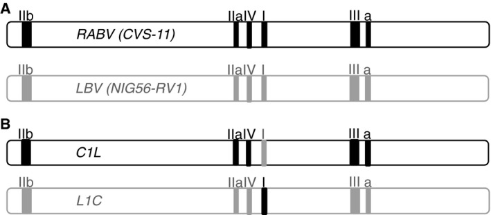
Figure EV1 Schematic showing generation of epitope swapped G proteins A Wild‐type G genes from RABV (CVS‐11) and LBV (NIG56‐RV1) isolates are shown with known epitopes highlighted by the solid boxes. Using overlap extension PCR (Heckman & Pease, 2007), we were able to generate matching sets of G genes for which each epitope in turn had been swapped from CVS‐11 to LBV and vice versa.Examples of the chimeric G genes generated when antigenic site I was swapped.
Figure EV1 Schematic showing generation of epitope swapped G proteins A Wild‐type G genes from RABV (CVS‐11) and LBV (NIG56‐RV1) isolates are shown with known epitopes highlighted by the solid boxes. Using overlap extension PCR (Heckman & Pease, 2007), we were able to generate matching sets of G genes for which each epitope in turn had been swapped from CVS‐11 to LBV and vice versa.Examples of the chimeric G genes generated when antigenic site I was swapped.CC BY · PubMed Central
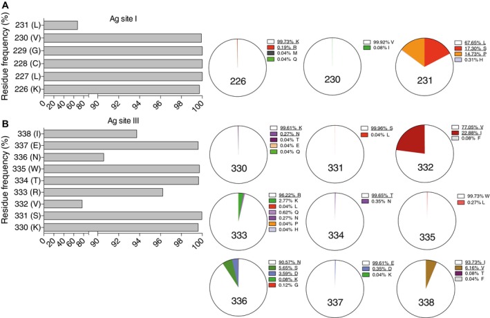
Figure 6 RVC20 and RVC58 target highly conserved epitopes in antigenic sites I and III Level of amino acid residue conservation in antigenic sites I and III as calculated by the analysis of the G protein sequences from 2,566 RABVs. Pie charts show the detailed distribution of amino acid usage at each position. Underlined residues indicate that viruses carrying the corresponding residue in that position are neutralized by either RVC20 or RVC58. Frequency of amino acid residues in antigenic site I.Frequency of amino acid residues in antigenic site III. Source data are available online for this figure.
Figure 6 RVC20 and RVC58 target highly conserved epitopes in antigenic sites I and III Level of amino acid residue conservation in antigenic sites I and III as calculated by the analysis of the G protein sequences from 2,566 RABVs. Pie charts show the detailed distribution of amino acid usage at each position. Underlined residues indicate that viruses carrying the corresponding residue in that position are neutralized by either RVC20 or RVC58. Frequency of amino acid residues in antigenic site I.Frequency of amino acid residues in antigenic site III. Source data are available online for this figure.CC BY · PubMed Central
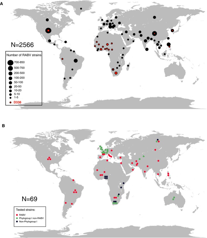
Figure EV2 World maps showing the geographic distribution of the analyzed lyssaviruses Geographic origin of the 2,566 RABV strains used for the analysis of RVC58 and RVC20 epitope conservation. Dot size is proportional to the number of isolates as indicated in the legend.Geographical origin of the viruses tested in this study.
Figure EV2 World maps showing the geographic distribution of the analyzed lyssaviruses Geographic origin of the 2,566 RABV strains used for the analysis of RVC58 and RVC20 epitope conservation. Dot size is proportional to the number of isolates as indicated in the legend.Geographical origin of the viruses tested in this study.CC BY · PubMed Central
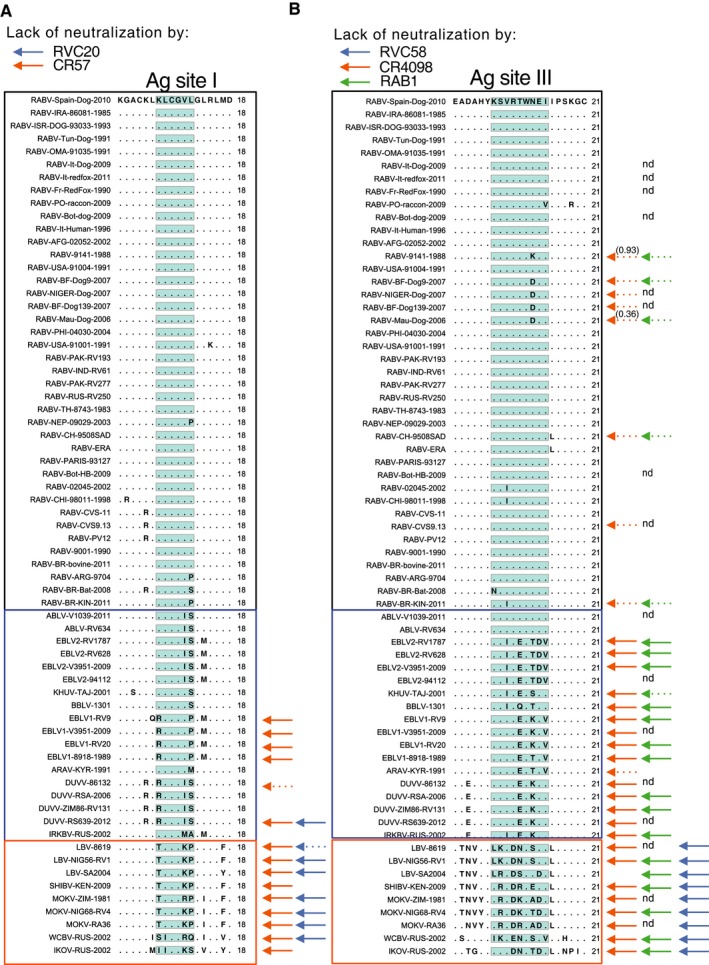
Figure EV3 Alignment of antigenic site sequences of the G protein from all the tested lyssaviruses Highlighted in blue and orange are phylogroup I non‐RABV and phylogroup II‐IV non‐RABV lyssaviruses, respectively. Nd, not tested. Antigenic site I.Antigenic site III.
Figure EV3 Alignment of antigenic site sequences of the G protein from all the tested lyssaviruses Highlighted in blue and orange are phylogroup I non‐RABV and phylogroup II‐IV non‐RABV lyssaviruses, respectively. Nd, not tested. Antigenic site I.Antigenic site III.CC BY · PubMed Central
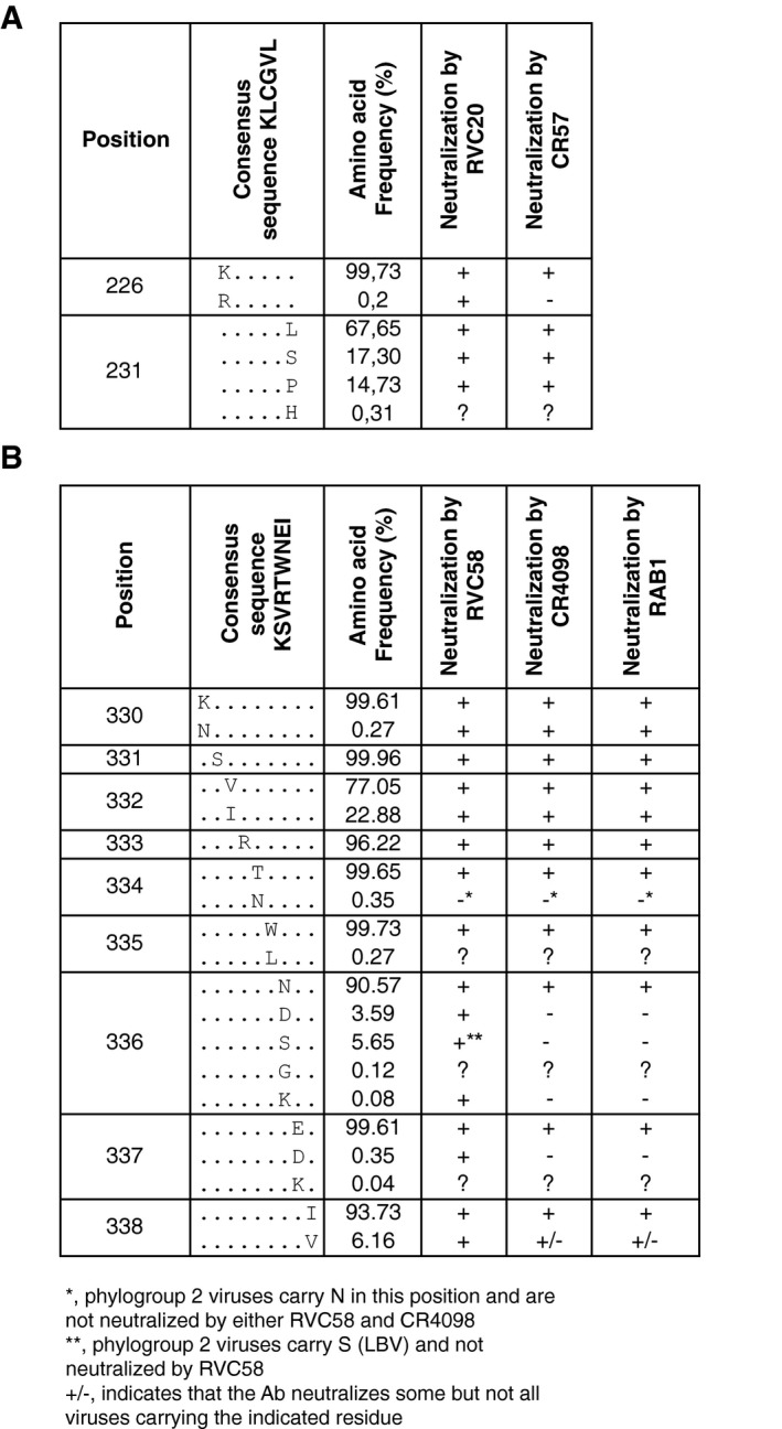
Figure EV4 Level of amino acid residue conservation in antigenic site I (A) and antigenic site III (B) calculated by the analysis of the G‐protein sequences from 2,566 RABV isolates as shown in Fig 6 A, B Shown is a summary of the viruses neutralized by the indicated monoclonal antibodies.
Figure EV4 Level of amino acid residue conservation in antigenic site I (A) and antigenic site III (B) calculated by the analysis of the G‐protein sequences from 2,566 RABV isolates as shown in Fig 6 A, B Shown is a summary of the viruses neutralized by the indicated monoclonal antibodies.CC BY · PubMed Central
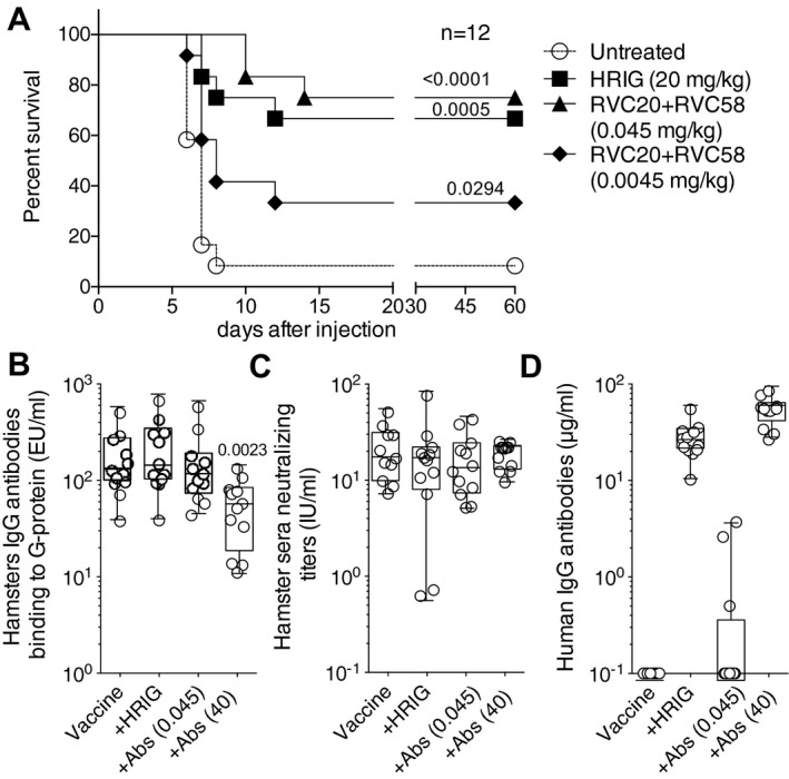
Figure 7 RVC20 and RVC58 antibodies protect Syrian hamsters from lethal RABV infection Percent survival in Syrian hamsters infected with RABV CVS‐11 isolate and then left untreated or treated with the standard PEP (HRIG and vaccination) or with two experimental PEP protocols replacing HRIG with different doses of a cocktail of RVC20 and RVC58 monoclonal antibodies (and vaccination). Kaplan–Meier survival curves are shown by plotting percent survival against time (in days). Mantel–Cox test performed to compare treated groups versus untreated group.Titers of hamster IgG antibodies binding to G protein measured in sera collected 42 days after immunization. The P‐value of a Mann–Whitney test comparing the vaccine alone group with all others is shown. RABV‐neutralizing antibodies measured in sera collected 42 days after immunization.Residuals human IgG antibodies measured in sera collected 42 days after immunization.Data information: Box and whiskers plot (B‐C): box containing 50% with median and whiskers minimum to maximum values. Source data are available online for this figure.
Figure 7 RVC20 and RVC58 antibodies protect Syrian hamsters from lethal RABV infection Percent survival in Syrian hamsters infected with RABV CVS‐11 isolate and then left untreated or treated with the standard PEP (HRIG and vaccination) or with two experimental PEP protocols replacing HRIG with different doses of a cocktail of RVC20 and RVC58 monoclonal antibodies (and vaccination). Kaplan–Meier survival curves are shown by plotting percent survival against time (in days). Mantel–Cox test performed to compare treated groups versus untreated group.Titers of hamster IgG antibodies binding to G protein measured in sera collected 42 days after immunization. The P‐value of a Mann–Whitney test comparing the vaccine alone group with all others is shown. RABV‐neutralizing antibodies measured in sera collected 42 days after immunization.Residuals human IgG antibodies measured in sera collected 42 days after immunization.Data information: Box and whiskers plot (B‐C): box containing 50% with median and whiskers minimum to maximum values. Source data are available online for this figure.CC BY · PubMed Central

Verification

Claims the paper makes

The claims and statistics of this paper haven’t been checked yet.

Run the check — free, no account needed. It reads the open-access text and checks the sources it cites.

A few free checks a day · papers already verified open instantly · results are public

Claims other papers make about this one

Each is checked against this paper’s own text: does it say what the citing paper says it does?

Self-check

Percentages, sums and reported differences re-computed from the paper’s own numbers.

The self-check needs the paper’s full text.

Citations

Cites

Show all 63 references

Research works printed but not linked: 4

  1. Atanasiu P, Cannon DA, Dean DJ, Fox JP, Habel K, Kaplan MM, Kissling RE, Koprowski H, Lepine P, Perez Gallardo F (1961) Rabies neutralizing antibody response to different schedules of serum and vaccine inoculations in non‐exposed persons. 3. Bull World Health Organ 25: 103–114
  2. Atanasiu P, Dean DJ, Habel K, Kaplan MM, Koprowski H, Lepine P, Serie C (1967) Rabies neutralizing antibody response to different schedules of serum and vaccine inoculations in non‐exposed persons. 4. Bull World Health Organ 36: 361–365
  3. Babes V, Lepp M (1889) Recherches sur la vaccination antirabique. Ann Inst Pasteur 3: 384–390
  4. Dietzgen R, Calisher C, Kurath G, Kuzmin I, Rodriguez L, Stone D, Tesh R, Tordo N, Walker P, Wetzel T et al (2011) Rhabdoviridae. In Virus Taxonomy: Ninth Report of the International Committee on Taxonomy of Viruses, King AMQ, Adams MJ, Carstens EB, Lefkowitz EJ (eds), pp 686–714. Oxford, UK: Elsevier

Cited by

Sorted by citations

See all 103

Metadata

Authors
De Benedictis, P., Minola, A., Rota Nodari, E., Aiello, R., Zecchin, B., Salomoni, A., Foglierini, M., Agatic, G., Vanzetta, F., Lavenir, R., Lepelletier, A., Bentley, E., Weiss, R., Cattoli, G., Capua, I., Sallusto, F., Wright, E., Lanzavecchia, A., Bourhy, H., & Corti, D.
Affiliations
  • Food and Agriculture Organization of the United Nations (IT)
  • Institute for Research in Biomedicine (CH)
  • Institut Pasteur (FR)
  • University of Westminster (GB)
  • University College London (GB)
  • ETH Zurich (CH)
Corresponding author
Corti, Davide
Venue
EMBO Molecular Medicine
Published
2016-03-18
Volume
8
Issue
4
Pages
407-421
ISSN
  • 1757-4676 (ISSN-L)
  • 1757-4684
OpenAlex
W2298668359
Cited by
103
References
63
Citations by year
2026: 11 · 2025: 13 · 2024: 7 · 2023: 13 · 2022: 10 · 2021: 5 · 2020: 14 · 2019: 2 · 2018: 16 · 2017: 9 · 2016: 3
FWCI
6.90
Type
journal article
Language
en
Open access
Locations
  • EMBO Molecular Medicine (publishedVersion, cc-by)
  • Europe PMC (PubMed Central) (publishedVersion, cc-by)
  • PubMed Central (publishedVersion, cc-by)
  • PubMed (publishedVersion)
  • WestminsterResearch (University of Westminster) (acceptedVersion)
  • WestminsterResearch (University of Westminster) (acceptedVersion, cc-by)
  • reroDoc Digital Library (submittedVersion)
  • Sussex Research Online (University of Sussex) (submittedVersion, cc-by)
  • HAL (submittedVersion, cc-by)
  • DOAJ (DOAJ: Directory of Open Access Journals) (submittedVersion, cc-by-sa)
  • Figshare (submittedVersion, cc-by)
Funders
  • Italian Ministry of Health (RC IZSVe 08/2009)
  • Helmut Horten Foundation
  • Swiss Vaccine Research Institute
  • FAO/National RC for Rabies, IZSVe‐Italy
  • IAEA Laboratories‐Austria
Topics
  • Life Sciences › Immunology and Microbiology › Virology › Rabies epidemiology and control
  • Life Sciences › Immunology and Microbiology › Immunology › Monoclonal and Polyclonal Antibodies Research
  • Health Sciences › Medicine › Epidemiology › Virology and Viral Diseases
Keywords
rabies post-exposure prophylaxis, human monoclonal antibodies, broadly neutralizing antibodies, rabies virus, lyssaviruses, memory B cells, antibody epitope specificity, neutralizing activity
MeSH terms
Animals, Antibodies, Monoclonal, Antibodies, Viral, Disease Models, Animal, Humans, Immunization, Passive, Immunologic Factors, Mesocricetus, Rabies, Rabies virus, Survival Analysis, Treatment Outcome, Post-Exposure Prophylaxis, Antibodies, Neutralizing

About these checks

Registry, retraction, Cites and statistics results are computed, not judged, with one exception: a printed reference that no index lists is read once by a model before it counts as a citation error. Claims are extracted by a model and kept only when their quote appears verbatim in the text; each cited source is then checked, with a model’s help, for whether it says what the claim relies on. Those verdicts can be wrong — read the claim’s quote and the cited source before relying on one.

Statistics and arithmetic are recomputed, not judged.

Confidence bands are shown only for fields calibrated on our annotated test set. None is calibrated yet, so no band is shown.

What counts as a claim ›How we measure confidence ›How we check this ›

Are you an author, or do you see a problem with this page? Request a review