Cited by 324Neuroinflammation and Neurodegeneration MechanismsAlzheimer's disease research and treatmentsInflammasome and immune disorders

The DOI matches its Crossref record; 63 of the 63 references it cites were found in a registry or an index.

Enhancing protective microglial activities with a dual function TREM2 antibody to the stalk region

Open access

Abstract

Triggering receptor expressed on myeloid cells 2 (TREM2) is essential for the transition of homeostatic microglia to a disease-associated microglial state. To enhance TREM2 activity, we sought to selectively increase the full-length protein on the cell surface via reducing its proteolytic shedding by A Disintegrin And Metalloproteinase (i.e., α-secretase) 10/17. We screened a panel of monoclonal antibodies against TREM2, with the aim to selectively compete for α-secretase-mediated shedding. Monoclonal antibody 4D9, which has a stalk region epitope close to the cleavage site, demonstrated dual mechanisms of action by stabilizing TREM2 on the cell surface and reducing its shedding, and concomitantly activating phospho-SYK signaling. 4D9 stimulated survival of macrophages and increased microglial uptake of myelin debris and amyloid β-peptide in vitro. In vivo target engagement was demonstrated in cerebrospinal fluid, where nearly all soluble TREM2 was 4D9-bound. Moreover, in a mouse model for Alzheimer's disease-related pathology, 4D9 reduced amyloidogenesis, enhanced microglial TREM2 expression, and reduced a homeostatic marker, suggesting a protective function by driving microglia toward a disease-associated state.

Overview

The claims and statistics of this paper haven’t been checked yet.

Run the check — free, no account needed. It reads the open-access text and checks the sources it cites.

A few free checks a day · papers already verified open instantly · results are public

Integrity signals

Registry

Crossref · 10.15252/emmm.201911227

Cites

63 of 63 references verified0 citation errors · 0 not checkable

Statistics

Figures

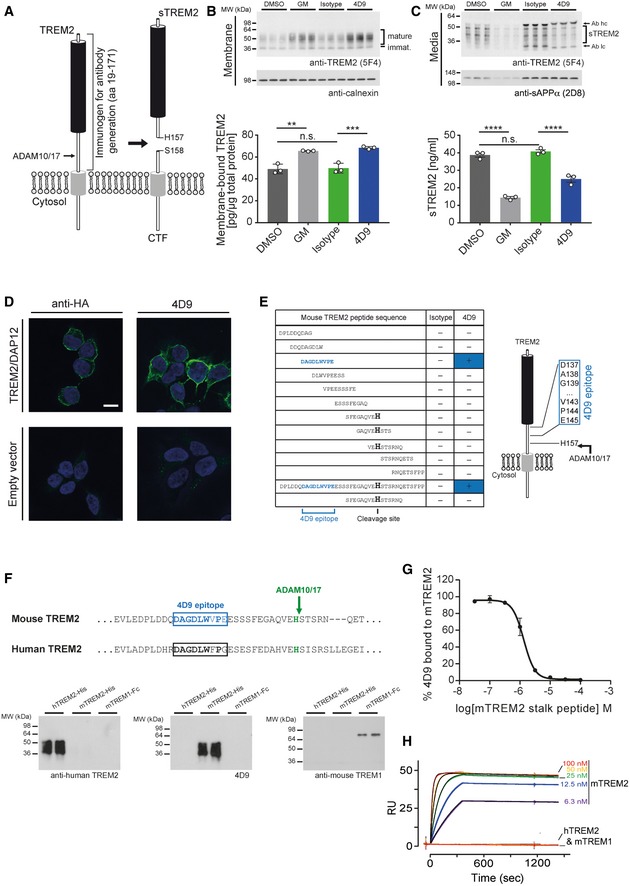
Figure 1 Screening and molecular characterization of anti‐mouse TREM2 antibodies Schematic representation of TREM2 processing by ADAM10/17. Cleavage occurs C‐terminal of residue His 157. The entire ectodomain (residues 19–171) was used for immunization of rats to generate TREM2 antibodies. CTF, C‐terminal fragment; sTREM2, soluble TREM2.Immunoblot analysis of membrane fractions of HEK293 Flp‐In cells stably overexpressing both mouse TREM2 and mouse DAP12 upon treatment with 4D9 antibody reveals increased levels of membrane‐bound TREM2 similar to what can be achieved by ADAM protease inhibition using the GM6001 inhibitor. An isotype antibody was used as a negative control. Calnexin served as a loading control. Levels of membrane‐bound TREM2 were quantified by MSD ELISA. Data represent the mean ± SEM (n = 3). One‐way ANOVA, Tukey's post hoc test; P (DMSO vs GM) = 0.0011; P (DMSO vs isotype) = 0.992; P (isotype vs 4D9) = 0.0005; n.s., not significant.Immunoblot analysis of conditioned media from HEK293 Flp‐In cells stably overexpressing both mouse TREM2 and mouse DAP12 upon treatment with 4D9 antibody reveals decreased levels of sTREM2 similar to what can be achieved by ADAM protease inhibition using the GM6001 inhibitor. An isotype antibody was used as a negative control. sAPPα served as a loading control. Note that heavy and light chains of the antibodies used for treatment are also detected and annotated. Levels of sTREM2 were quantified by MSD ELISA. Data represent the mean ± SEM (n = 3). One‐way ANOVA, Tukey's post hoc test; P (DMSO vs GM) < 0.0001; P (DMSO vs isotype) = 0.6372; P (isotype vs 4D9) < 0.0001; n.s., not significant.4D9 antibody selectively detects TREM2 on the cell surface of HEK293 Flp‐In cells stably overexpressing mouse TREM2 and mouse DAP12. An anti‐HA antibody was used as a positive control, while empty vector‐transfected HEK293 Flp‐In cells were used as a negative control. Scale bar = 10 μm.Peptide ELISAs detect anti‐mouse TREM2 antibody binding to tiled stalk region peptides, full‐length stalk peptide, or a truncated ADAM cleavage site peptide. The binding epitope of 4D9 antibody is located 12‐amino acids N‐terminal of the ADAM cleavage site at His 157.Sequence comparison of mouse TREM2 and human TREM2 shows substantial sequence conservation around the 4D9 epitope (upper panel). Immunoblot analysis demonstrates that antibody 4D9 is highly specific for mouse TREM2 and does not detect human TREM2 or mouse TREM1 (lower panel).4D9 binding to the mouse TREM2 ECD is competed off by a stalk region peptide. A competition ELISA demonstrates that a dose titration of stalk peptide reduces binding of 4D9 to TREM2 ECD with an EC50 of 1.3 μM. Data represent the mean ± SEM (n = 3). ECD, extracellular domain.Surface plasmon resonance binding kinetics of increasing concentrations of 4D9 antibody to mouse TREM2 ECD evaluated by Biacore, k on = 5.9 × 10⁵ M−1 s−1, k off = 4.0 × 10⁻⁵ s−1, K D = 68 pM. 4D9 binding to human TREM2 or mouse TREM1 was undetectable.Data information: Statistical evaluations are displayed as follows: **P < 0.01; ***P < 0.001; ****P < 0.0001.Source data are available online for this figure.
Figure 1 Screening and molecular characterization of anti‐mouse TREM2 antibodies Schematic representation of TREM2 processing by ADAM10/17. Cleavage occurs C‐terminal of residue His 157. The entire ectodomain (residues 19–171) was used for immunization of rats to generate TREM2 antibodies. CTF, C‐terminal fragment; sTREM2, soluble TREM2.Immunoblot analysis of membrane fractions of HEK293 Flp‐In cells stably overexpressing both mouse TREM2 and mouse DAP12 upon treatment with 4D9 antibody reveals increased levels of membrane‐bound TREM2 similar to what can be achieved by ADAM protease inhibition using the GM6001 inhibitor. An isotype antibody was used as a negative control. Calnexin served as a loading control. Levels of membrane‐bound TREM2 were quantified by MSD ELISA. Data represent the mean ± SEM (n = 3). One‐way ANOVA, Tukey's post hoc test; P (DMSO vs GM) = 0.0011; P (DMSO vs isotype) = 0.992; P (isotype vs 4D9) = 0.0005; n.s., not significant.Immunoblot analysis of conditioned media from HEK293 Flp‐In cells stably overexpressing both mouse TREM2 and mouse DAP12 upon treatment with 4D9 antibody reveals decreased levels of sTREM2 similar to what can be achieved by ADAM protease inhibition using the GM6001 inhibitor. An isotype antibody was used as a negative control. sAPPα served as a loading control. Note that heavy and light chains of the antibodies used for treatment are also detected and annotated. Levels of sTREM2 were quantified by MSD ELISA. Data represent the mean ± SEM (n = 3). One‐way ANOVA, Tukey's post hoc test; P (DMSO vs GM) < 0.0001; P (DMSO vs isotype) = 0.6372; P (isotype vs 4D9) < 0.0001; n.s., not significant.4D9 antibody selectively detects TREM2 on the cell surface of HEK293 Flp‐In cells stably overexpressing mouse TREM2 and mouse DAP12. An anti‐HA antibody was used as a positive control, while empty vector‐transfected HEK293 Flp‐In cells were used as a negative control. Scale bar = 10 μm.Peptide ELISAs detect anti‐mouse TREM2 antibody binding to tiled stalk region peptides, full‐length stalk peptide, or a truncated ADAM cleavage site peptide. The binding epitope of 4D9 antibody is located 12‐amino acids N‐terminal of the ADAM cleavage site at His 157.Sequence comparison of mouse TREM2 and human TREM2 shows substantial sequence conservation around the 4D9 epitope (upper panel). Immunoblot analysis demonstrates that antibody 4D9 is highly specific for mouse TREM2 and does not detect human TREM2 or mouse TREM1 (lower panel).4D9 binding to the mouse TREM2 ECD is competed off by a stalk region peptide. A competition ELISA demonstrates that a dose titration of stalk peptide reduces binding of 4D9 to TREM2 ECD with an EC50 of 1.3 μM. Data represent the mean ± SEM (n = 3). ECD, extracellular domain.Surface plasmon resonance binding kinetics of increasing concentrations of 4D9 antibody to mouse TREM2 ECD evaluated by Biacore, k on = 5.9 × 10⁵ M−1 s−1, k off = 4.0 × 10⁻⁵ s−1, K D = 68 pM. 4D9 binding to human TREM2 or mouse TREM1 was undetectable.Data information: Statistical evaluations are displayed as follows: **P < 0.01; ***P < 0.001; ****P < 0.0001.Source data are available online for this figure.CC BY · PubMed Central
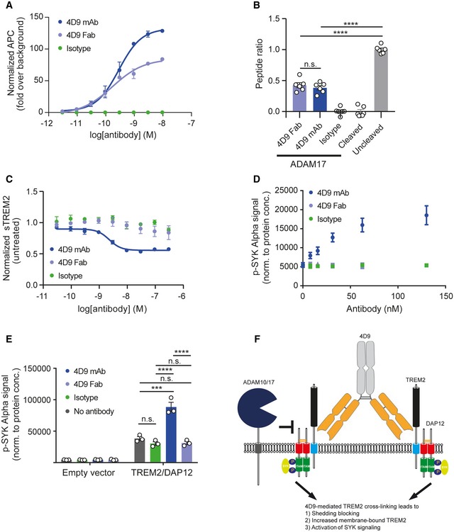
Figure 2 4D9 antibody stimulates TREM2‐dependent SYK signaling and blocks ADAM17‐mediated TREM2 shedding Flow cytometry dose–response curve for cell binding of 4D9 mAb (EC50 = 0.29 nM), 4D9 Fab (EC50 = 0.17 nM), and isotype to HEK cells stably overexpressing mouse TREM2. Data represent the mean ± SEM (n = 2). In vitro ADAM17 sheddase activity is blocked by 4D9‐effectorless mAb and 4D9 Fab fragment but not an isotype control. Fluorescence polarization of FAM‐conjugated TREM2 stalk peptide was detected in the presence or absence of ADAM17 and 4D9 mAb, 4D9 Fab, and isotype control. Data represent the mean ± SEM (n = 6). One‐way ANOVA, Tukey's post hoc test; P (4D9 Fab vs 4D9 mAb) = 0.8855; P (4D9 Fab vs uncleaved) < 0.0001; P (4D9 mAb vs uncleaved) < 0.0001; n.s., not significant.ELISA‐mediated quantification of sTREM2 in conditioned media from HEK293 cells stably overexpressing mouse TREM2 treated with a dose titration of 4D9 mAb (EC50 = 2.3 nM), 4D9 Fab, or isotype for 18 h. Data represent the mean ± SEM (n = 3).AlphaLISA‐mediated quantification of p‐SYK levels in HEK293 Flp‐In cells stably overexpressing mouse TREM2 and mouse DAP12 treated with a dose titration of 4D9 mAb, 4D9 Fab, or isotype for 5 min. Data represent the mean ± SEM (n = 3).AlphaLISA‐mediated quantification of p‐SYK levels in HEK293 Flp‐In cells stably overexpressing mouse TREM2 and mouse DAP12 or empty vector treated with 1 mg/ml POPC/POPS liposomes and 20 μg/ml 4D9 mAb, 4D9 Fab, or isotype for 5 min. p‐SYK levels were also determined for cells treated with liposomes only. Data represent the mean ± SEM (n = 3). Two‐way ANOVA, Tukey's post hoc test (cell line effect: F 1,16 = 365.7, P ≤ 0.0001; treatment effect: F 3,16 = 39.35, P < 0.0001; cell line × treatment effect: F 3,16 = 38.75, P < 0.0001); P (no Ab vs isotype) = 0.6218; P (no Ab vs 4D9 mAb) < 0.0001; P (no Ab vs 4D9 Fab) = 0.7301; P (isotype vs 4D9 mAb) < 0.0001; P (isotype vs 4D9 Fab) > 0.9999; P (4D9 mAb vs 4D9 Fab) < 0.0001; n.s., not significant.Schematic representation of the proposed mechanism of action of antibody 4D9. Binding of 4D9 to TREM2 leads to receptor clustering on the cell surface, thereby driving downstream p‐SYK signaling. At the same time, cell‐surface levels are enhanced by inhibition of ectodomain shedding, potentially because of the inability of proteases to cleave dimeric substrates. Data information: Statistical evaluations are displayed as follows: ***P < 0.001; ****P < 0.0001.
Figure 2 4D9 antibody stimulates TREM2‐dependent SYK signaling and blocks ADAM17‐mediated TREM2 shedding Flow cytometry dose–response curve for cell binding of 4D9 mAb (EC50 = 0.29 nM), 4D9 Fab (EC50 = 0.17 nM), and isotype to HEK cells stably overexpressing mouse TREM2. Data represent the mean ± SEM (n = 2). In vitro ADAM17 sheddase activity is blocked by 4D9‐effectorless mAb and 4D9 Fab fragment but not an isotype control. Fluorescence polarization of FAM‐conjugated TREM2 stalk peptide was detected in the presence or absence of ADAM17 and 4D9 mAb, 4D9 Fab, and isotype control. Data represent the mean ± SEM (n = 6). One‐way ANOVA, Tukey's post hoc test; P (4D9 Fab vs 4D9 mAb) = 0.8855; P (4D9 Fab vs uncleaved) < 0.0001; P (4D9 mAb vs uncleaved) < 0.0001; n.s., not significant.ELISA‐mediated quantification of sTREM2 in conditioned media from HEK293 cells stably overexpressing mouse TREM2 treated with a dose titration of 4D9 mAb (EC50 = 2.3 nM), 4D9 Fab, or isotype for 18 h. Data represent the mean ± SEM (n = 3).AlphaLISA‐mediated quantification of p‐SYK levels in HEK293 Flp‐In cells stably overexpressing mouse TREM2 and mouse DAP12 treated with a dose titration of 4D9 mAb, 4D9 Fab, or isotype for 5 min. Data represent the mean ± SEM (n = 3).AlphaLISA‐mediated quantification of p‐SYK levels in HEK293 Flp‐In cells stably overexpressing mouse TREM2 and mouse DAP12 or empty vector treated with 1 mg/ml POPC/POPS liposomes and 20 μg/ml 4D9 mAb, 4D9 Fab, or isotype for 5 min. p‐SYK levels were also determined for cells treated with liposomes only. Data represent the mean ± SEM (n = 3). Two‐way ANOVA, Tukey's post hoc test (cell line effect: F 1,16 = 365.7, P ≤ 0.0001; treatment effect: F 3,16 = 39.35, P < 0.0001; cell line × treatment effect: F 3,16 = 38.75, P < 0.0001); P (no Ab vs isotype) = 0.6218; P (no Ab vs 4D9 mAb) < 0.0001; P (no Ab vs 4D9 Fab) = 0.7301; P (isotype vs 4D9 mAb) < 0.0001; P (isotype vs 4D9 Fab) > 0.9999; P (4D9 mAb vs 4D9 Fab) < 0.0001; n.s., not significant.Schematic representation of the proposed mechanism of action of antibody 4D9. Binding of 4D9 to TREM2 leads to receptor clustering on the cell surface, thereby driving downstream p‐SYK signaling. At the same time, cell‐surface levels are enhanced by inhibition of ectodomain shedding, potentially because of the inability of proteases to cleave dimeric substrates. Data information: Statistical evaluations are displayed as follows: ***P < 0.001; ****P < 0.0001.CC BY · PubMed Central
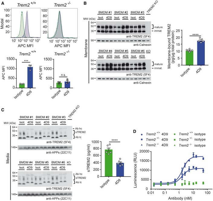
Figure 3 4D9 antibody modulates TREM2 expression in macrophages and stimulates survival Flow cytometry detection of cell‐surface binding of 4D9 (blue) and isotype (green) to wild‐type and Trem2 −/− mouse bone marrow‐derived macrophages. Representative histograms are shown as MFI (mean fluorescent intensity). Graphs represent the median signal intensity ± SEM (n = 3). Student's t‐test (two‐tailed); Trem2 +/+: P = 0.0005; Trem2 −/−: P = 0.4435; n.s., not significant.Immunoblot analysis of membrane fractions of bone marrow‐derived macrophages (BMDMs) upon treatment with 4D9 antibody reveals increased levels of membrane‐bound TREM2 relative to an isotype antibody. BMDMs from TREM2 knockout mice were included to show the specificity of the anti‐TREM2 antibody. Calnexin served as a loading control. Levels of membrane‐bound TREM2 were quantified by MSD ELISA. TREM2 antibody clone 5F4 was used for detection. Data represent the mean ± SEM (n = 5–6). Unpaired t‐test (two‐tailed) with Welch's correction; P < 0.0001. No gender‐specific differences could be observed.Immunoblot analysis of sTREM2 in conditioned media from BMDM upon treatment with 4D9 and isotype antibodies. BMDMs from TREM2 knockout mice were included to show the specificity of the anti‐TREM2 antibody. Soluble APP served as a loading control. Note that heavy and light chains of the antibodies used for treatment are also detected and annotated as follows: Ab hc, antibody heavy chain; Ab lc, antibody light chain. Levels of sTREM2 were quantified by MSD ELISA. TREM2 antibody clone 5F4 was used for detection. Data represent the mean ± SEM (n = 6). Unpaired t‐test (two‐tailed) with Welch's correction; P < 0.0001. No gender‐specific differences could be observed.Wild‐type, Trem2 +/−, and Trem2 −/− BMDMs were plated in low concentration of M‐CSF on 4D9 or isotype‐coated wells for 5 days, and cellular ATP levels were measured by luminescence detection to indicate cell viability. 4D9 and isotype data for each genotype represent the mean ± SEM (n = 2 and n = 1, respectively).Data information: Statistical evaluations are displayed as follows: ***P < 0.001; ****P < 0.0001.Source data are available online for this figure.
Figure 3 4D9 antibody modulates TREM2 expression in macrophages and stimulates survival Flow cytometry detection of cell‐surface binding of 4D9 (blue) and isotype (green) to wild‐type and Trem2 −/− mouse bone marrow‐derived macrophages. Representative histograms are shown as MFI (mean fluorescent intensity). Graphs represent the median signal intensity ± SEM (n = 3). Student's t‐test (two‐tailed); Trem2 +/+: P = 0.0005; Trem2 −/−: P = 0.4435; n.s., not significant.Immunoblot analysis of membrane fractions of bone marrow‐derived macrophages (BMDMs) upon treatment with 4D9 antibody reveals increased levels of membrane‐bound TREM2 relative to an isotype antibody. BMDMs from TREM2 knockout mice were included to show the specificity of the anti‐TREM2 antibody. Calnexin served as a loading control. Levels of membrane‐bound TREM2 were quantified by MSD ELISA. TREM2 antibody clone 5F4 was used for detection. Data represent the mean ± SEM (n = 5–6). Unpaired t‐test (two‐tailed) with Welch's correction; P < 0.0001. No gender‐specific differences could be observed.Immunoblot analysis of sTREM2 in conditioned media from BMDM upon treatment with 4D9 and isotype antibodies. BMDMs from TREM2 knockout mice were included to show the specificity of the anti‐TREM2 antibody. Soluble APP served as a loading control. Note that heavy and light chains of the antibodies used for treatment are also detected and annotated as follows: Ab hc, antibody heavy chain; Ab lc, antibody light chain. Levels of sTREM2 were quantified by MSD ELISA. TREM2 antibody clone 5F4 was used for detection. Data represent the mean ± SEM (n = 6). Unpaired t‐test (two‐tailed) with Welch's correction; P < 0.0001. No gender‐specific differences could be observed.Wild‐type, Trem2 +/−, and Trem2 −/− BMDMs were plated in low concentration of M‐CSF on 4D9 or isotype‐coated wells for 5 days, and cellular ATP levels were measured by luminescence detection to indicate cell viability. 4D9 and isotype data for each genotype represent the mean ± SEM (n = 2 and n = 1, respectively).Data information: Statistical evaluations are displayed as follows: ***P < 0.001; ****P < 0.0001.Source data are available online for this figure.CC BY · PubMed Central
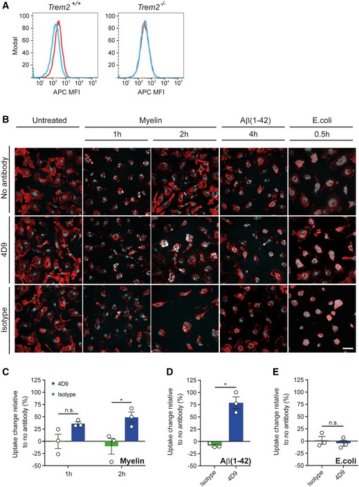
Figure 4 4D9 antibody promotes myelin and Aβ(1–42) phagocytosis in primary microglia AFlow cytometry detection of cell‐surface 4D9 (red) binding and isotype control (blue) on primary wild‐type and Trem2 −/− mouse microglia. Data are shown as mean fluorescent intensity (MFI).BUptake assay for fluorescently labeled myelin, Aβ(1–42), and inactivated Escherichia coli particles. The top row shows that myelin and Aβ (pseudocolored in white) accumulate within the plasma membrane of primary microglia (labeled with DyLight 649 isolectin and pseudocolored in red). Overnight, pre‐treatment with antibody 4D9 significantly increased the percentage of substrate‐positive cells (middle row). Pre‐treatment with the isotope control did not affect the uptake rate (bottom row). No changes in the uptake of E. coli particles were observed. Substrate uptake in the absence of antibody is shown in the top row. Hoechst 33342 was used to counter stain the nuclei (in cyan) and to assess cell density. Scale bar = 20 μm.C–EQuantification of the change in uptake of myelin (C), Aβ(1–42) (D), and E. coli (E) upon antibody treatment relative to the uptake of the respective substrate in the absence of antibody. The number of substrate‐positive cells relative to the total number of cells was quantified in each condition. Data represent the mean ± SEM (n = 3). Two‐way ANOVA (myelin), Sidak's multiple comparisons test (time effect: F 1,4 = 0.01123, P = 0.9207; treatment effect: F 1,4 = 27.98, P = 0.0061; time x treatment effect: F 1,4 = 0.5948, P = 0.4836); P (1 h) = 0.1362; P (2 h) = 0.0185; unpaired t‐test (two‐tailed) with Welch's correction (Aβ(1–42) and E. coli); P (Aβ(1–42)) = 0.0151; P (E. coli) = 0.5751; n.s., not significant.Data information: Statistical evaluations are displayed as follows: *P < 0.05.
Figure 4 4D9 antibody promotes myelin and Aβ(1–42) phagocytosis in primary microglia AFlow cytometry detection of cell‐surface 4D9 (red) binding and isotype control (blue) on primary wild‐type and Trem2 −/− mouse microglia. Data are shown as mean fluorescent intensity (MFI).BUptake assay for fluorescently labeled myelin, Aβ(1–42), and inactivated Escherichia coli particles. The top row shows that myelin and Aβ (pseudocolored in white) accumulate within the plasma membrane of primary microglia (labeled with DyLight 649 isolectin and pseudocolored in red). Overnight, pre‐treatment with antibody 4D9 significantly increased the percentage of substrate‐positive cells (middle row). Pre‐treatment with the isotope control did not affect the uptake rate (bottom row). No changes in the uptake of E. coli particles were observed. Substrate uptake in the absence of antibody is shown in the top row. Hoechst 33342 was used to counter stain the nuclei (in cyan) and to assess cell density. Scale bar = 20 μm.C–EQuantification of the change in uptake of myelin (C), Aβ(1–42) (D), and E. coli (E) upon antibody treatment relative to the uptake of the respective substrate in the absence of antibody. The number of substrate‐positive cells relative to the total number of cells was quantified in each condition. Data represent the mean ± SEM (n = 3). Two‐way ANOVA (myelin), Sidak's multiple comparisons test (time effect: F 1,4 = 0.01123, P = 0.9207; treatment effect: F 1,4 = 27.98, P = 0.0061; time x treatment effect: F 1,4 = 0.5948, P = 0.4836); P (1 h) = 0.1362; P (2 h) = 0.0185; unpaired t‐test (two‐tailed) with Welch's correction (Aβ(1–42) and E. coli); P (Aβ(1–42)) = 0.0151; P (E. coli) = 0.5751; n.s., not significant.Data information: Statistical evaluations are displayed as follows: *P < 0.05.CC BY · PubMed Central
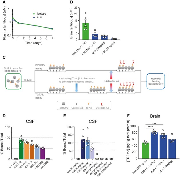
Figure 5 Pharmacokinetics and in vivo target engagement (TE) with 4D9 antibody 4D9 demonstrates standard IgG pharmacokinetics in vivo. Peripheral clearance rates of 4D9 on a human IgG‐effectorless backbone compared with an isotype control were determined in wild‐type mice by hIgG ELISA detection of plasma antibody concentrations 1 and 24 h, and 4 and 7 days after 10 mg/kg intravenous injection of antibody.Brain antibody concentration was measured 24 h post‐intravenous dosing of isotype hIgG at 100 mg/kg, and 4D9‐hIgG dosed intravenously at 100, 50, and 10 mg/kg. Detection of hIgG levels by ELISA demonstrated dose‐dependent brain concentrations of 4D9 in the single digit nM range. Animals were perfused to minimize IgG contribution from the plasma. Data represent the mean ± SEM (n = 5).Schematic depicting the TE assay setup for bound and total sTREM2 detection. For the bound assay, a secondary anti‐human IgG detects 4D9 antibody bound to soluble TREM2 in CSF and plasma. For the total assay, a saturating amount of 4D9 antibody is added to eliminate unbound sTREM2. The ratio of 4D9‐bound sTREM2 to total sTREM2 is calculated to determine the level of TE achieved by the concentration of antibody present.Target engagement time course demonstrated near 100% 4D9‐bound sTREM2 in CSF of wild‐type mice at 24 h post‐dose. Over a 10‐day time course with time points at days 1, 2, 4, and 7 and study termination at day 10, the bound/total sTREM2 reduces gradually to reach ˜ 50% by day 10. Animals were dosed intravenously with 50 mg/kg of isotype and 4D9 antibodies. Data represent the mean ± SEM (n = 5).Target engagement dose response demonstrated saturated bound sTREM2 at 50 and 10 mg/kg with > 50% bound sTREM2 at 1 mg/kg of antibody. 4D9‐bound sTREM2 was undetectable at 0.1 mg/kg and lower. 4D9 was IV‐dosed at 50, 10, 1, 0.1, 0.01, 0.001, and 0.0001 mg/kg, and isotype‐dosed at 50 mg/kg. CSF bound: total sTREM2 in wild‐type mice was measured at 24 h. Data represent the mean ± SEM (n = 5).4D9 antibody demonstrates a dose‐dependent increase in total brain TREM2 levels. Quantification of total TREM2 in brain lysates from wild‐type mice dosed with 4D9 or isotype control was performed by a MSD‐platform‐based ELISA. Data represent the mean ± SEM (n = 5) and are shown as pg TREM2 per ug of total protein. One‐way ANOVA, Dunnett's post hoc test, P (isotype vs 4D9 [100 mg/kg]) < 0.0001; P (isotype vs 4D9 [50 mg/kg]) = 0.0007.Data information: Only male mice were used. Statistical evaluations are displayed as follows: ***P < 0.001; ****P < 0.0001.
Figure 5 Pharmacokinetics and in vivo target engagement (TE) with 4D9 antibody 4D9 demonstrates standard IgG pharmacokinetics in vivo. Peripheral clearance rates of 4D9 on a human IgG‐effectorless backbone compared with an isotype control were determined in wild‐type mice by hIgG ELISA detection of plasma antibody concentrations 1 and 24 h, and 4 and 7 days after 10 mg/kg intravenous injection of antibody.Brain antibody concentration was measured 24 h post‐intravenous dosing of isotype hIgG at 100 mg/kg, and 4D9‐hIgG dosed intravenously at 100, 50, and 10 mg/kg. Detection of hIgG levels by ELISA demonstrated dose‐dependent brain concentrations of 4D9 in the single digit nM range. Animals were perfused to minimize IgG contribution from the plasma. Data represent the mean ± SEM (n = 5).Schematic depicting the TE assay setup for bound and total sTREM2 detection. For the bound assay, a secondary anti‐human IgG detects 4D9 antibody bound to soluble TREM2 in CSF and plasma. For the total assay, a saturating amount of 4D9 antibody is added to eliminate unbound sTREM2. The ratio of 4D9‐bound sTREM2 to total sTREM2 is calculated to determine the level of TE achieved by the concentration of antibody present.Target engagement time course demonstrated near 100% 4D9‐bound sTREM2 in CSF of wild‐type mice at 24 h post‐dose. Over a 10‐day time course with time points at days 1, 2, 4, and 7 and study termination at day 10, the bound/total sTREM2 reduces gradually to reach ˜ 50% by day 10. Animals were dosed intravenously with 50 mg/kg of isotype and 4D9 antibodies. Data represent the mean ± SEM (n = 5).Target engagement dose response demonstrated saturated bound sTREM2 at 50 and 10 mg/kg with > 50% bound sTREM2 at 1 mg/kg of antibody. 4D9‐bound sTREM2 was undetectable at 0.1 mg/kg and lower. 4D9 was IV‐dosed at 50, 10, 1, 0.1, 0.01, 0.001, and 0.0001 mg/kg, and isotype‐dosed at 50 mg/kg. CSF bound: total sTREM2 in wild‐type mice was measured at 24 h. Data represent the mean ± SEM (n = 5).4D9 antibody demonstrates a dose‐dependent increase in total brain TREM2 levels. Quantification of total TREM2 in brain lysates from wild‐type mice dosed with 4D9 or isotype control was performed by a MSD‐platform‐based ELISA. Data represent the mean ± SEM (n = 5) and are shown as pg TREM2 per ug of total protein. One‐way ANOVA, Dunnett's post hoc test, P (isotype vs 4D9 [100 mg/kg]) < 0.0001; P (isotype vs 4D9 [50 mg/kg]) = 0.0007.Data information: Only male mice were used. Statistical evaluations are displayed as follows: ***P < 0.001; ****P < 0.0001.CC BY · PubMed Central
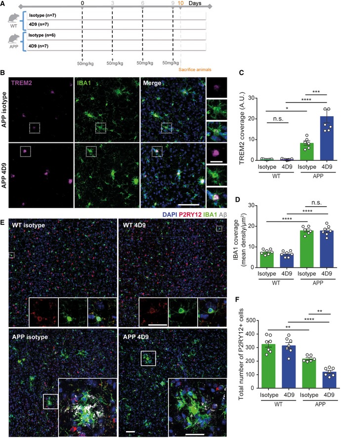
Figure 6 4D9 antibody modulates TREM2 functions within the brain Schematic outlining study design and timeline of intraperitoneal injections of either isotype control of 4D9 antibody in 6‐month‐old APP‐NL‐G‐F and age‐matched WT mice.Immunohistochemical costainings of TREM2 (magenta) and IBA1 (green) microglia in the cortex of isotype control and 4D9‐injected WT and APP‐NL‐G‐F mice. Side panel shows images from each staining at a larger magnification indicated by the dotted white boxes. Scale bar = 10 μm; scale bar (inset) = 2.5 μm.Quantification of cortical TREM2 stainings shown in (B). Two‐way ANOVA, Tukey's multiple comparison test (genotype effect: F 1,23 = 70.63, P ≤ 0.0001; treatment effect: F 1,23 = 14.33, P = 0.0010; genotype × treatment effect: F 1,23 = 14.51, P = 0.0009); P (WT isotype vs WT 4D9) > 0.9999; P (WT isotype vs APP isotype) = 0.02; P (WT 4D9 vs APP 4D9) < 0.0001; P (APP isotype vs APP 4D9) = 0.0001; n.s., not significant.Quantification of cortical IBA1 staining shown in B. Two‐way ANOVA, Tukey's multiple comparison test (genotype effect: F 1,21 = 215.4, P ≤ 0.0001; treatment effect: F 1,21 = 0.5994, P = 0.4475; genotype × treatment effect: F 1,21 = 0.2992, P = 0.5902); P (WT isotype vs APP isotype) < 0.0001; P (WT 4D9 vs APP 4D9) < 0.0001; P (APP isotype vs APP 4D9) = 0.9986; n.s., not significant.Confocal images of P2RY12 (red), IBA1 (green), and Aβ (gray) costainings from cortex. Top panel: Dotted white boxes indicate the areas in P2RY12 and IBA1 costainings that are magnified as inset. Bottom panel: Insets show P2RY12, IBA1, and Aβ costainings at a larger magnification. Of note, we did not observe a complete co‐localization of P2RY12 and IBA1 suggesting different microglial populations. Scale bar = 10 μm; scale bar (insets) = 5 μm.Quantification of P2RY12‐stained cells in the cortex shown in (E). Two‐way ANOVA, Tukey's multiple comparison test (genotype effect: F 1,23 = 71.99, P ≤ 0.0001; treatment effect: F 1,23 = 9.029, P = 0.0063; genotype × treatment effect: F 1,23 = 5.93, P = 0.0231); P (WT isotype vs APP isotype) = 0.0018; P (WT 4D9 vs APP 4D9) < 0.0001; P (APP isotype vs APP 4D9) = 0.0051.Data information: Each data point for all quantifications represents an average of n = 3 replicates per mouse. All data represent the mean ± SEM (n = 6–7). No gender‐specific differences could be observed. Statistical evaluations are displayed as follows: *P < 0.05; **P < 0.01; ***P < 0.001; ****P < 0.0001.
Figure 6 4D9 antibody modulates TREM2 functions within the brain Schematic outlining study design and timeline of intraperitoneal injections of either isotype control of 4D9 antibody in 6‐month‐old APP‐NL‐G‐F and age‐matched WT mice.Immunohistochemical costainings of TREM2 (magenta) and IBA1 (green) microglia in the cortex of isotype control and 4D9‐injected WT and APP‐NL‐G‐F mice. Side panel shows images from each staining at a larger magnification indicated by the dotted white boxes. Scale bar = 10 μm; scale bar (inset) = 2.5 μm.Quantification of cortical TREM2 stainings shown in (B). Two‐way ANOVA, Tukey's multiple comparison test (genotype effect: F 1,23 = 70.63, P ≤ 0.0001; treatment effect: F 1,23 = 14.33, P = 0.0010; genotype × treatment effect: F 1,23 = 14.51, P = 0.0009); P (WT isotype vs WT 4D9) > 0.9999; P (WT isotype vs APP isotype) = 0.02; P (WT 4D9 vs APP 4D9) < 0.0001; P (APP isotype vs APP 4D9) = 0.0001; n.s., not significant.Quantification of cortical IBA1 staining shown in B. Two‐way ANOVA, Tukey's multiple comparison test (genotype effect: F 1,21 = 215.4, P ≤ 0.0001; treatment effect: F 1,21 = 0.5994, P = 0.4475; genotype × treatment effect: F 1,21 = 0.2992, P = 0.5902); P (WT isotype vs APP isotype) < 0.0001; P (WT 4D9 vs APP 4D9) < 0.0001; P (APP isotype vs APP 4D9) = 0.9986; n.s., not significant.Confocal images of P2RY12 (red), IBA1 (green), and Aβ (gray) costainings from cortex. Top panel: Dotted white boxes indicate the areas in P2RY12 and IBA1 costainings that are magnified as inset. Bottom panel: Insets show P2RY12, IBA1, and Aβ costainings at a larger magnification. Of note, we did not observe a complete co‐localization of P2RY12 and IBA1 suggesting different microglial populations. Scale bar = 10 μm; scale bar (insets) = 5 μm.Quantification of P2RY12‐stained cells in the cortex shown in (E). Two‐way ANOVA, Tukey's multiple comparison test (genotype effect: F 1,23 = 71.99, P ≤ 0.0001; treatment effect: F 1,23 = 9.029, P = 0.0063; genotype × treatment effect: F 1,23 = 5.93, P = 0.0231); P (WT isotype vs APP isotype) = 0.0018; P (WT 4D9 vs APP 4D9) < 0.0001; P (APP isotype vs APP 4D9) = 0.0051.Data information: Each data point for all quantifications represents an average of n = 3 replicates per mouse. All data represent the mean ± SEM (n = 6–7). No gender‐specific differences could be observed. Statistical evaluations are displayed as follows: *P < 0.05; **P < 0.01; ***P < 0.001; ****P < 0.0001.CC BY · PubMed Central
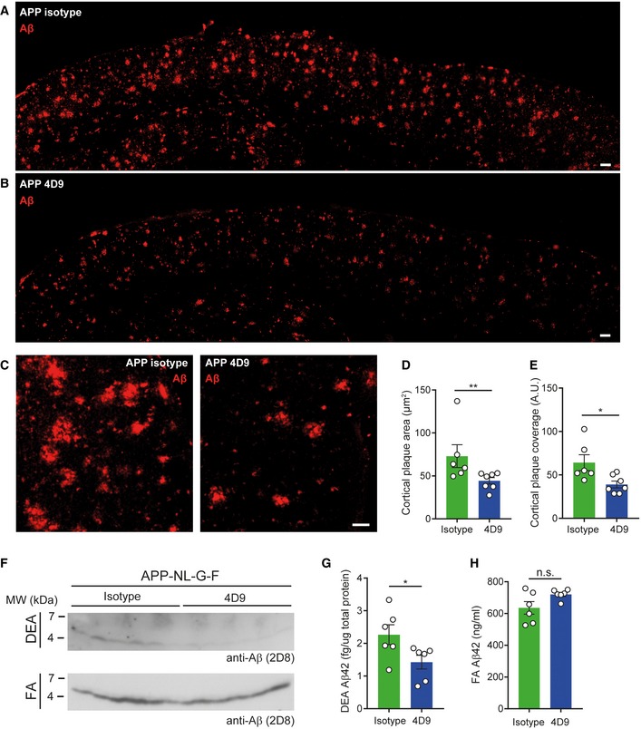
Figure 7 4D9 antibody reduces plaque burden A, BOverview of the cortex immunohistochemically stained for Aβ plaques in 6‐month‐old APP‐NL‐G‐F mice treated with isotype control (A) and 4D9 antibody (B). Scale bar = 100 μm.CHigher magnification images of cortical Aβ plaques in APP isotype and 4D9‐injected mice. Scale bar = 10 μm.DQuantification of cortical plaque area from stainings shown in (C). Mann–Whitney U‐test, P = 0.0082.EQuantification of total plaque coverage of Aβ stainings in the cortex shown in (C). Mann–Whitney U‐test, P = 0.014.FImmunoblot analysis of soluble (DEA fraction, top) and insoluble Aβ levels (FA fraction, bottom) in brains of APP‐NL‐G‐F mice treated with either isotype control or 4D9 antibody. While a reduction in levels of soluble Aβ is evident from the DEA fractions, no change in levels of insoluble Aβ could be detected in the FA fractions. Note that the higher background in the DEA immunoblot is due to a much longer exposure time, which was needed to visualize soluble Aβ.GMSD ELISA quantification of soluble Aβ42 in DEA extracts as shown in (F). Mann–Whitney U‐test, P = 0.026.HMSD ELISA quantification of insoluble Aβ42 in FA extracts as shown in (F). Unpaired t‐test (two‐tailed) with Welch's correction; P = 0.0877.Data information: Each data point for both quantifications represents an average of n = 3 replicates per mouse. All data represent the mean ± SEM (n = 6–7). No gender‐specific differences could be observed. Statistical evaluations are displayed as follows: *P < 0.05; **P < 0.01.Source data are available online for this figure.
Figure 7 4D9 antibody reduces plaque burden A, BOverview of the cortex immunohistochemically stained for Aβ plaques in 6‐month‐old APP‐NL‐G‐F mice treated with isotype control (A) and 4D9 antibody (B). Scale bar = 100 μm.CHigher magnification images of cortical Aβ plaques in APP isotype and 4D9‐injected mice. Scale bar = 10 μm.DQuantification of cortical plaque area from stainings shown in (C). Mann–Whitney U‐test, P = 0.0082.EQuantification of total plaque coverage of Aβ stainings in the cortex shown in (C). Mann–Whitney U‐test, P = 0.014.FImmunoblot analysis of soluble (DEA fraction, top) and insoluble Aβ levels (FA fraction, bottom) in brains of APP‐NL‐G‐F mice treated with either isotype control or 4D9 antibody. While a reduction in levels of soluble Aβ is evident from the DEA fractions, no change in levels of insoluble Aβ could be detected in the FA fractions. Note that the higher background in the DEA immunoblot is due to a much longer exposure time, which was needed to visualize soluble Aβ.GMSD ELISA quantification of soluble Aβ42 in DEA extracts as shown in (F). Mann–Whitney U‐test, P = 0.026.HMSD ELISA quantification of insoluble Aβ42 in FA extracts as shown in (F). Unpaired t‐test (two‐tailed) with Welch's correction; P = 0.0877.Data information: Each data point for both quantifications represents an average of n = 3 replicates per mouse. All data represent the mean ± SEM (n = 6–7). No gender‐specific differences could be observed. Statistical evaluations are displayed as follows: *P < 0.05; **P < 0.01.Source data are available online for this figure.CC BY · PubMed Central

Verification

Claims the paper makes

The claims and statistics of this paper haven’t been checked yet.

Run the check — free, no account needed. It reads the open-access text and checks the sources it cites.

A few free checks a day · papers already verified open instantly · results are public

Claims other papers make about this one

Each is checked against this paper’s own text: does it say what the citing paper says it does?

Self-check

Percentages, sums and reported differences re-computed from the paper’s own numbers.

The self-check needs the paper’s full text.

Citations

Cites

Show all 63 references

Research works printed but not linked: 2

  1. Bradbury A (2010) Antibody engineering, Vol. 1. Berlin Heidelberg: Springer‐Verlag
  2. Jonsson T, Stefansson K (2013) TREM2 and neurodegenerative disease. N Engl J Med 369: 1568–1569

Cited by

Sorted by citations

See all 324

Metadata

Authors
Schlepckow, K., Monroe, K. M., Kleinberger, G., Cantuti‐Castelvetri, L., Parhizkar, S., Xia, D., Willem, M., Werner, G., Pettkus, N., Brunner, B., Sülzen, A., Nuscher, B., Hampel, H., Xiang, X., Feederle, R., Tahirovic, S., Park, J. I., Prorok, R., Mahon, C., Liang, C., Shi, J., Kim, D. J., Sabelström, H., Huang, F., Di Paolo, G., Simons, M., Lewcock, J. W., & Haass, C.
Affiliations
  • German Center for Neurodegenerative Diseases (DE)
  • Denali Therapeutics (United States) (US)
  • Ludwig-Maximilians-Universität München (DE)
Corresponding author
  • Lewcock, Joseph W
  • Haass, Christian
Venue
EMBO Molecular Medicine
Published
2020-03-10
Volume
12
Issue
4
Pages
e11227-e11227
ISSN
  • 1757-4676 (ISSN-L)
  • 1757-4684
OpenAlex
W3012091890
Cited by
324
References
63
Citations by year
2026: 42 · 2025: 55 · 2024: 52 · 2023: 58 · 2022: 50 · 2021: 39 · 2020: 28
FWCI
16.91
Type
journal article
Language
en
Open access
Locations
  • EMBO Molecular Medicine (publishedVersion, cc-by)
  • PubMed Central (publishedVersion, cc-by)
  • PubMed (publishedVersion)
  • DOAJ (DOAJ: Directory of Open Access Journals) (submittedVersion, cc-by-sa)
  • Open access LMU (Ludwid Maxmilian's Universitat Munchen) (submittedVersion, cc-by)
  • mediaTUM (Technical University of Munich) (submittedVersion)
  • DZNE Pub (submittedVersion, other-oa)
  • Zenodo (CERN European Organization for Nuclear Research) (submittedVersion, cc-by)
Funders
  • Deutsche Forschungsgemeinschaft (EXC 2145; ID 390857198 SyNergy, HA1737/16‐1)
  • Helmholtz Association (ZT‐0027)
  • Dr. Miriam and Sheldon G. Adelson Medical Research Foundation
  • Innovative Medicines Initiative (115976)
Topics
  • Life Sciences › Neuroscience › Neurology › Neuroinflammation and Neurodegeneration Mechanisms
  • Life Sciences › Neuroscience › Cellular and Molecular Neuroscience › Alzheimer's disease research and treatments
  • Life Sciences › Biochemistry, Genetics and Molecular Biology › Molecular Biology › Inflammasome and immune disorders
Keywords
TREM2, microglia, antibody therapy, Syk signaling, disease-associated microglia, macrophage survival, amyloid-beta, Alzheimer's disease, soluble TREM2, cerebrospinal fluid
MeSH terms
Animals, Antibodies, Monoclonal, Female, Macrophages, Membrane Glycoproteins, Multiple Myeloma, Receptors, Immunologic, Amyloid beta-Peptides, Rats, Wistar, Microglia, Cell Line, Tumor, Mice, Rats

About these checks

Registry, retraction, Cites and statistics results are computed, not judged, with one exception: a printed reference that no index lists is read once by a model before it counts as a citation error. Claims are extracted by a model and kept only when their quote appears verbatim in the text; each cited source is then checked, with a model’s help, for whether it says what the claim relies on. Those verdicts can be wrong — read the claim’s quote and the cited source before relying on one.

Statistics and arithmetic are recomputed, not judged.

Confidence bands are shown only for fields calibrated on our annotated test set. None is calibrated yet, so no band is shown.

What counts as a claim ›How we measure confidence ›How we check this ›

Are you an author, or do you see a problem with this page? Request a review