

The DOI matches its Crossref record; 2 of the 2 references it cites were found in a registry or an index.
Identification and Characterization of Breakpoints and Mutations on Drosophila melanogaster Balancer Chromosomes
Abstract
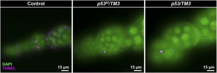
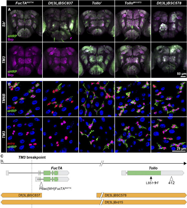
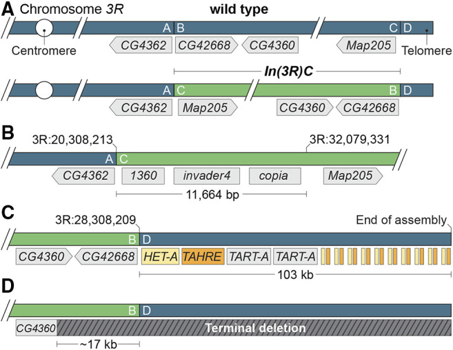
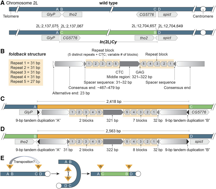
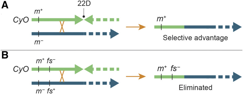
Balancers are rearranged chromosomes used in Drosophila melanogaster to maintain deleterious mutations in stable populations, preserve sets of linked genetic elements and construct complex experimental stocks. Here, we assess the phenotypes associated with breakpoint-induced mutations on commonly used third chromosome balancers and show remarkably few deleterious effects. We demonstrate that a breakpoint in p53 causes loss of radiation-induced apoptosis and a breakpoint in Fucosyltransferase A causes loss of fucosylation in nervous and intestinal tissue—the latter study providing new markers for intestinal cell identity and challenging previous conclusions about the regulation of fucosylation. We also describe thousands of potentially harmful mutations shared among X or third chromosome balancers, or unique to specific balancers, including an Ankyrin 2 mutation present on most TM3 balancers, and reiterate the risks of using balancers as experimental controls. We used long-read sequencing to confirm or refine the positions of two inversions with breakpoints lying in repetitive sequences and provide evidence that one of the inversions, In(2L)Cy, arose by ectopic recombination between foldback transposon insertions and the other, In(3R)C, cleanly separates subtelomeric and telomeric sequences and moves the subtelomeric sequences to an internal chromosome position. In addition, our characterization of In(3R)C shows that balancers may be polymorphic for terminal deletions. Finally, we present evidence that extremely distal mutations on balancers can add to the stability of stocks whose purpose is to maintain homologous chromosomes carrying mutations in distal genes. Overall, these studies add to our understanding of the structure, diversity and effectiveness of balancer chromosomes.
Overview
The claims and statistics of this paper haven’t been checked yet.
Run the check — free, no account needed. It reads the open-access text and checks the sources it cites.
Integrity signals
Registry
Cites
Statistics
Figures
Verification
Claims the paper makes
The claims and statistics of this paper haven’t been checked yet.
Run the check — free, no account needed. It reads the open-access text and checks the sources it cites.
Claims other papers make about this one
Each is checked against this paper’s own text: does it say what the citing paper says it does?
The cited source says what the claim attributes to it.
In the citing paper
“Of note, the known In(3R)C inversion 36 is present in the pangenome, along with 23 other smaller inversions.”
From Pangenome graph construction from genome alignments with Minigraph-Cactus (2024) · cites this paper as [36]
Self-check
Percentages, sums and reported differences re-computed from the paper’s own numbers.
The self-check needs the paper’s full text.
Citations
Cites
Highly rearranged chromosomes reveal uncoupling between genome topology and gene expression
Ghavi-Helm, Y., Jankowski, A., Meiers, S., Viales, R. R., Korbel, J. O., & Furlong, E. E. M. (2019) · Nature Genetics · doi:10.1038/s41588-019-0462-3 · Cited by 354
P53 and The Immune Response: 40 Years of Exploration—A Plan for the Future
Levine, A. J. (2020) · International Journal of Molecular Sciences · doi:10.3390/ijms21020541 · Cited by 97
Research works printed but not linked: 83
- Anderson, 2008, Molecular Population Genetics of Drosophila Subtelomeric DNA., Genetics, 178, 477, 10.1534/genetics.107.083196 · DOI
- Asahina, 2014, Tachykinin-expressing neurons control male-specific aggressive arousal in Drosophila., Cell, 156, 221, 10.1016/j.cell.2013.11.045 · DOI
- Ashburner, 1972, New Mutants Report., Drosoph. Inf. Serv., 49, 34
- Asif-Laidin, 2017, Short and long-term evolutionary dynamics of subtelomeric piRNA clusters in Drosophila., DNA Res., 24, 459, 10.1093/dnares/dsx017 · DOI
- Baas, 2011, Sugar-free frosting, a homolog of SAD kinase, drives neural-specific glycan expression in the Drosophila embryo., Development, 138, 553, 10.1242/dev.055376 · DOI
- Badal, 2006, An FB-NOF mediated duplication of the white gene is responsible for the zeste 1 phenotype in some Drosophila melanogaster unstable strains., Mol. Genet. Genomics, 275, 35, 10.1007/s00438-005-0068-6 · DOI
- Badal, 2006, Molecular and bioinformatic analysis of the FB-NOF transposable element., Gene, 371, 130, 10.1016/j.gene.2005.11.020 · DOI
- Badal, 2013, FB-NOF is a non-autonomous transposable element, expressed in Drosophila melanogaster and present only in the melanogaster group., Gene, 526, 459, 10.1016/j.gene.2013.04.082 · DOI
- Bardin, 2010, Transcriptional control of stem cell maintenance in the Drosophila intestine., Development, 137, 705, 10.1242/dev.039404 · DOI
- Besse, 2003, Apoptosis-mediated cell death within the ovarian polar cell lineage of Drosophila melanogaster., Development, 130, 1017, 10.1242/dev.00313 · DOI
- Biessmann, 1988, Progressive loss of DNA sequences from terminal chromosome deficiencies in Drosophila melanogaster., EMBO J., 7, 1081, 10.1002/j.1460-2075.1988.tb02916.x · DOI
- Brodsky, 2004, Drosophila melanogaster MNK/Chk2 and p53 Regulate Multiple DNA Repair and Apoptotic Pathways following DNA Damage., Mol. Cell. Biol., 24, 1219, 10.1128/MCB.24.3.1219-1231.2004 · DOI
- Buddika, 2020, Canonical nucleators are dispensable for stress granule assembly in Drosophila intestinal progenitors., J. Cell Sci., 133, jcs243451, 10.1242/jcs.243451 · DOI
- Cáceres, 1999, Generation of a Widespread Drosophila Inversion by a Transposable Element., Science, 285, 415, 10.1126/science.285.5426.415 · DOI
- Casals, 2003, The Foldback-like Transposon Galileo Is Involved in the Generation of Two Different Natural Chromosomal Inversions of Drosophila buzzatii., Mol. Biol. Evol., 20, 674, 10.1093/molbev/msg070 · DOI
- de Celis, 1995, Ventral veinless, the gene encoding the Cf1a transcription factor, links positional information and cell differentiation during embryonic and imaginal development in Drosophila melanogaster., Development, 121, 3405, 10.1242/dev.121.10.3405 · DOI
- Cingolani, 2012, A program for annotating and predicting the effects of single nucleotide polymorphisms, SnpEff: SNPs in the genome of Drosophila melanogaster strain w1118; iso-2; iso-3., Fly (Austin), 6, 80, 10.4161/fly.19695 · DOI · Paper page
- Comeron, 2012, The many landscapes of recombination in Drosophila melanogaster., PLoS Genet., 8, e1002905, 10.1371/journal.pgen.1002905 · DOI
- Danecek, 2011, The variant call format and VCFtools., Bioinformatics, 27, 2156, 10.1093/bioinformatics/btr330 · DOI
- Delprat, 2009, The transposon Galileo generates natural chromosomal inversions in Drosophila by ectopic recombination., PLoS One, 4, e7883, 10.1371/journal.pone.0007883 · DOI
- Dexter, 1914, The Analysis of a Case of Continuous Variation in Drosophila by a Study of Its Linkage Relations., Am. Nat., 48, 712, 10.1086/279446 · DOI
- Diaz-Benjumea, 1990, Genetic analysis of the wing vein pattern of Drosophila., Roux’s Archives Dev Biology Official Organ Edbo, 198, 336, 10.1007/BF00383772 · DOI
- Duncan, 1998, Control of distal antennal identity and tarsal development in Drosophila by spineless-aristapedia, a homolog of the mammalian dioxin receptor., Genes Dev., 12, 1290, 10.1101/gad.12.9.1290 · DOI
- Fabini, 2001, Identification of core alpha 1,3-fucosylated glycans and cloning of the requisite fucosyltransferase cDNA from Drosophila melanogaster. Potential basis of the neural anti-horseadish peroxidase epitope., J. Biol. Chem., 276, 28058, 10.1074/jbc.M100573200 · DOI
- Gray, 1996, P-element-induced recombination in Drosophila melanogaster: hybrid element insertion., Genetics, 144, 1601, 10.1093/genetics/144.4.1601 · DOI
- Gutzwiller, 2015, Dynamics of Wolbachia pipientis Gene Expression Across the Drosophila melanogaster Life Cycle., G3 (Bethesda), 5, 2843, 10.1534/g3.115.021931 · DOI
- Haase, 2001, A tissue-specific marker of Ecdysozoa., Dev. Genes Evol., 211, 428, 10.1007/s004270100173 · DOI
- Hawley, 1980, Chromosomal sites necessary for normal levels of meiotic recombination in Drosophila melanogaster. I. Evidence for and mapping of the sites., Genetics, 94, 625, 10.1093/genetics/94.3.625 · DOI
- Inoue, 1994, On the category of naturally occurring inversions of Drosophila melanogaster., Jpn. J. Genet., 69, 105, 10.1266/jjg.69.105 · DOI
- Jan, 1982, Antibodies to horseradish peroxidase as specific neuronal markers in Drosophila and in grasshopper embryos., Proc. Natl. Acad. Sci. USA, 79, 2700, 10.1073/pnas.79.8.2700 · DOI
- Kahsai, 2018, Mapping Second Chromosome Mutations to Defined Genomic Regions in Drosophila melanogaster., G3 (Bethesda), 8, 9, 10.1534/g3.117.300289 · DOI
- Kahsai, 2010, Neuropeptides in the Drosophila central complex in modulation of locomotor behavior., J. Exp. Biol., 213, 2256, 10.1242/jeb.043190 · DOI
- Koch, 2008, Drosophila ankyrin 2 is required for synaptic stability., Neuron, 58, 210, 10.1016/j.neuron.2008.03.019 · DOI
- Kolmogorov, 2019, Assembly of long, error-prone reads using repeat graphs., Nat. Biotechnol., 37, 540, 10.1038/s41587-019-0072-8 · DOI · Paper page
- Kurzhals, 2011, Chk2 and p53 are haploinsufficient with dependent and independent functions to eliminate cells after telomere loss., PLoS Genet., 7, e1002103, 10.1371/journal.pgen.1002103 · DOI
- Larsson, 1996, Mutations in the Drosophila melanogaster gene encoding S-adenosylmethionine synthetase suppress position-effect variegation., Genetics, 143, 887, 10.1093/genetics/143.2.887 · DOI
- Lee, 2007, Loss of LRRK2/PARK8 induces degeneration of dopaminergic neurons in Drosophila., Biochem. Biophys. Res. Commun., 358, 534, 10.1016/j.bbrc.2007.04.156 · DOI
- Lee, 2003, In vivo p53 function is indispensable for DNA damage-induced apoptotic signaling in Drosophila., FEBS Lett., 550, 5, 10.1016/S0014-5793(03)00771-3 · DOI
- Levis, 1993, Transposons in place of telomeric repeats at a Drosophila telomere., Cell, 75, 1083, 10.1016/0092-8674(93)90318-K · DOI
- Li, 2009, Fast and accurate short read alignment with Burrows–Wheeler transform., Bioinformatics, 25, 1754, 10.1093/bioinformatics/btp324 · DOI · Paper page
- Li, 2009, The Sequence Alignment/Map format and SAMtools., Bioinformatics, 25, 2078, 10.1093/bioinformatics/btp352 · DOI · Paper page
- Mason, 2004, A Deficiency Screen for Dominant Suppressors of Telomeric Silencing in Drosophila., Genetics, 168, 1353, 10.1534/genetics.104.030676 · DOI
- Mehrotra, 2008, Endocycling cells do not apoptose in response to DNA rereplication genotoxic stress., Genes Dev., 22, 3158, 10.1101/gad.1710208 · DOI
- Micchelli, 2006, Evidence that stem cells reside in the adult Drosophila midgut epithelium., Nature, 439, 475, 10.1038/nature04371 · DOI
- Miller, 2016, Third Chromosome Balancer Inversions Disrupt Protein-Coding Genes and Influence Distal Recombination Events in Drosophila melanogaster., G3 (Bethesda), 6, 1959, 10.1534/g3.116.029330 · DOI
- Miller, 2019, The joy of balancers., PLoS Genet., 15, e1008421, 10.1371/journal.pgen.1008421 · DOI
- Miller, 2018, The Molecular and Genetic Characterization of Second Chromosome Balancers in Drosophila melanogaster, G3 (Bethesda), 8, 1161, 10.1534/g3.118.200021 · DOI
- Miller, 2016, Rare recombination events generate sequence diversity among balancer chromosomes in Drosophila melanogaster., Proc. Natl. Acad. Sci. USA, 113, E1352, 10.1073/pnas.1601232113 · DOI
- Miller, 2018, Highly Contiguous Genome Assemblies of 15 Drosophila Species Generated Using Nanopore Sequencing., G3 (Bethesda), 8, 3131, 10.1534/g3.118.200160 · DOI
- Morata, 1978, Anterior and posterior compartments in the head of Drosophila., Nature, 274, 473, 10.1038/274473a0 · DOI
- Morgan, 1937, Constitution of the germinal material in relation to heredity., Yearb. Carnegie Instn, 36, 298
- Moschetti, 2004, FB elements can promote exon shuffling: a promoter-less white allele can be reactivated by FB mediated transposition in Drosophila melanogaster., Mol. Genet. Genomics, 271, 394, 10.1007/s00438-004-1007-7 · DOI
- Muller, 1918, Genetic Variability, Twin Hybrids and Constant Hybrids, in a Case of Balanced Lethal Factors., Genetics, 3, 422, 10.1093/genetics/3.5.422 · DOI
- Nüsslein-Volhard, 1984, Mutations affecting the pattern of the larval cuticle in Drosophila melanogaster: I. Zygotic loci on the second chromosome., Wihelm Roux Arch. Dev. Biol., 193, 267, 10.1007/BF00848156 · DOI
- O’Brien, 2011, Altered modes of stem cell division drive adaptive intestinal growth., Cell, 147, 603, 10.1016/j.cell.2011.08.048 · DOI
- Orengo, 2019, The molecular characterization of fixed inversions breakpoints unveils the ancestral character of the Drosophila guanche chromosomal arrangements., Sci Rep-uk, 9, 1706, 10.1038/s41598-018-37121-5 · DOI
- Payne, 1924, Crossover Modifiers in the Third Chromosome of Drosophila melanogaster., Genetics, 9, 327, 10.1093/genetics/9.4.327 · DOI
- Phalke, 2009, Retrotransposon silencing and telomere integrity in somatic cells of Drosophila depends on the cytosine-5 methyltransferase DNMT2., Nat. Genet., 41, 696, 10.1038/ng.360 · DOI
- Qi, 2016, Different cell cycle modifications repress apoptosis at different steps independent of developmental signaling in Drosophila., Mol. Biol. Cell, 27, 1885, 10.1091/mbc.e16-03-0139 · DOI
- Radion, 2018, Key role of piRNAs in telomeric chromatin maintenance and telomere nuclear positioning in Drosophila germline., Epigenetics Chromatin, 11, 40, 10.1186/s13072-018-0210-4 · DOI
- Ranz, 2007, Principles of Genome Evolution in the Drosophila melanogaster Species Group., PLoS Biol., 5, e152, 10.1371/journal.pbio.0050152 · DOI
- Rein, 1999, A quantitative three-dimensional model of the Drosophila optic lobes., Curr. Biol., 9, 93, 10.1016/S0960-9822(99)80021-9 · DOI
- Reis, 2018, Origin and consequences of chromosomal inversions in the virilis group of Drosophila., Genome Biol. Evol., 10, 3152, 10.1093/gbe/evy239 · DOI
- Roegiers, 2009, Frequent unanticipated alleles of lethal giant larvae in Drosophila second chromosome stocks., Genetics, 182, 407, 10.1534/genetics.109.101808 · DOI
- Rong, 2002, Targeted mutagenesis by homologous recombination in D. melanogaster., Genes Dev., 16, 1568, 10.1101/gad.986602 · DOI
- Schwartz, 2008, 10.4159/9780674043336 · DOI
- Seppo, 2003, Induction of neuron-specific glycosylation by Tollo/Toll-8, a Drosophila Toll-like receptor expressed in non-neural cells., Development, 130, 1439, 10.1242/dev.00347 · DOI
- Shearin, 2014, Hexameric GFP and mCherry reporters for the Drosophila GAL4, Q, and LexA transcription systems., Genetics, 196, 951, 10.1534/genetics.113.161141 · DOI
- Snow, 1987, Neural-specific carbohydrate moiety shared by many surface glycoproteins in Drosophila and grasshopper embryos., J. Neurosci., 7, 4137, 10.1523/JNEUROSCI.07-12-04137.1987 · DOI
- Sogame, 2003, Drosophila p53 preserves genomic stability by regulating cell death., Proc. Natl. Acad. Sci. USA, 100, 4696, 10.1073/pnas.0736384100 · DOI
- Struhl, 1982, Spineless-aristapedia: a homeotic gene that does not control the development of specific compartments in Drosophila., Genetics, 102, 737, 10.1093/genetics/102.4.737 · DOI
- Sturtevant, 1926, A crossover reducer in Drosophila melanogaster due to inversion of a section of the third chromosome., Biol. Zent. Bl., 697, 702
- Sturtevant, 1931, Known and probable inverted sections of the autosomes of Drosophila melanogaster., Publications of the Carnegie Institution, 421, 1
- Tang, 2020, Mutant p53 on the Path to Metastasis., Trends Cancer, 6, 62, 10.1016/j.trecan.2019.11.004 · DOI
- Tearle, 1987, Tübingen mutants and stock list., Drosoph. Inf. Serv., 66, 209
- Tinderholt, 1960, New Mutants Report., Drosoph. Inf. Serv., 34, 53
- Wagh, 2006, Bruchpilot, a Protein with Homology to ELKS/CAST, Is Required for Structural Integrity and Function of Synaptic Active Zones in Drosophila., Neuron, 49, 833, 10.1016/j.neuron.2006.02.008 · DOI · Paper page
- Ward, 1923, The Genetics of Curly Wing in Drosophila. Another Case of Balanced Lethal Factors., Genetics, 8, 276, 10.1093/genetics/8.3.276 · DOI
- Xie, 2004, Gene Deletions by Ends-In Targeting in Drosophila melanogaster., Genetics, 168, 1477, 10.1534/genetics.104.030882 · DOI
- Yagi, 2010, Functional analysis of Toll-related genes in Drosophila., Dev. Growth Differ., 52, 771, 10.1111/j.1440-169X.2010.01213.x · DOI
- Yamamoto-Hino, 2010, Identification of genes required for neural-specific glycosylation using functional genomics., PLoS Genet., 6, e1001254, 10.1371/journal.pgen.1001254 · DOI
- Yazdani, 2008, The glucose transporter (GLUT4) enhancer factor is required for normal wing positioning in Drosophila., Genetics, 178, 919, 10.1534/genetics.107.078030 · DOI
- Zhou, 2019, Advances in Experimental Medicine and Biology., Adv. Exp. Med. Biol., 1167, 105, 10.1007/978-3-030-23629-8_6 · DOI
Cited by
Sorted by citations
Pangenome graph construction from genome alignments with Minigraph-Cactus
Hickey, G., Monlong, J., Ebler, J., Novak, A. M., Eizenga, J. M., Gao, Y., Human Pangenome Reference Consortium, Abel, H. J., Antonacci-Fulton, L. L., Asri, M., et al. (2024) · Nature Biotechnology · doi:10.1038/s41587-023-01793-w · Cited by 343
Pangenome Graph Construction from Genome Alignment with Minigraph-Cactus
Hickey, G., Monlong, J., Ebler, J., Novak, A., Eizenga, J. M., Gao, Y., Human Pangenome Reference Consortium, Marschall, T., Li, H., & Paten, B. (2022) · doi:10.1101/2022.10.06.511217 · Cited by 345
Metadata
- Authors
- Miller, D. E., Kahsai, L., Buddika, K., Dixon, M. J., Kim, B. Y., Calvi, B. R., Sokol, N. S., Hawley, R. S., & Cook, K. R.
- Affiliations
- Department of Pediatrics, Division of Genetic Medicine, University of Washington, and Seattle Children’s Hospital, Seattle, WA 98105
- Department of Biology, Indiana University, Bloomington, IN 47405
- Department of Biology, Stanford University, Stanford, CA 94305
- Department of Molecular and Integrative Physiology, University of Kansas Medical Center, Kansas City, KS 66160
- Venue
- G3 Genes|Genomes|Genetics
- Publisher
- Oxford University Press (OUP)
- Published
- 2020-11-01
- Volume
- 10
- Issue
- 11
- Pages
- 4271-4285
- ISSN
- 2160-1836
- PMC
- PMC7642927
- Cited by
- 16
- References
- 2
- Language
- en
About these checks
Registry, retraction, Cites and statistics results are computed, not judged, with one exception: a printed reference that no index lists is read once by a model before it counts as a citation error. Claims are extracted by a model and kept only when their quote appears verbatim in the text; each cited source is then checked, with a model’s help, for whether it says what the claim relies on. Those verdicts can be wrong — read the claim’s quote and the cited source before relying on one.
Statistics and arithmetic are recomputed, not judged.
Confidence bands are shown only for fields calibrated on our annotated test set. None is calibrated yet, so no band is shown.
What counts as a claim ›How we measure confidence ›How we check this ›
Are you an author, or do you see a problem with this page? Request a review
