

The DOI matches its Crossref record; 7 of the 7 references it cites were found in a registry or an index.
p38β MAPK mediates ULK1-dependent induction of autophagy in skeletal muscle of tumor-bearing mice
Overview
The claims and statistics of this paper haven’t been checked yet.
Run the check — free, no account needed. It reads the open-access text and checks the sources it cites.
Integrity signals
Registry
Cites
Statistics
Figures
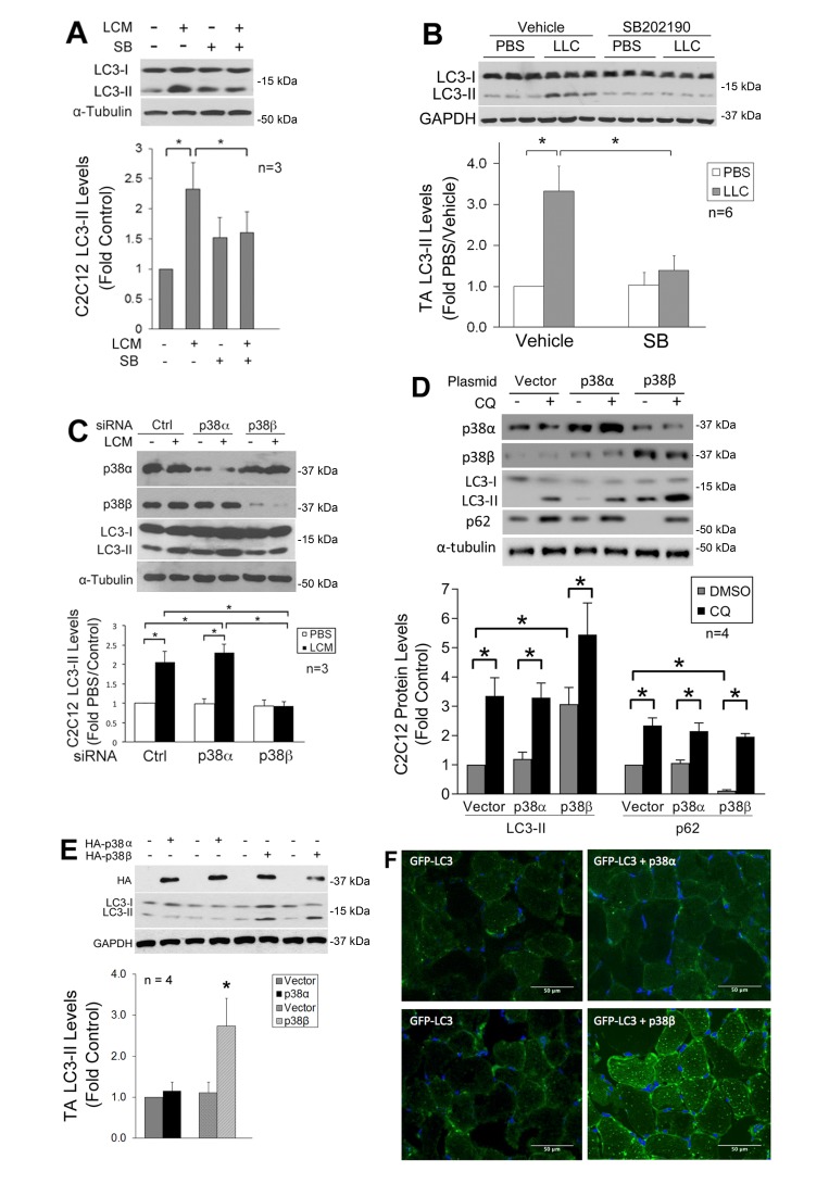
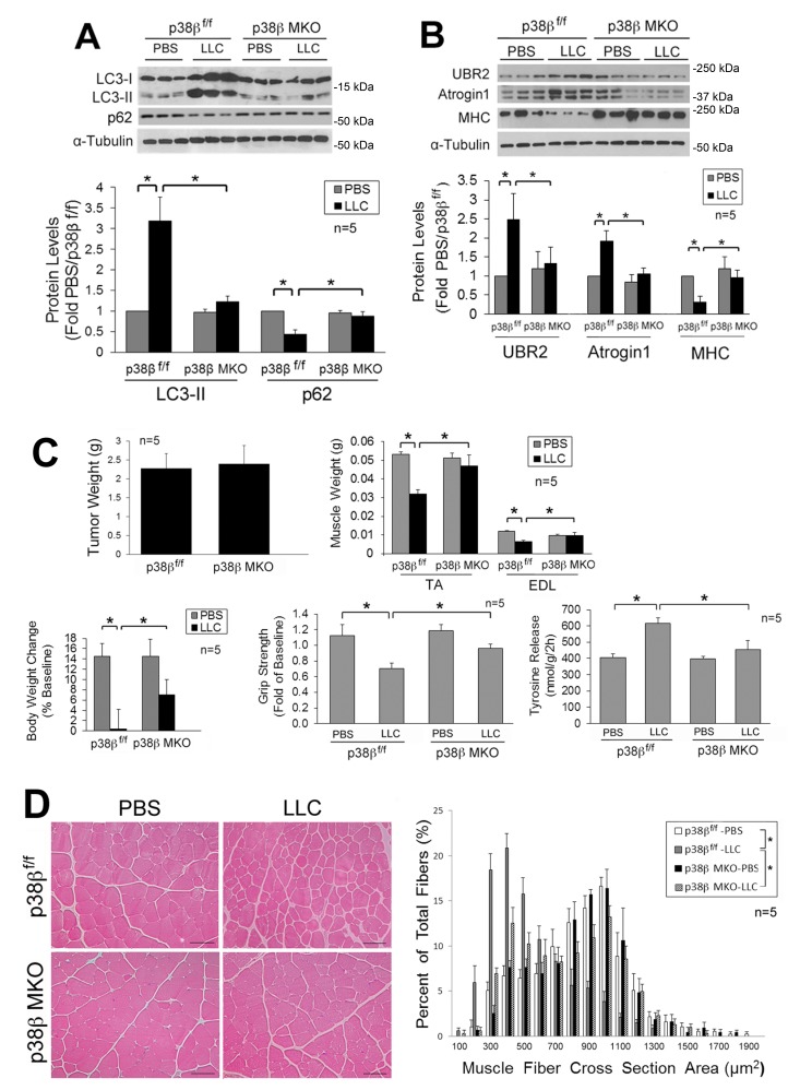
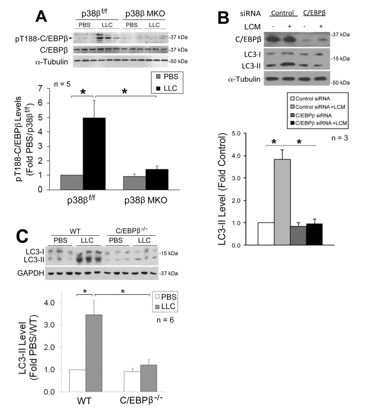
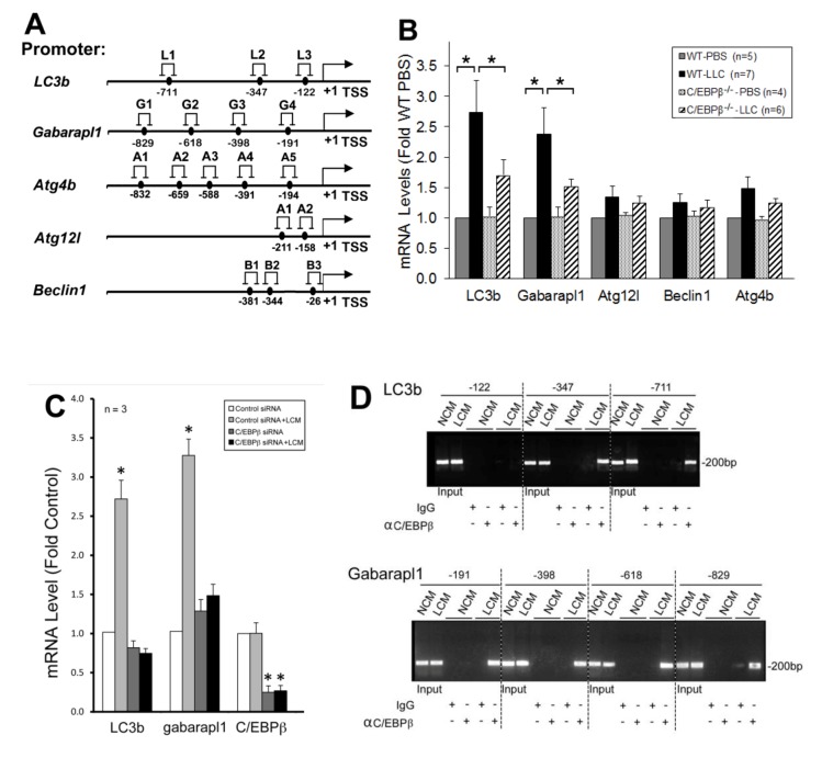
Verification
Claims the paper makes
The claims and statistics of this paper haven’t been checked yet.
Run the check — free, no account needed. It reads the open-access text and checks the sources it cites.
Claims other papers make about this one
Each is checked against this paper’s own text: does it say what the citing paper says it does?
The check wasn’t confident either way, so it gives no verdict — never disputed.
In the citing paper
“that ULK1 autophosphorylates and transphosphorylates its binding partners ATG13, RB1CC1, and ATG101 [644, 645, 692-700].”
From Guidelines for the use and interpretation of assays for monitoring autophagy (4th edition) 1 (2021) · cites this paper as [692]
Experimental — accuracy not yet measured
Self-check
Percentages, sums and reported differences re-computed from the paper’s own numbers.
The self-check needs the paper’s full text.
Citations
Cites
Multiple types of skeletal muscle atrophy involve a common program of changes in gene expression
Lecker, S. H., Jagoe, R. T., Gilbert, A., Gomes, M., Baracos, V., Bailey, J., Price, S. R., Mitch, W. E., & Goldberg, A. L. (2004) · The FASEB Journal · doi:10.1096/fj.03-0610com · Cited by 1,539 · cited 1×
“It has been well-established that the ubiquitin proteasome pathway (UPP) plays an important role in cancer-induced muscle wasting by degrading myofibrillar proteins [3-5].”
BackgroundMilan, G., Romanello, V., Pescatore, F., Armani, A., Paik, J.-H., Frasson, L., Seydel, A., Zhao, J., Abraham, R., Goldberg, A. L., et al. (2015) · Nature Communications · doi:10.1038/ncomms7670 · Cited by 709
Tumor induces muscle wasting in mice through releasing extracellular Hsp70 and Hsp90
Zhang, G., Liu, Z., Ding, H., Zhou, Y., Doan, H. A., Sin, K. W. T., Zhu, Z. J., Flores, R., Wen, Y., Gong, X., et al. (2017) · Nature Communications · doi:10.1038/s41467-017-00726-x · Cited by 248
Self-citationPogozelski, A. R., Geng, T., Li, P., Yin, X., Lira, V. A., Zhang, M., Chi, J.-T., & Yan, Z. (2009) · PLoS ONE · doi:10.1371/journal.pone.0007934 · Cited by 164
P38-γ–dependent gene silencing restricts entry into the myogenic differentiation program
Gillespie, M. A., Le Grand, F., Scimè, A., Kuang, S., von Maltzahn, J., Seale, V., Cuenda, A., Ranish, J. A., & Rudnicki, M. A. (2009) · Journal of Cell Biology · doi:10.1083/jcb.200907037 · Cited by 131 · cited 1×
“…for most of the known roles of p38 MAPK including mediating inflammation [44, 45] and promoting myogenesis [46, 47]. p38 γ MAPK regulates the expansion of myogenic precursor cells [48], endurance exercise-induced mitochondrial biogenesis and angiogenesis [30], as well as glucose uptake [49].”
Epigenetic targeting of bromodomain protein BRD4 counteracts cancer cachexia and prolongs survival
Segatto, M., Fittipaldi, R., Pin, F., Sartori, R., Dae Ko, K., Zare, H., Fenizia, C., Zanchettin, G., Pierobon, E. S., Hatakeyama, S., et al. (2017) · Nature Communications · doi:10.1038/s41467-017-01645-7 · Cited by 101
P38β MAPK Upregulates Atrogin1/MAFbx by Specific Phosphorylation of C/EBPβ
Zhang, G., & Li, Y.-P. (2012) · Skeletal Muscle · doi:10.1186/2044-5040-2-20 · Cited by 53
Self-citation
Research works printed but not linked: 51
- [1]Fearon K Strasser F Anker SD Bosaeus I Bruera E Fainsinger RL Jatoi A Loprinzi C Macdonald N Mantovani G Davis M Muscaritoli M Ottery F Radbruch L Ravasco P Walsh D Wilcock A Kaasa S Baracos VE Definition and classification of cancer cachexia: an international consensus. Lancet Oncol 2011 12 5 489 495 10.1016/S1470-2045(10)70218-7 21296615 · DOI
- [2]Fearon KC Glass DJ Guttridge DC Cancer cachexia: mediators, signaling, and metabolic pathways. Cell Metab 2012 16 2 153 166 10.1016/j.cmet.2012.06.011 22795476 · DOI
- [5]Zhou X Wang JL Lu J Song Y Kwak KS Jiao Q Rosenfeld R Chen Q Boone T Simonet WS Lacey DL Goldberg AL Han HQ Reversal of cancer cachexia and muscle wasting by ActRIIB antagonism leads to prolonged survival. Cell 2010 142 4 531 543 10.1016/j.cell.2010.07.011 20723755 · DOI
- [6]Penna F Costamagna D Pin F Camperi A Fanzani A Chiarpotto EM Cavallini G Bonelli G Baccino FM Costelli P Autophagic degradation contributes to muscle wasting in cancer cachexia. Am J Pathol 2013 182 4 1367 1378 10.1016/j.ajpath.2012.12.023 23395093 · DOI
- [7]Bohnert KR Gallot YS Sato S Xiong G Hindi SM Kumar A Inhibition of ER stress and unfolding protein response pathways causes skeletal muscle wasting during cancer cachexia. FASEB J 2016 30 9 3053 3068 10.1096/fj.201600250RR 27206451 PMC5001510 · DOI
- [8]Talbert EE Metzger GA He WA Guttridge DC Modeling human cancer cachexia in colon 26 tumor-bearing adult mice. J Cachexia Sarcopenia Muscle 2014 5 4 321 328 10.1007/s13539-014-0141-2 24668658 PMC4248405 · DOI
- [9]Zhang G Liu Z Ding H Miao H Garcia JM Li YP Toll-like receptor 4 mediates Lewis lung carcinoma-induced muscle wasting via coordinate activation of protein degradation pathways. Sci Rep 2017 7 1 2273 10.1038/s41598-017-02347-2 28536426 PMC5442131 · DOI
- [10]Aversa Z Pin F Lucia S Penna F Verzaro R Fazi M Colasante G Tirone A Rossi Fanelli F Ramaccini C Costelli P Muscaritoli M Autophagy is induced in the skeletal muscle of cachectic cancer patients. Sci Rep 2016 6 30340 10.1038/srep30340 27459917 PMC4962093 · DOI
- [11]Stephens NA Skipworth RJ Gallagher IJ Greig CA Guttridge DC Ross JA Fearon KC Evaluating potential biomarkers of cachexia and survival in skeletal muscle of upper gastrointestinal cancer patients. J Cachexia Sarcopenia Muscle 2015 6 1 53 61 10.1002/jcsm.12005 26136412 PMC4435097 · DOI
- [12]Op den Kamp CM Langen RC Snepvangers FJ de Theije CC Schellekens JM Laugs F Dingemans AM Schols AM Nuclear transcription factor kappa B activation and protein turnover adaptations in skeletal muscle of patients with progressive stages of lung cancer cachexia. Am J Clin Nutr 2013 98 3 738 748 10.3945/ajcn.113.058388 23902785 · DOI
- [13]Bechet D Tassa A Taillandier D Combaret L Attaix D Lysosomal proteolysis in skeletal muscle. Int J Biochem Cell Biol 2005 37 10 2098 2114 10.1016/j.biocel.2005.02.029 16125113 · DOI
- [14]Kirkin V McEwan DG Novak I Dikic I A role for ubiquitin in selective autophagy. Mol Cell 2009 34 3 259 269 10.1016/j.molcel.2009.04.026 19450525 · DOI
- [15]Doyle A Zhang G Abdel Fattah EA Eissa NT Li YP Toll-like receptor 4 mediates lipopolysaccharide-induced muscle catabolism via coordinate activation of ubiquitin-proteasome and autophagy-lysosome pathways. FASEB J 2011 25 1 99 110 10.1096/fj.10-164152 20826541 PMC3005430 · DOI · Paper page
- [16]Bianchi ME DAMPs, PAMPs and alarmins: all we need to know about danger. J Leukoc Biol 2007 81 1 1 5 10.1189/jlb.0306164 17032697 · DOI · Paper page
- [17]Cannon TY Guttridge D Dahlman J George JR Lai V Shores C Buzkova P Couch ME The effect of altered Toll-like receptor 4 signaling on cancer cachexia. Arch Otolaryngol Head Neck Surg 2007 133 12 1263 1269 10.1001/archotol.133.12.1263 18086970 · DOI
- [18]Zhao J Brault JJ Schild A Cao P Sandri M Schiaffino S Lecker SH Goldberg AL FoxO3 coordinately activates protein degradation by the autophagic/lysosomal and proteasomal pathways in atrophying muscle cells. Cell Metab 2007 6 6 472 483 10.1016/j.cmet.2007.11.004 18054316 · DOI · Paper page
- [20]Penna F Bonetto A Muscaritoli M Costamagna D Minero VG Bonelli G Rossi Fanelli F Baccino FM Costelli P Muscle atrophy in experimental cancer cachexia: is the IGF-1 signaling pathway involved? Int J Cancer 2010 127 7 1706 1717 10.1002/ijc.25146 20039316 · DOI
- [21]Zhang G Jin B Li YP C/EBPbeta mediates tumour-induced ubiquitin ligase atrogin1/MAFbx upregulation and muscle wasting. EMBO J 2011 30 20 4323 4335 10.1038/emboj.2011.292 21847090 PMC3199382 · DOI
- [22]Sandri M Sandri C Gilbert A Skurk C Calabria E Picard A Walsh K Schiaffino S Lecker SH Goldberg AL Foxo transcription factors induce the atrophy-related ubiquitin ligase atrogin-1 and cause skeletal muscle atrophy. Cell 2004 117 3 399 412 10.1016/s0092-8674(04)00400-3 15109499 PMC3619734 · DOI · Paper page
- [23]McClung JM Judge AR Powers SK Yan Z p38 MAPK links oxidative stress to autophagy-related gene expression in cachectic muscle wasting. Am J Physiol Cell Physiol 2010 298 3 C542 C549 10.1152/ajpcell.00192.2009 19955483 PMC2838571 · DOI · Paper page
- [24]Li YP Chen Y John J Moylan J Jin B Mann DL Reid MB TNF-alpha acts via p38 MAPK to stimulate expression of the ubiquitin ligase atrogin1/MAFbx in skeletal muscle. FASEB J 2005 19 3 362 370 10.1096/fj.04-2364com 15746179 PMC3099533 · DOI
- [25]Puppa MJ Gao S Narsale AA Carson JA Skeletal muscle glycoprotein 130's role in Lewis lung carcinoma-induced cachexia. FASEB J 2014 28 2 998 1009 10.1096/fj.13-240580 24145720 PMC3898653 · DOI · Paper page
- [26]Li W Moylan JS Chambers MA Smith J Reid MB Interleukin-1 stimulates catabolism in C2C12 myotubes. Am J Physiol Cell Physiol 2009 297 3 C706 C714 10.1152/ajpcell.00626.2008 19625606 PMC2740393 · DOI
- [27]Li H Mittal A Paul PK Kumar M Srivastava DS Tyagi SC Kumar A Tumor necrosis factor-related weak inducer of apoptosis augments matrix metalloproteinase 9 (MMP-9) production in skeletal muscle through the activation of nuclear factor-kappaB-inducing kinase and p38 mitogen-activated protein kinase: a potential role of MMP-9 in myopathy. J Biol Chem 2009 284 7 4439 4450 10.1074/jbc.M805546200 19074147 PMC2640955 · DOI
- [28]Ding H Zhang G Sin KW Liu Z Lin RK Li M Li YP Activin A induces skeletal muscle catabolism via p38beta mitogen-activated protein kinase. J Cachexia Sarcopenia Muscle 2016 10.1002/jcsm.12145 PMC5377410 27897407 · DOI
- [31]Beardmore VA Hinton HJ Eftychi C Apostolaki M Armaka M Darragh J McIlrath J Carr JM Armit LJ Clacher C Malone L Kollias G Arthur JS Generation and characterization of p38beta (MAPK11) gene-targeted mice. Mol Cell Biol 2005 25 23 10454 10464 10.1128/MCB.25.23.10454-10464.2005 16287858 PMC1291241 · DOI
- [32]Zhang G Lin RK Kwon YT Li YP Signaling mechanism of tumor cell-induced up-regulation of E3 ubiquitin ligase UBR2. FASEB J 2013 27 7 2893 2901 10.1096/fj.12-222711 23568773 PMC3688742 · DOI
- [33]Mizushima N Yoshimori T Ohsumi Y The role of Atg proteins in autophagosome formation. Annu Rev Cell Dev Biol 2011 27 107 132 10.1146/annurev-cellbio-092910-154005 21801009 · DOI · Paper page
- [34]Lin MG Hurley JH Structure and function of the ULK1 complex in autophagy. Curr Opin Cell Biol 2016 39 61-68 10.1016/j.ceb.2016.02.010 PMC4828305 26921696 · DOI
- [35]Bach M Larance M James DE Ramm G The serine/threonine kinase ULK1 is a target of multiple phosphorylation events. Biochem J 2011 440 2 283 291 10.1042/BJ20101894 21819378 · DOI · Paper page
- [36]Egan DF Shackelford DB Mihaylova MM Gelino S Kohnz RA Mair W Vasquez DS Joshi A Gwinn DM Taylor R Asara JM Fitzpatrick J Dillin A Viollet B Kundu M Hansen M Shaw RJ Phosphorylation of ULK1 (hATG1) by AMP-activated protein kinase connects energy sensing to mitophagy. Science 2011 331 6016 456 461 10.1126/science.1196371 21205641 PMC3030664 · DOI · Paper page
- [37]Puppa MJ Murphy EA Fayad R Hand GA Carson JA Cachectic skeletal muscle response to a novel bout of low-frequency stimulation. J Appl Physiol 2014 116 8 1078 1087 10.1152/japplphysiol.01270.2013 24610533 PMC4035786 · DOI
- [38]Zhou G Myers R Li Y Chen Y Shen X Fenyk-Melody J Wu M Ventre J Doebber T Fujii N Musi N Hirshman MF Goodyear LJ Moller DE Role of AMP-activated protein kinase in mechanism of metformin action. J Clin Invest 2001 108 8 1167 1174 10.1172/JCI13505 11602624 PMC209533 · DOI · Paper page
- [39]Kim J Kundu M Viollet B Guan KL AMPK and mTOR regulate autophagy through direct phosphorylation of Ulk1. Nat Cell Biol 2011 13 2 132 141 10.1038/ncb2152 21258367 PMC3987946 · DOI · Paper page
- [40]Tanaka A Jin Y Lee SJ Zhang M Kim HP Stolz DB Ryter SW Choi AM Hyperoxia-induced LC3B interacts with the Fas apoptotic pathway in epithelial cell death. Am J Respir Cell Mol Biol 2012 46 4 507 514 10.1165/rcmb.2009-0415OC 22095627 PMC3359946 · DOI
- [41]Tsukahara T Matsuda Y Usui Y Haniu H Highly purified, multi-wall carbon nanotubes induce light-chain 3B expression in human lung cells. Biochem Biophys Res Commun 2013 440 2 348 353 10.1016/j.bbrc.2013.09.089 24076389 · DOI
- [42]Mammucari C Milan G Romanello V Masiero E Rudolf R Del Piccolo P Burden SJ Di Lisi R Sandri C Zhao J Goldberg AL Schiaffino S Sandri M FoxO3 controls autophagy in skeletal muscle in vivo. Cell Metab 2007 6 6 458 471 10.1016/j.cmet.2007.11.001 18054315 · DOI · Paper page
- [44]O'Keefe SJ Mudgett JS Cupo S Parsons JN Chartrain NA Fitzgerald C Chen SL Lowitz K Rasa C Visco D Luell S Carballo-Jane E Owens K Zaller DM Chemical genetics define the roles of p38alpha and p38beta in acute and chronic inflammation. J Biol Chem 2007 282 48 34663 34671 10.1074/jbc.M704236200 17855341 · DOI
- [45]Sicard P Clark JE Jacquet S Mohammadi S Arthur JS O'Keefe SJ Marber MS The activation of p38 alpha, and not p38 beta, mitogen-activated protein kinase is required for ischemic preconditioning. J Mol Cell Cardiol 2010 48 6 1324 1328 10.1016/j.yjmcc.2010.02.013 20188737 PMC2877771 · DOI
- [46]Perdiguero E Ruiz-Bonilla V Gresh L Hui L Ballestar E Sousa-Victor P Baeza-Raja B Jardi M Bosch-Comas A Esteller M Caelles C Serrano AL Wagner EF Munoz-Canoves P Genetic analysis of p38 MAP kinases in myogenesis: fundamental role of p38alpha in abrogating myoblast proliferation. EMBO J 2007 26 5 1245 1256 10.1038/sj.emboj.7601587 17304211 PMC1817635 · DOI
- [47]Palacios D Mozzetta C Consalvi S Caretti G Saccone V Proserpio V Marquez VE Valente S Mai A Forcales SV Sartorelli V Puri PL TNF/p38alpha/polycomb signaling to Pax7 locus in satellite cells links inflammation to the epigenetic control of muscle regeneration. Cell Stem Cell 2010 7 4 455 469 10.1016/j.stem.2010.08.013 20887952 PMC2951277 · DOI · Paper page
- [49]Ho RC Alcazar O Fujii N Hirshman MF Goodyear LJ p38gamma MAPK regulation of glucose transporter expression and glucose uptake in L6 myotubes and mouse skeletal muscle. Am J Physiol Regul Integr Comp Physiol 2004 286 2 R342 R349 10.1152/ajpregu.00563.2003 14592936 · DOI
- [50]Xu Y Liu XD Gong X Eissa NT Signaling pathway of autophagy associated with innate immunity. Autophagy 2008 4 1 110 112 10.4161/auto.5225 18059159 · DOI
- [51]Zhong W Zhu H Sheng F Tian Y Zhou J Chen Y Li S Lin J Activation of the MAPK11/12/13/14 (p38 MAPK) pathway regulates the transcription of autophagy genes in response to oxidative stress induced by a novel copper complex in HeLa cells. Autophagy 2014 10 7 1285 1300 10.4161/auto.28789 24905917 PMC4203553 · DOI
- [52]Matsuzawa T Kim BH Shenoy AR Kamitani S Miyake M Macmicking JD IFN-gamma elicits macrophage autophagy via the p38 MAPK signaling pathway. J Immunol 2012 189 2 813 818 10.4049/jimmunol.1102041 22675202 PMC3392356 · DOI
- [53]Yu Y Li S Wang Z He J Ding Y Zhang H Yu W Shi Y Cui Z Wang X Wang Z Sun L Zhang R Du H Zhu Z Interferon regulatory factor-1 activates autophagy to aggravate hepatic ischemia-reperfusion injury via the P38/P62 pathway in mice. Sci Rep 2017 7 43684 10.1038/srep43684 28266555 PMC5339805 · DOI
- [54]Moruno-Manchon JF Perez-Jimenez E Knecht E Glucose induces autophagy under starvation conditions by a p38 MAPK-dependent pathway. Biochem J 2013 449 2 497 506 10.1042/BJ20121122 23116132 · DOI
- [55]Xu Y Jagannath C Liu XD Sharafkhaneh A Kolodziejska KE Eissa NT Toll-like receptor 4 is a sensor for autophagy associated with innate immunity. Immunity 2007 27 1 135 144 10.1016/j.immuni.2007.05.022 17658277 PMC2680670 · DOI · Paper page
- [56]Sterneck E Tessarollo L Johnson PF An essential role for C/EBPbeta in female reproduction. Genes Dev 1997 11 17 2153 2162 10.1101/gad.11.17.2153 9303532 PMC275394 · DOI
- [57]Askari N Beenstock J Livnah O Engelberg D p38 is Active in vitro and in vivo When Monophosphorylated on Thr180. Biochemistry 2009 24 48 10.1021/bi900024v 19209848 · DOI
- [58]Avitzour M Diskin R Raboy B Askari N Engelberg D Livnah O Intrinsically active variants of all human p38 isoforms. Febs J 2007 274 4 963 975 10.1111/j.1742-4658.2007.05644.x 17241234 · DOI
Cited by
Sorted by citations
Guidelines for the use and interpretation of assays for monitoring autophagy (4th edition) 1
Klionsky, D. J., Abdel-Aziz, A. K., Abdelfatah, S., Abdellatif, M., Abdoli, A., Abel, S., Abeliovich, H., Abildgaard, M. H., Abudu, Y. P., Acevedo-Arozena, A., et al. (2021) · Autophagy · doi:10.1080/15548627.2020.1797280 · Cited by 2,897
Self-citation
Metadata
- Authors
- Liu, Z., Sin, K. W. T., Ding, H., Doan, H. A., Gao, S., Miao, H., Wei, Y., Wang, Y., Zhang, G., & Li, Y.-P.
- Venue
- Cell Stress
- Publisher
- Shared Science Publishers OG
- Published
- 2018-11-12
- Volume
- 2
- Issue
- 11
- Pages
- 311-324
- ISSN
- 2523-0204
- PMC
- PMC6551802
- Cited by
- 46
- References
- 7
About these checks
Registry, retraction, Cites and statistics results are computed, not judged, with one exception: a printed reference that no index lists is read once by a model before it counts as a citation error. Claims are extracted by a model and kept only when their quote appears verbatim in the text; each cited source is then checked, with a model’s help, for whether it says what the claim relies on. Those verdicts can be wrong — read the claim’s quote and the cited source before relying on one.
Statistics and arithmetic are recomputed, not judged.
Confidence bands are shown only for fields calibrated on our annotated test set. None is calibrated yet, so no band is shown.
What counts as a claim ›How we measure confidence ›How we check this ›
Are you an author, or do you see a problem with this page? Request a review
