

The DOI matches its Crossref record; 1 of the 1 references it cites were found in a registry or an index.
Interaction between human mucins and parasite glycoproteins: the role of lectins and glycosidases in colonization by intestinal protozoa
Overview
The claims and statistics of this paper haven’t been checked yet.
Run the check — free, no account needed. It reads the open-access text and checks the sources it cites.
Integrity signals
Registry
Cites
Statistics
Figures
Verification
Claims the paper makes
The claims and statistics of this paper haven’t been checked yet.
Run the check — free, no account needed. It reads the open-access text and checks the sources it cites.
Claims other papers make about this one
Each is checked against this paper’s own text: does it say what the citing paper says it does?
In the citing paper
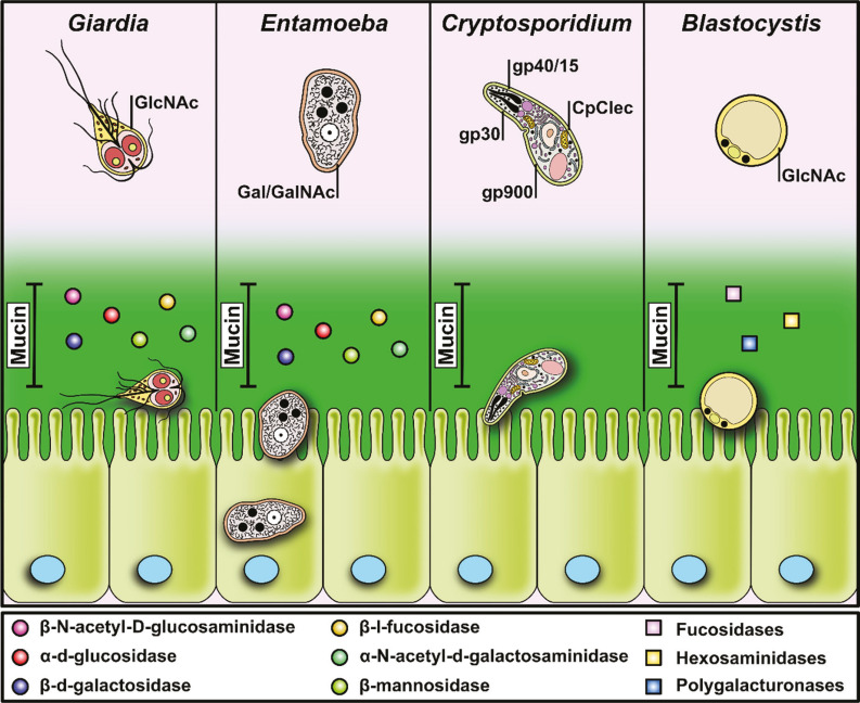
“Increased intestinal permeability may facilitate the penetration of parasite-derived products, microbial components, and food antigens, thereby promoting mucosal inflammation and potentially enhancing food allergy-related symptoms in predisposed individuals (Fig. 1) (Allain et al. 2017; Martinez-Ocana et al. 2020; Troeger et al. 2007).”
From Parasites and allergies: a complex bidirectional relationship from evolutionary origins to modern therapeutics (2026) · cites this paper as Martinez-Ocana et al. 2020
Self-check
Percentages, sums and reported differences re-computed from the paper’s own numbers.
The self-check needs the paper’s full text.
Citations
Cites
Mucin dynamics and enteric pathogens
McGuckin, M. A., Lindén, S. K., Sutton, P., & Florin, T. H. (2011) · Nature Reviews Microbiology · doi:10.1038/nrmicro2538 · Cited by 1,428
Research works printed but not linked: 57
- Linden SK, 2008, Mucins in the mucosal barrier to infection, Mucosal Immunol, 1, 183, 10.1038/mi.2008.5 · DOI
- Theodoropoulos G, 2001, The role of mucins in host-parasite interactions: Part II - helminth parasites, Trends Parasitol, 17, 130, 10.1016/S1471-4922(00)01775-X · DOI
- Johansson ME, 2013, The gastrointestinal mucus system in health and disease, Nat Rev Gastroenterol Hepatol, 10, 352, 10.1038/nrgastro.2013.35 · DOI · Paper page
- Kim YS, 2010, Intestinal goblet cells and mucins in health and disease: recent insights and progress, Curr Gastroenterol Rep, 12, 319, 10.1007/s11894-010-0131-2 · DOI
- Dhanisha SS, 2018, Mucins: structural diversity, biosynthesis, its role in pathogenesis and as possible therapeutic targets, Crit Rev Oncol Hematol, 122, 98, 10.1016/j.critrevonc.2017.12.006 · DOI · Paper page
- Singh RS, 2011, Characteristics of yeast lectins and their role in cell-cell interactions, Biotechnol Adv, 29, 726, 10.1016/j.biotechadv.2011.06.002 · DOI
- Singh RS, 2016, Protozoa lectins and their role in host-pathogen interactions, Biotechnol Adv, 34, 1018, 10.1016/j.biotechadv.2016.06.002 · DOI
- Moody-Haupt S, 2000, The major surface antigens of Entamoeba histolytica trophozoites are GPI-anchored proteophosphoglycans, J Mol Biol, 297, 409, 10.1006/jmbi.2000.3577 · DOI
- Barnes DA, 1998, A novel multi-domain mucin-like glycoprotein of Cryptosporidium parvum mediates invasion, Mol Biochem Parasitol, 96, 93
- Cevallos AM, 2000, Molecular cloning and expression of a gene encoding Cryptosporidium parvum glycoproteins gp40 and gp15, Infect Immun, 68, 4108, 10.1128/IAI.68.7.4108-4116.2000 · DOI
- Hicks SJ, 2000, The role of mucins in host-parasite interactions: Part I - Protozoan parasites, Parasitol Today, 16, 476, 10.1016/S0169-4758(00)01773-7 · DOI
- Samuelson J, 2011, A simple fibril and lectin model for cyst walls of Entamoeba and perhaps Giardia, Trends Parasitol, 27, 17, 10.1016/j.pt.2010.09.002 · DOI
- Chatterjee A, 2010, Giardia cyst wall protein 1 is a lectin that binds to curled fibrils of the GalNAc homopolymer, PLoS Pathog, 19
- Sousa MC, 2001, Adherence of Giardia lamblia trophozoites to Int-407 human intestinal cells, Clin Diagn Lab Immunol, 8, 258, 10.1128/CDLI.8.2.258-265.2001 · DOI
- Connaris S, 1997, Glycosidases in mucin-dwelling protozoans, Glycoconj J, 14, 879, 10.1023/A:1018554408558 · DOI
- Li E, 2016, Complement activation by Giardia duodenalis parasites through the lectin pathway contributes to mast cell responses and parasite control, Infect Immun, 84, 1092, 10.1128/IAI.00074-16 · DOI
- Jiménez JC, 2007, Excreted/secreted glycoproteins of G. intestinalis play an essential role in the antibody response, Parasitol Res, 100, 715, 10.1007/s00436-006-0339-0 · DOI
- Chadee K, 1988, Binding and internalization of rat colonic mucins by the galactose/N-acetyl-D-galactosamine adherence lectin of Entamoeba histolytica, J Infect Dis, 158, 398, 10.1093/infdis/158.2.398 · DOI
- Aguirre-García M, 2015, Entamoeba histolytica: adhesins and lectins in the trophozoite surface, Molecules, 20, 2802, 10.3390/molecules20022802 · DOI
- Chadee K, 1987, Rat and human colonic mucins bind to and inhibit the adherence lectin of Entamoeba histolytica, J Clin Invest, 80, 1245, 10.1172/JCI113199 · DOI
- Coudrier E, 2005, Myosin II and the Gal-GalNAc lectin play a crucial role in tissue invasion by Entamoeba histolytica, Cell Microbiol, 7, 19, 10.1111/j.1462-5822.2004.00426.x · DOI
- Petri Jr WA, 2002, The bittersweet interface of parasite and host: lectin-carbohydrate interactions during human invasion by the parasite Entamoeba histolytica, Annu Rev Microbiol, 56, 39, 10.1146/annurev.micro.56.012302.160959 · DOI
- Katz U, 2002, Entamoeba histolytica expressing a dominant negative N-truncated light subunit of its gal-lectin are less virulent, Mol Biol Cell, 13, 4256, 10.1091/mbc.e02-06-0344 · DOI
- Cheng XJ, 2001, Intermediate subunit of the Gal/GalNAc lectin of Entamoeba histolytica is a member of a gene family containing multiple CXXC sequence motifs, Infect Immun, 69, 5892, 10.1128/IAI.69.9.5892-5898.2001 · DOI
- Min X, 2016, Evaluation of the C-terminal fragment of Entamoeba histolytica Gal/GalNAc lectin intermediate subunit as a vaccine candidate against amebic liver abscess, PLoS Negl Trop Dis, 10
- Galván-Moroyoqui JM, 2011, Pathogenic bacteria prime the induction of Toll-like receptor signalling in human colonic cells by the Gal/GalNAc lectin Carbohydrate Recognition Domain of Entamoeba histolytica, Int J Parasitol, 41, 1101, 10.1016/j.ijpara.2011.06.003 · DOI
- Kato K, 2017, Comparison of hemolytic activity of the intermediate subunit of Entamoeba histolytica and Entamoeba dispar lectins, PLoS One, 12, 10.1371/journal.pone.0181864 · DOI
- Ocádiz R, 2005, EhCP112 is an Entamoeba histolytica secreted cysteine protease that may be involved in the parasite-virulence, Cell Microbiol, 7, 221, 10.1111/j.1462-5822.2004.00453.x · DOI
- Betanzos A, 2019, Host invasion by pathogenic amoebae: epithelial disruption by parasite proteins, Genes, 10, 10.3390/genes10080618 · DOI
- Nok AJ, 2003, Characterization of sialidase from Entamoaeba hystolitica and possible pathogenic role in amebiasis, Parasitol Res, 89, 302, 10.1007/s00436-002-0646-z · DOI
- Espinosa-Cantellano M, 2000, Pathogenesis of intestinal amebiasis: from molecules to disease, Clin Microbiol Rev, 13, 318, 10.1128/CMR.13.2.318 · DOI
- Thibeaux R, 2013, Identification of the virulence landscape essential for Entamoeba histolytica invasion of the human colon, PLoS Pathog, 9, 10.1371/journal.ppat.1003824 · DOI
- Que X, 2000, Cysteine proteinases and the pathogenesis of amebiasis, Clin Microbiol Rev, 13, 196, 10.1128/CMR.13.2.196 · DOI
- Gibson AR, 2018, Cryptosporidium, Curr Biol, 28, R193, 10.1016/j.cub.2017.11.070 · DOI
- Peng MM, 1997, Genetic polymorphism among Cryptosporidium parvum isolates: evidence of two distinct human transmission cycles, Emerg Infect Dis, 3, 567, 10.3201/eid0304.970423 · DOI
- Bouzid M, 2013, Cryptosporidium pathogenicity and virulence, Clin Microbiol Rev, 26, 115, 10.1128/CMR.00076-12 · DOI
- Wanyiri J, 2006, Molecular basis of Cryptosporidium-host cell interactions: recent advances and future prospects, Future Microbiol, 1, 201, 10.2217/17460913.1.2.201 · DOI
- Yao L, 2007, Cryptosporidium parvum: identification of a new surface adhesion protein on sporozoite and oocyst by screening of a phage-display cDNA library, Exp Parasitol, 115, 333, 10.1016/j.exppara.2006.09.018 · DOI
- Bhalchandra S, 2013, Identification and characterization of Cryptosporidium parvum Clec, a novel C-type lectin domain-containing mucin-like glycoprotein, Infect Immun, 81, 3356, 10.1128/IAI.00436-13 · DOI
- Ludington JG, 2016, The Cryptosporidium parvum C-type lectin CpClec mediates infection of intestinal epithelial cells via interactions with sulfated proteoglycans, Infect Immun, 84, 1593, 10.1128/IAI.01410-15 · DOI
- Bhat N, 2007, Cryptosporidium p30, a galactose/N-acetylgalactosamine-specific lectin, mediates infection in vitro, J Biol Chem, 282, 34877, 10.1074/jbc.M706950200 · DOI
- Tosini F, 2004, A new modular protein of Cryptosporidium parvum, with ricin B and LCCL domains, expressed in the sporozoite invasive stage, Mol Biochem Parasitol, 134, 137, 10.1016/j.molbiopara.2003.11.014 · DOI
- Langer RC, 2001, Characterization of an intestinal epithelial cell receptor recognized by the Cryptosporidium parvum sporozoite ligand CSL, Infect Immun, 69, 1661, 10.1128/IAI.69.3.1661-1670.2001 · DOI
- Deng M, 2004, Host intestinal epithelial response to Cryptosporidium parvum, Adv Drug Deliv Rev, 56, 869, 10.1016/j.addr.2003.10.034 · DOI
- Boulter-Bitzer JI, 2007, Molecular targets for detection and immunotherapy in Cryptosporidium parvum, Biotechnol Adv, 25, 13, 10.1016/j.biotechadv.2006.08.003 · DOI
- Naitza S, 1998, The thrombospondin-related protein family of Apicomplexan parasites: the gears of the cell invasion machinery, Parasitol Today, 14, 479, 10.1016/S0169-4758(98)01346-5 · DOI
- Kappe S, 1999, Conservation of a gliding motility and cell invasion machinery in Apicomplexan parasites, J Cell Biol, 147, 937, 10.1083/jcb.147.5.937 · DOI
- Roberts T, 2014, Update on the pathogenic potential and treatment options for Blastocystis sp, Gut Pathog, 6, 10.1186/1757-4749-6-17 · DOI
- Tan KS, 2008, New insights on classification, identification, and clinical relevance of Blastocystis spp, Clin Microbiol Rev, 21, 639, 10.1128/CMR.00022-08 · DOI
- Stensvold CR, 2016, Current status of Blastocystis: a personal view, Parasitol Int, 65, 763, 10.1016/j.parint.2016.05.015 · DOI
- Boorom KF, 2008, Oh my aching gut: irritable bowel syndrome, Blastocystis, and asymptomatic infection, Parasit Vectors, 1, 10.1186/1756-3305-1-40 · DOI
- Lanuza MD, 1996, Identification of surface coat carbohydrates in Blastocystis hominis by lectin probes, Int J Parasitol, 26, 527, 10.1016/0020-7519(96)00010-0 · DOI
- Wu B, 2010, Blastocystis legumain is localized on the cell surface, and specific inhibition of its activity implicates a pro-survival role for the enzyme, J Biol Chem, 285, 1790, 10.1074/jbc.M109.049064 · DOI
- Wawrzyniak I, 2012, Characterization of two cysteine proteases secreted by Blastocystis ST7, a human intestinal parasite, Parasitol Int, 61, 437, 10.1016/j.parint.2012.02.007 · DOI
- Denoeud F, 2011, Genome sequence of the stramenopile Blastocystis, a human anaerobic parasite, Genome Biol, 12, 10.1186/gb-2011-12-3-r29 · DOI
- Yason JA, 2018, Membrane surface features of Blastocystis subtypes, Genes, 9, 10.3390/genes9080417 · DOI
- Eme L, 2017, Lateral gene transfer in the adaptation of the anaerobic parasite Blastocystis to the gut, Curr Biol, 27, 807, 10.1016/j.cub.2017.02.003 · DOI
Cited by
Sorted by citations
Packi, K., Świerczewski, D., Klimczak, S., Ochab, D., Gałęziewska, J., Gromek, P., Kruczkowska, W., Płuciennik, E., Kałuzińska-Kołat, Ż., & Kołat, D. (2026) · Antonie van Leeuwenhoek · doi:10.1007/s10482-026-02425-4
Metadata
- Authors
- Martínez-Ocaña, J., Maravilla, P., & Olivo-Díaz, A.
- Venue
- Revista do Instituto de Medicina Tropical de São Paulo
- Publisher
- FapUNIFESP (SciELO)
- Published
- 2020
- Volume
- 62
- ISSN
- 1678-9946
- PMC
- PMC7477959
- Cited by
- 16
- References
- 1
About these checks
Registry, retraction, Cites and statistics results are computed, not judged, with one exception: a printed reference that no index lists is read once by a model before it counts as a citation error. Claims are extracted by a model and kept only when their quote appears verbatim in the text; each cited source is then checked, with a model’s help, for whether it says what the claim relies on. Those verdicts can be wrong — read the claim’s quote and the cited source before relying on one.
Statistics and arithmetic are recomputed, not judged.
Confidence bands are shown only for fields calibrated on our annotated test set. None is calibrated yet, so no band is shown.
What counts as a claim ›How we measure confidence ›How we check this ›
Are you an author, or do you see a problem with this page? Request a review
