Cited by 26

The DOI matches its Crossref record; 1 of the 1 references it cites were found in a registry or an index.

Microtubule inhibitor, SP-6-27 inhibits angiogenesis and induces apoptosis in ovarian cancer cells

Kulshrestha, Arpita, Katara, Gajendra K., Ibrahim, Safaa A., Patil, Renukadevi, Patil, Shivaputra A., Beaman, Kenneth D.·2017·Oncotarget

Overview

The claims and statistics of this paper haven’t been checked yet.

Run the check — free, no account needed. It reads the open-access text and checks the sources it cites.

A few free checks a day · papers already verified open instantly · results are public

Integrity signals

Registry

Crossref · 10.18632/oncotarget.17549

Cites

1 of 1 references verified0 citation errors · 0 not checkable

Statistics

Figures

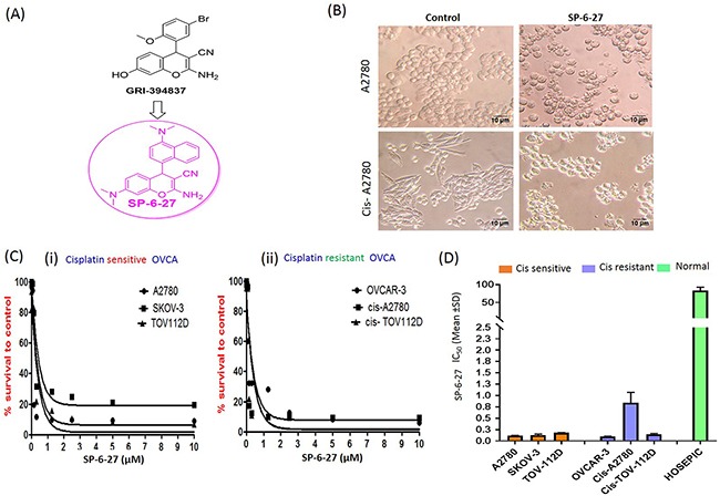
Figure 1 Microtubule inhibitor SP-6-27 exhibits cytotoxicity towards human ovarian cancer cells (A) Structure of SP-6-27 as derived from the parent compound GRI-394837. (B) Changes in the cell morphology of the ovarian cancer cells upon treatment with SP-6-27. Cisplatin sensitive (A2780) and resistant (cis-A2780) ovarian carcinoma cells were incubated with vehicle control (DMSO) or SP-6-27 (0.5 μM, 24 h). Cell morphology was assessed by phase-contrast microscopy and representative images (20X magnification; scale bar- 10 μm) are shown here. (C) Dose–response curves of SP-6-27 in a panel of (i) cisplatin sensitive (A2780, SKOV-3, TOV-112D) and (ii) cisplatin resistant (OVCAR-3, cis-A2780, cis-TOV-112D) ovarian cancer cell lines following 72h treatment. (D) IC50 values (mean± SD) for SP-6-27 in different human ovarian cancer cell lines compared to normal ovarian epithelial cells (HOSEpiC). The cell viability was assessed by Alamar Blue assay. Data represents mean of 3 independent experiments performed at least in triplicate.
Figure 1 Microtubule inhibitor SP-6-27 exhibits cytotoxicity towards human ovarian cancer cells (A) Structure of SP-6-27 as derived from the parent compound GRI-394837. (B) Changes in the cell morphology of the ovarian cancer cells upon treatment with SP-6-27. Cisplatin sensitive (A2780) and resistant (cis-A2780) ovarian carcinoma cells were incubated with vehicle control (DMSO) or SP-6-27 (0.5 μM, 24 h). Cell morphology was assessed by phase-contrast microscopy and representative images (20X magnification; scale bar- 10 μm) are shown here. (C) Dose–response curves of SP-6-27 in a panel of (i) cisplatin sensitive (A2780, SKOV-3, TOV-112D) and (ii) cisplatin resistant (OVCAR-3, cis-A2780, cis-TOV-112D) ovarian cancer cell lines following 72h treatment. (D) IC50 values (mean± SD) for SP-6-27 in different human ovarian cancer cell lines compared to normal ovarian epithelial cells (HOSEpiC). The cell viability was assessed by Alamar Blue assay. Data represents mean of 3 independent experiments performed at least in triplicate.CC BY · PubMed Central
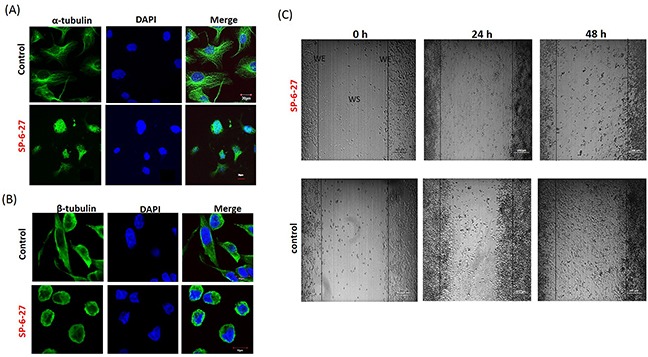
Figure 2 Chromene analog SP-6-27 disrupts the microtubular dynamics and inhibits migration in ovarian cancer cells Ovarian cancer cells (A2780) were treated with control (DMSO, vehicle) or SP-6-27 (0.5μM) for 24 h and stained with DAPI (blue) and an (A) anti–α-tubulin or (B) anti-β-tubulin antibody (green). Images were captured using an Olympus FluoView confocal microscope. Representative confocal micrographs (original magnification: 80X) are shown. (C) Effect of SP-6-27 on OVCA cell migration. In vitro wound healing assay was performed using A2780 cells cultured in 6 well plates. Confluent cultures were scratched with a 1 mL pipette tip as described in the Methods section. Representative phase-contrast images of cells migrating into the wounded area in SP-6-27 treated and control wells (0, 24 and 48 h) are shown here. W: wound space, WE: wound edge (magnification- 4X, scale bar-200 μm).
Figure 2 Chromene analog SP-6-27 disrupts the microtubular dynamics and inhibits migration in ovarian cancer cells Ovarian cancer cells (A2780) were treated with control (DMSO, vehicle) or SP-6-27 (0.5μM) for 24 h and stained with DAPI (blue) and an (A) anti–α-tubulin or (B) anti-β-tubulin antibody (green). Images were captured using an Olympus FluoView confocal microscope. Representative confocal micrographs (original magnification: 80X) are shown. (C) Effect of SP-6-27 on OVCA cell migration. In vitro wound healing assay was performed using A2780 cells cultured in 6 well plates. Confluent cultures were scratched with a 1 mL pipette tip as described in the Methods section. Representative phase-contrast images of cells migrating into the wounded area in SP-6-27 treated and control wells (0, 24 and 48 h) are shown here. W: wound space, WE: wound edge (magnification- 4X, scale bar-200 μm).CC BY · PubMed Central
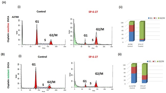
Figure 3 Cisplatin sensitive and resistant ovarian cancer cells arrest in G2 and M phase following SP-6-27 treatment Cisplatin sensitive A2780 or cisplatin resistant cis-A2780 ovarian cancer cells were treated with 0.5μM SP-6-27 or DMSO vehicle control for 24 hours. The cells were evaluated for effects on cell cycle using PI staining and analyzed by flow cytometry using ModFit software. (Ai) Representative cell cycle micrographs of cisplatin sensitive cells depicting G1, S and G2/M cell populations in control and SP-6-27 treated cells. (Aii) Stacked bar graph illustrating the phase distribution of cisplatin sensitive cells in control and SP-6-27 treated groups determined as percentage of the total number of cells in cycle. (Bi) Representative cell cycle micrographs of cisplatin resistant cells depicting G1, S and G2/M cell populations in control and SP-6-27 treated cells. (Bii) Stacked bar graph illustrating the phase distribution of cisplatin resistant cells in control and SP-6-27 treated groups. The data indicates ovarian cancer cell cycle arrest in G2/M phase upon SP-6-27 treatment. Average of three experiments is shown.
Figure 3 Cisplatin sensitive and resistant ovarian cancer cells arrest in G2 and M phase following SP-6-27 treatment Cisplatin sensitive A2780 or cisplatin resistant cis-A2780 ovarian cancer cells were treated with 0.5μM SP-6-27 or DMSO vehicle control for 24 hours. The cells were evaluated for effects on cell cycle using PI staining and analyzed by flow cytometry using ModFit software. (Ai) Representative cell cycle micrographs of cisplatin sensitive cells depicting G1, S and G2/M cell populations in control and SP-6-27 treated cells. (Aii) Stacked bar graph illustrating the phase distribution of cisplatin sensitive cells in control and SP-6-27 treated groups determined as percentage of the total number of cells in cycle. (Bi) Representative cell cycle micrographs of cisplatin resistant cells depicting G1, S and G2/M cell populations in control and SP-6-27 treated cells. (Bii) Stacked bar graph illustrating the phase distribution of cisplatin resistant cells in control and SP-6-27 treated groups. The data indicates ovarian cancer cell cycle arrest in G2/M phase upon SP-6-27 treatment. Average of three experiments is shown.CC BY · PubMed Central
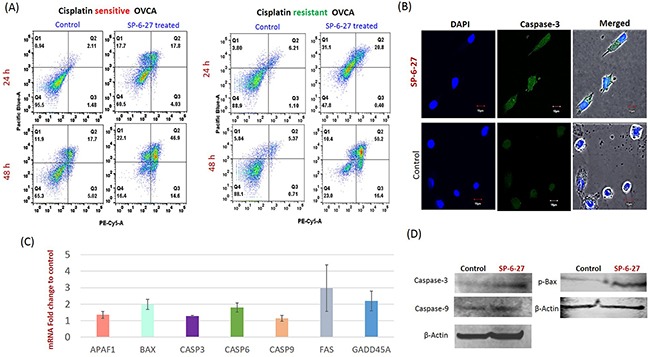
Figure 4 SP-6-27 induces apoptotic cell death in chemo-resistant and sensitive ovarian cancer cells (A) Cisplatin sensitive (A2780) and cisplatin resistant (cis-A2780) ovarian cancer cells were treated with 0.5 μM SP-6-27 (24, 48 h) followed by Annexin V/7-AAD assay. Early apoptotic [first (Q1) quadrant], late apoptotic [second (Q2) quadrant] and dead cells [(Q3) quadrant] are shown. (B) Confocal microscopy analysis for cleaved caspase-3 in SP-6-27 treated (0.5μM, 24h) ovarian cancer cells. Images show active caspase-3 expression in (i) SP-6-27 treated cells compared to (ii) control cells. Nuclei staining was performed using DAPI (blue). Merged phase-contrast-fluorescence microscopy images displaying cellular expression of active Caspase-3 and DAPI are shown here. Original magnification-× 80; Scale bars-10μm. (C) Q-RT-PCR array was performed for identification of the cell death pathway associated genes altered in ovarian cancer cells upon SP-6-27 treatment. mean ±SD of the mRNA fold change using two endogenous controls (BACT and HPRT) is depicted here. Data are the average of triplicate experiments [mean ± (SD)]. (D) Western blot analysis of the intrinsic apoptotic molecules in SP-6-27 treated A2780 ovarian cancer cells show enhanced expression compared to control cells.
Figure 4 SP-6-27 induces apoptotic cell death in chemo-resistant and sensitive ovarian cancer cells (A) Cisplatin sensitive (A2780) and cisplatin resistant (cis-A2780) ovarian cancer cells were treated with 0.5 μM SP-6-27 (24, 48 h) followed by Annexin V/7-AAD assay. Early apoptotic [first (Q1) quadrant], late apoptotic [second (Q2) quadrant] and dead cells [(Q3) quadrant] are shown. (B) Confocal microscopy analysis for cleaved caspase-3 in SP-6-27 treated (0.5μM, 24h) ovarian cancer cells. Images show active caspase-3 expression in (i) SP-6-27 treated cells compared to (ii) control cells. Nuclei staining was performed using DAPI (blue). Merged phase-contrast-fluorescence microscopy images displaying cellular expression of active Caspase-3 and DAPI are shown here. Original magnification-× 80; Scale bars-10μm. (C) Q-RT-PCR array was performed for identification of the cell death pathway associated genes altered in ovarian cancer cells upon SP-6-27 treatment. mean ±SD of the mRNA fold change using two endogenous controls (BACT and HPRT) is depicted here. Data are the average of triplicate experiments [mean ± (SD)]. (D) Western blot analysis of the intrinsic apoptotic molecules in SP-6-27 treated A2780 ovarian cancer cells show enhanced expression compared to control cells.CC BY · PubMed Central
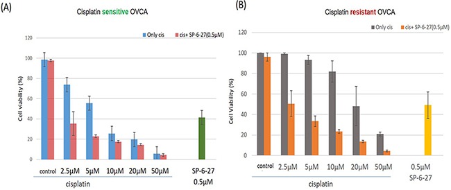
Figure 5 SP-6-27 and cisplatin combination treatment exhibits enhanced cytotoxicity towards ovarian cancer cells (A) The cell viability of cisplatin sensitive cells (A2780) following simultaneous treatment with a combination of SP-6-27 and cisplatin. Blue columns, cells treated with cisplatin only (0-50μM, 48h); red columns, cells treated with combination of cisplatin (0-50μM) and 0.5 μg SP-6-27 for 48h; green column shows cells treated with SP-6-27 alone (0.5 μM, 48h). (B) Sequential combinatorial treatment with SP-6-27 (0.5μM; 24h) followed by cisplatin (0- 50μM; 24h) enhanced cytotoxicity in the of cisplatin resistant cells (cis-A2780) cells. Gray columns, cells treated with cisplatin only; orange column, cells treated with a combination of cisplatin and SP-6-27; yellow column shows cells treated with SP-6-27 (0.5 μM, 48h) alone. The experiments were performed twice in triplicate.
Figure 5 SP-6-27 and cisplatin combination treatment exhibits enhanced cytotoxicity towards ovarian cancer cells (A) The cell viability of cisplatin sensitive cells (A2780) following simultaneous treatment with a combination of SP-6-27 and cisplatin. Blue columns, cells treated with cisplatin only (0-50μM, 48h); red columns, cells treated with combination of cisplatin (0-50μM) and 0.5 μg SP-6-27 for 48h; green column shows cells treated with SP-6-27 alone (0.5 μM, 48h). (B) Sequential combinatorial treatment with SP-6-27 (0.5μM; 24h) followed by cisplatin (0- 50μM; 24h) enhanced cytotoxicity in the of cisplatin resistant cells (cis-A2780) cells. Gray columns, cells treated with cisplatin only; orange column, cells treated with a combination of cisplatin and SP-6-27; yellow column shows cells treated with SP-6-27 (0.5 μM, 48h) alone. The experiments were performed twice in triplicate.CC BY · PubMed Central
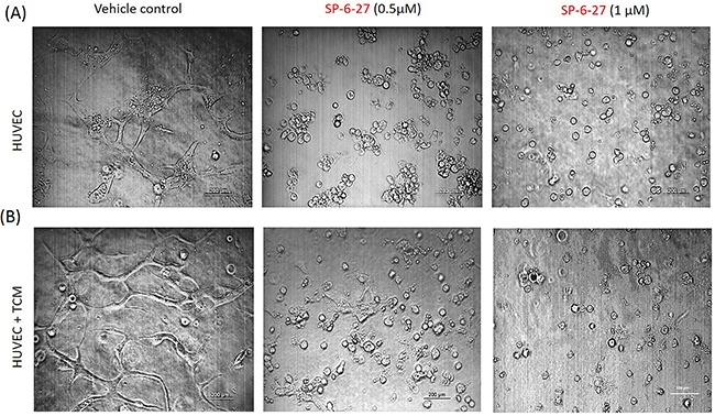
Figure 6 SP-6-27 inhibits capillary tube formation by endothelial cells HUVECs endothelial cells were grown on matrigel-coated 96-well plates for 24 h. The cells were treated with SP-6-27 (0.5, 1μM) or DMSO vehicle, in the (A) absence/(B) presence of tumor conditioned media from ovarian cells. The ability of the HUVEC cells to form capillary tube like structures was analyzed using phase contrast microscopy at a magnification of ×4, scale bars- 200μm. Images shown are representative of three independent experiments.
Figure 6 SP-6-27 inhibits capillary tube formation by endothelial cells HUVECs endothelial cells were grown on matrigel-coated 96-well plates for 24 h. The cells were treated with SP-6-27 (0.5, 1μM) or DMSO vehicle, in the (A) absence/(B) presence of tumor conditioned media from ovarian cells. The ability of the HUVEC cells to form capillary tube like structures was analyzed using phase contrast microscopy at a magnification of ×4, scale bars- 200μm. Images shown are representative of three independent experiments.CC BY · PubMed Central
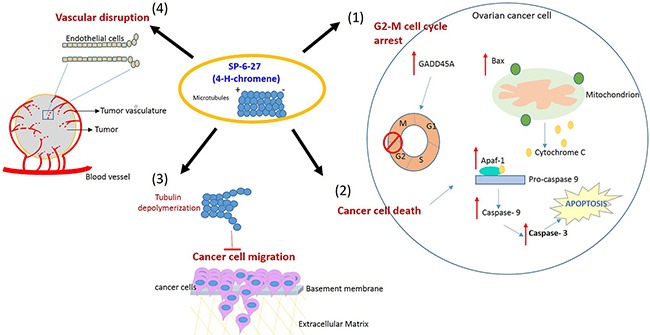
Figure 7 Proposed model illustrating the effects of SP-6-27 mediated microtubule inhibition on ovarian cancer cells and endothelial cells Schematic of the pleotropic effects of SP-6-27 on ovarian cancer growth and angiogenesis. Tubulin de-polymerization caused by SP-6-27 leads to (1) G2-M phase cell cycle arrest in these cells. (2) Enhanced apoptotic cell death- The elevated expression of Bax contributes to the mitochondrial cytochrome c mediated activation of caspase -9 involving Apaf-1. The activated caspases-9 further cleaves procaspase-3 and subsequently leads to apoptosis of the cell. (3) Reduced cancer cell migration- SP-6-27 mediated tubulin de-polymerization suppresses ovarian cancer cell migration. (4) Vascular disruption- SP-6-26 causes vascular disruption of the endothelial cells, thus inhibiting angiogenesis. The endothelial vascular disruption is evident in the presence of ovarian cancer tumor conditioned medium.
Figure 7 Proposed model illustrating the effects of SP-6-27 mediated microtubule inhibition on ovarian cancer cells and endothelial cells Schematic of the pleotropic effects of SP-6-27 on ovarian cancer growth and angiogenesis. Tubulin de-polymerization caused by SP-6-27 leads to (1) G2-M phase cell cycle arrest in these cells. (2) Enhanced apoptotic cell death- The elevated expression of Bax contributes to the mitochondrial cytochrome c mediated activation of caspase -9 involving Apaf-1. The activated caspases-9 further cleaves procaspase-3 and subsequently leads to apoptosis of the cell. (3) Reduced cancer cell migration- SP-6-27 mediated tubulin de-polymerization suppresses ovarian cancer cell migration. (4) Vascular disruption- SP-6-26 causes vascular disruption of the endothelial cells, thus inhibiting angiogenesis. The endothelial vascular disruption is evident in the presence of ovarian cancer tumor conditioned medium.CC BY · PubMed Central

Verification

Claims the paper makes

The claims and statistics of this paper haven’t been checked yet.

Run the check — free, no account needed. It reads the open-access text and checks the sources it cites.

A few free checks a day · papers already verified open instantly · results are public

Claims other papers make about this one

Each is checked against this paper’s own text: does it say what the citing paper says it does?

Self-check

Percentages, sums and reported differences re-computed from the paper’s own numbers.

The self-check needs the paper’s full text.

Citations

Cites

Research works printed but not linked: 58

  1. Jemal, 2016, Cancer statistics, 2016, CA Cancer J Clin, 66, 7, 10.3322/caac.21332 · DOI · Paper page
  2. Kaye, 2011, Rethinking ovarian cancer: recommendations for improving outcomes, Nat Rev Cancer, 11, 719, 10.1038/nrc3144 · DOI
  3. Ledermann, 2014, Ovarian cancer, Lancet, 384, 1376, 10.1016/S0140-6736(13)62146-7 · DOI
  4. Ozols, 2006, Challenges for chemotherapy in ovarian cancer, Ann Oncol, 17, v181, 10.1093/annonc/mdj978 · DOI
  5. Ledermann, 2013, Update on first-line treatment of advanced ovarian carcinoma, Int J Womens Health, 5, 45, 10.2147/IJWH.S30231 · DOI
  6. Essigmann, 2001, Mechanisms of resistance to cisplatin, Mutat Res, 478, 23, 10.1016/S0027-5107(01)00141-5 · DOI
  7. Lee, 2016, An update on current and emerging therapies for epithelial ovarian cancer: focus on poly (adenosine diphosphate-ribose) polymerase inhibition and antiangiogenesis, J Oncol Pharm Pract
  8. Nogales, 2000, Structural insights into microtubule function, Ann Rev Biochem, 69, 277, 10.1146/annurev.biochem.69.1.277 · DOI
  9. Jordan, 2010, Microtubule-binding agents: a dynamic field of cancer therapeutics, Nat Rev Drug Discover, 9, 790, 10.1038/nrd3253 · DOI
  10. Gergely, 2015, Hitting the brakes: targeting microtubule motors in cancer, Br J Cancer, 113, 693, 10.1038/bjc.2015.264 · DOI
  11. McGown, 2003, Tubulin and microtubules as targets for anticancer drugs, Prog Cell Cycle Res, 5, 309
  12. Fornier, 2008, Microtubule active agents: beyond the taxane frontier, Clin Cancer Res, 14, 7167, 10.1158/1078-0432.CCR-08-0169 · DOI
  13. Mukhtar, 2014, Targeting microtubules by natural agents for cancer therapy, Mol Cancer Ther, 13, 275, 10.1158/1535-7163.MCT-13-0791 · DOI
  14. Kavallaris, 2010, Microtubules and resistance to tubulin-binding agents, Nature reviews Cancer, 10, 194, 10.1038/nrc2803 · DOI
  15. Perez, 2009, Microtubule inhibitors: differentiating tubulin-inhibiting agents based on mechanisms of action, clinical activity, and resistance, Mol Cancer Ther, 8, 2086, 10.1158/1535-7163.MCT-09-0366 · DOI
  16. Yumoto, 2009, Paclitaxel-resistance conferred by altered expression of efflux and influx transporters for paclitaxel in the human hepatoma cell line, HepG2, Drug Metab Pharmacokinet, 24, 418, 10.2133/dmpk.24.418 · DOI
  17. Miller, 2012, An overview of tubulin inhibitors that interact with the colchicine binding site, Pharm Res, 29, 2943, 10.1007/s11095-012-0828-z · DOI · Paper page
  18. Tian, 2009, A review on hemisynthesis, biosynthesis, biological activities, mode of action, and structure-activity relationship of podophyllotoxins: 2003-2007, Curr Med Chem, 16, 327, 10.2174/092986709787002682 · DOI
  19. Aneja, 2011, Drugs that target dynamic microtubules: a new molecular perspective, Med Res Rev, 31, 443, 10.1002/med.20242 · DOI
  20. Zachariah, 2013, Pharmacological activities of chromene derivatives: an overview, Asian J Pharm Clin Res, 6, 11
  21. Adamson, 2014, “Gliomas: Classification, symptoms, treatment and prognosis.”, 211
  22. Miller, 2015, Identification of imidazoquinoline derivatives as potent antiglioma agents, Med Chem, 11, 400, 10.2174/1573406410666140914162701 · DOI
  23. Miller, 2012, New substituted 4H-chromenes as anticancer agents, Bioorg Med Chem Lett, 22, 4458, 10.1016/j.bmcl.2012.04.074 · DOI
  24. Goldman, 1971, The role of three cytoplasmic fibers in BHK-21 cell motility. I. Microtubules and the effects of colchicine, J Cell Biol, 51, 752, 10.1083/jcb.51.3.752 · DOI
  25. Kavallaris, 2014, Movers and shakers: cell cytoskeleton in cancer metastasis, Br J Pharmacol, 171, 5507, 10.1111/bph.12704 · DOI
  26. Chang, 2007, A novel oral indoline-sulfonamide agent, N-[1-(4-methoxybenzenesulfonyl)-2,3-dihydro-1H-indol-7-yl]-isonicotinamide(J30), exhibits potent activity against human cancer cells in vitro and in vivo through the disruption of microtubule, J Pharmacol Exp Ther, 323, 398, 10.1124/jpet.107.126680 · DOI
  27. Cleveland, 2005, On the road to cancer: aneuploidy and the mitotic checkpoint, Nature reviews Cancer, 5, 773, 10.1038/nrc1714 · DOI
  28. Lim, 2014, Suprafenacine, an indazole-hydrazide agent, targets cancer cells through microtubule destabilization, PLoS One, 9, e110955, 10.1371/journal.pone.0110955 · DOI
  29. Kaufmann, 2002, G(1) and G(2) cell-cycle arrest following microtubule depolymerization in human breast cancer cells, J Clin Invest, 110, 91, 10.1172/JCI13275 · DOI · Paper page
  30. Haskill, 1999, Cisplatin inhibits paclitaxel-induced apoptosis in cisplatin-resistant ovarian cancer cell lines: possible explanation for failure of combination therapy, Cancer Res, 59, 2425
  31. Huot, 2007, Endothelial cell migration during angiogenesis, Circ Res, 100, 782, 10.1161/01.RES.0000259593.07661.1e · DOI
  32. Gewirtz, 2008, Colchicine site inhibitors of microtubule integrity as vascular disrupting agents, Drug Dev Res, 69, 352, 10.1002/ddr.20267 · DOI
  33. Hoskins, 1999, Ten-year follow-up of ovarian cancer patients after second-look laparotomy with negative findings, Obstet Gynecol, 93, 21
  34. An, 2013, Development of a novel class of tubulin inhibitors with promising anticancer activities, Mol Cancer Res, 11, 856, 10.1158/1541-7786.MCR-12-0177 · DOI
  35. Mann, 2003, Combretastatins: from natural products to drug discovery, Nat Prod Rep, 20, 558, 10.1039/b306797c · DOI
  36. Bompiani, 1981, The treatment of 48 patients affected by ovarian carcinoma at stages III and IV F.I.G.O, Eur J Gynaecol Oncol, 2, 89
  37. Coleman, 2014, Vintafolide: a novel targeted agent for epithelial ovarian cancer, Future Oncol, 10, 541, 10.2217/fon.14.8 · DOI
  38. Rej, 2004, Discovery of 4-aryl-4H-chromenes as a new series of apoptosis inducers using a cell- and caspase-based high-throughput screening assay. 1. Structure-activity relationships of the 4-aryl group, J Med Chem, 47, 6299, 10.1021/jm049640t · DOI
  39. Gajate, 2003, Microtubules, microtubule-interfering agents and apoptosis, Apoptosis, 8, 413, 10.1023/A:1025513106330 · DOI
  40. Al-Katib, 2002, Combretastatin-A4 prodrug induces mitotic catastrophe in chronic lymphocytic leukemia cell line independent of caspase activation and poly(ADP-ribose) polymerase cleavage, Clin Cancer Res, 8, 2735
  41. Cabral, 2010, Inhibition of cell migration and cell division correlates with distinct effects of microtubule inhibiting drugs, J Biol Chem, 285, 32242, 10.1074/jbc.M110.160820 · DOI
  42. Braguer, 2007, Microtubules in apoptosis induction: are they necessary?, Curr Cancer Drug Targets, 7, 713, 10.2174/156800907783220480 · DOI
  43. Giordano, 2000, Cell cycle and apoptosis, Neoplasia, 2, 291, 10.1038/sj.neo.7900101 · DOI
  44. Chang, 2001, If not apoptosis, then what? Treatment-induced senescence and mitotic catastrophe in tumor cells, Drug Resist Updat, 4, 303, 10.1054/drup.2001.0213 · DOI
  45. Shin, 1999, p53 negatively regulates cdc2 transcription via the CCAAT-binding NF-Y transcription factor, J Biol Chem, 274, 29677, 10.1074/jbc.274.42.29677 · DOI
  46. Stark, 2001, Regulation of the G2/M transition by p53, Oncogene, 20, 1803, 10.1038/sj.onc.1204252 · DOI
  47. Mak, 2013, Caspase functions in cell death and disease, Cold Spring Harb Perspect Biol, 5, a008656
  48. Dixit, 1997, Caspases: intracellular signaling by proteolysis, Cell, 91, 443, 10.1016/S0092-8674(00)80430-4 · DOI
  49. Arbuck, 1992, Taxol: the first of the taxanes, an important new class of antitumor agents, Semin Oncol, 19, 646
  50. Adams, 2002, PS-341, a novel proteasome inhibitor, induces Bcl-2 phosphorylation and cleavage in association with G2-M phase arrest and apoptosis, Mol Cancer Ther, 1, 841
  51. Thorpe, 2004, Vascular targeting agents as cancer therapeutics, Clin Cancer Res, 10, 415, 10.1158/1078-0432.CCR-0642-03 · DOI
  52. Folkman, 2003, Angiogenesis inhibitors: a new class of drugs, Cancer Biol Therap, 2, S127
  53. Bicknell, 2005, Tumour vascular targeting, Nature reviews Cancer, 5, 436, 10.1038/nrc1627 · DOI
  54. Baguley, 2005, Disrupting tumour blood vessels, Nature reviews Cancer, 5, 423, 10.1038/nrc1628 · DOI
  55. Tozer, 2007, Tumour targeting by microtubule-depolymerizing vascular disrupting agents, Expert Opin Ther Targets, 11, 1443, 10.1517/14728222.11.11.1443 · DOI
  56. Rustin, 2011, Phase II trial of combretastatin A4 phosphate, carboplatin, and paclitaxel in patients with platinum-resistant ovarian cancer, Ann Oncol, 22, 2036, 10.1093/annonc/mdq780 · DOI
  57. Beaman, 2016, Selective inhibition of tumor cell associated Vacuolar-ATPase ‘a2’ isoform overcomes cisplatin resistance in ovarian cancer cells, Mol Oncol, 10, 789, 10.1016/j.molonc.2016.01.003 · DOI
  58. Beaman, 2015, Breast cancer associated a2 isoform vacuolar ATPase immunomodulates neutrophils: potential role in tumor progression, Oncotarget, 6, 33033, 10.18632/oncotarget.5439 · DOI

Cited by

Sorted by citations

See all 26

Metadata

Authors
Kulshrestha, A., Katara, G. K., Ibrahim, S. A., Patil, R., Patil, S. A., & Beaman, K. D.
Affiliations
  • Department of Microbiology and Immunology, Chicago Medical School, Rosalind Franklin University of Medicine and Science, North Chicago, USA
  • Pharmaceutical Sciences Department, College of Pharmacy, Rosalind Franklin University of Medicine and Science, North Chicago, USA
Venue
Oncotarget
Published
2017-05-02
Volume
8
Issue
40
Pages
67017-67028
ISSN
1949-2553
Cited by
26
References
1
Language
en

About these checks

Registry, retraction, Cites and statistics results are computed, not judged, with one exception: a printed reference that no index lists is read once by a model before it counts as a citation error. Claims are extracted by a model and kept only when their quote appears verbatim in the text; each cited source is then checked, with a model’s help, for whether it says what the claim relies on. Those verdicts can be wrong — read the claim’s quote and the cited source before relying on one.

Statistics and arithmetic are recomputed, not judged.

Confidence bands are shown only for fields calibrated on our annotated test set. None is calibrated yet, so no band is shown.

What counts as a claim ›How we measure confidence ›How we check this ›

Are you an author, or do you see a problem with this page? Request a review