Student or educator? Sign up with your school email and get 6 months of Pro free.
PreprintCited by 9Gut microbiota and healthGlycosylation and Glycoproteins ResearchGenomics and Phylogenetic Studies

The DOI matches its Crossref record; 1 of the 1 references it cites were found in a registry or an index.

New carbohydrate binding domains identified by phage display based functional metagenomic screens of human gut microbiota

Open access

Preprint — published as 10.1038/s42003-023-04718-0 · Paper page

Abstract

Uncultured microbes represent a huge untapped biological resource of novel genes and gene products. Although recent genomic and metagenomic sequencing efforts have led to the identification of numerous genes that are homologous to existing annotated genes, there remains, yet, an enormous pool of unannotated genes that do not find significant sequence homology to existing annotated genes. Functional metagenomics offers a way to identify and annotate novel gene products. Here, we have constructed a metagenomic phage display library from healthy human fecal samples, and performed functional carbohydrate binding screens against dietary, microbial and host polysaccharides/glycoconjugates in order to mine novel carbohydrate binding domains that might aid gut commensals in the metabolism of complex carbohydrates and in adherence and colonization of the gut. We obtained several protein sequences that do not find a hit to any known Pfam domain but show carbohydrate binding module-like folds as per Alphafold2 prediction. We heterologously expressed, purified and biochemically characterized some of these protein domains and demonstrated their carbohydrate-binding function. Our study reveals several previously unannotated carbohydrate-binding domains, including a levan binding domain and four complex N-glycan binding domains that might be useful for the labeling, visualization, and isolation of these glycans.

Overview

The claims and statistics of this paper haven’t been checked yet.

Run the check — free, no account needed. It reads the open-access text and checks the sources it cites.

A few free checks a day · papers already verified open instantly · results are public

Integrity signals

Registry

Crossref · 10.21203/rs.3.rs-1898940/v1

Cites

1 of 1 references verified0 citation errors · 0 not checkable

Statistics

Figures

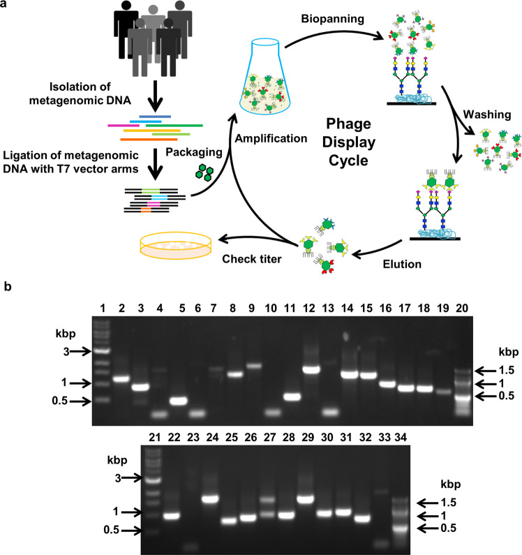
Fig. 1 Metagenomic phage display library. a Schematic representation of metagenomic phage display library preparation and biopanning to screen ligand binding recombinant phages. b Agarose gel electrophoresis of PCR amplicons indicating the presence or absence of any recombinant insert within randomly selected phages of the metagenomic phage display library. Results of a single screening experiment. Lanes 1 and 21: 1 kbp DNA marker (NEB). Lanes 20 and 34: 100 bp DNA marker (NEB). Lanes 2–19 and 22–33: PCR amplicons of the phages.
Fig. 1 Metagenomic phage display library. a Schematic representation of metagenomic phage display library preparation and biopanning to screen ligand binding recombinant phages. b Agarose gel electrophoresis of PCR amplicons indicating the presence or absence of any recombinant insert within randomly selected phages of the metagenomic phage display library. Results of a single screening experiment. Lanes 1 and 21: 1 kbp DNA marker (NEB). Lanes 20 and 34: 100 bp DNA marker (NEB). Lanes 2–19 and 22–33: PCR amplicons of the phages.CC BY · PubMed Central
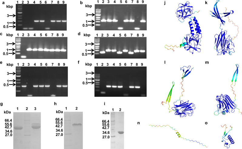
Fig. 2 Identification of mucin binding protein domains. a–f Agarose gel electrophoresis of PCR amplicons indicating the presence or absence of any recombinant insert following biopanning of the metagenomic phage display library against porcine stomach type II mucin. Results of a single screening experiment. a Lane 1: 1 kbp DNA marker. Lanes 2–9: Phages eluted with L-fucose, b Lane 1: 1 kbp DNA marker. Lanes 2–9: Phages eluted with D-galactose, c Lane 1: 1 kbp DNA marker. Lanes 2–9: Phages eluted with GalNAc, d Lane 1: 1 kbp DNA marker. Lanes 2–9: Phages eluted with GlcNAc, e Lane 1: 1 kbp DNA marker. Lanes 2–9: Phages eluted with D-mannose, f Lane 1: 1 kbp DNA marker. Lanes 2–9: Phages eluted with sialic acid (Neu5Ac), g Coomassie blue stained SDS-PAGE showing purified MG1 and MN3 proteins in the imidazole eluates following Ni-NTA metal ion affinity chromatography; Lane 1: Purified MG1 protein; Lane 2: Protein marker from NEB (2-212 kDa); Lane 3: Purified MN3 protein, h Coomassie blue stained SDS-PAGE showing purified MU1 protein in the imidazole eluates following Ni-NTA metal ion affinity chromatography; Lane 1: Protein marker from NEB (2–212 kDa); Lane 2: Purified MU1 protein, i Coomassie blue stained SDS-PAGE showing purified MU3 protein in the imidazole eluates following Ni-NTA metal ion affinity chromatography; Lane 1: Protein marker from NEB (2–212 kDa); Lane 2: Purified MU3 protein. j–n AlphaFold2 predicted structures of MG1 (j), MN3 (k), MU1 (l), MU3 (m), MF12 (n), and MF12F2 (o).
Fig. 2 Identification of mucin binding protein domains. a–f Agarose gel electrophoresis of PCR amplicons indicating the presence or absence of any recombinant insert following biopanning of the metagenomic phage display library against porcine stomach type II mucin. Results of a single screening experiment. a Lane 1: 1 kbp DNA marker. Lanes 2–9: Phages eluted with L-fucose, b Lane 1: 1 kbp DNA marker. Lanes 2–9: Phages eluted with D-galactose, c Lane 1: 1 kbp DNA marker. Lanes 2–9: Phages eluted with GalNAc, d Lane 1: 1 kbp DNA marker. Lanes 2–9: Phages eluted with GlcNAc, e Lane 1: 1 kbp DNA marker. Lanes 2–9: Phages eluted with D-mannose, f Lane 1: 1 kbp DNA marker. Lanes 2–9: Phages eluted with sialic acid (Neu5Ac), g Coomassie blue stained SDS-PAGE showing purified MG1 and MN3 proteins in the imidazole eluates following Ni-NTA metal ion affinity chromatography; Lane 1: Purified MG1 protein; Lane 2: Protein marker from NEB (2-212 kDa); Lane 3: Purified MN3 protein, h Coomassie blue stained SDS-PAGE showing purified MU1 protein in the imidazole eluates following Ni-NTA metal ion affinity chromatography; Lane 1: Protein marker from NEB (2–212 kDa); Lane 2: Purified MU1 protein, i Coomassie blue stained SDS-PAGE showing purified MU3 protein in the imidazole eluates following Ni-NTA metal ion affinity chromatography; Lane 1: Protein marker from NEB (2–212 kDa); Lane 2: Purified MU3 protein. j–n AlphaFold2 predicted structures of MG1 (j), MN3 (k), MU1 (l), MU3 (m), MF12 (n), and MF12F2 (o).CC BY · PubMed Central
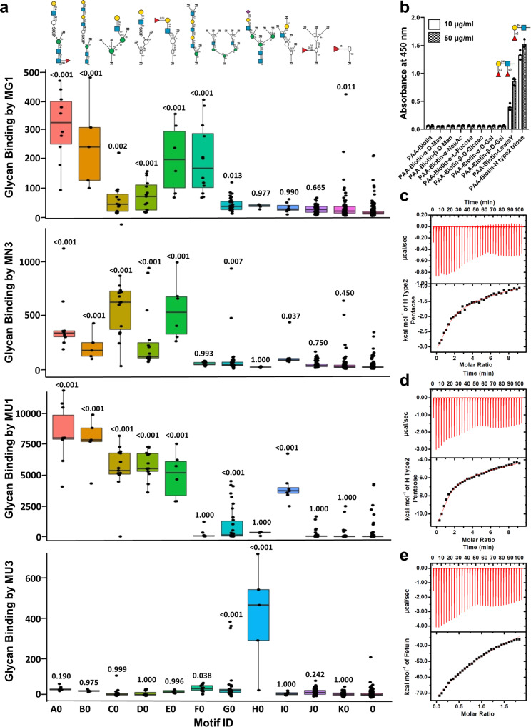
Fig. 3 Glycan binding by MG1, MN3, MU1, and MU3. a Bar plot indicating glycan binding intensities of motifs recognized by MG1, MN3, MU1, and MU3 (output of MotifFinder36,37). The motifs are represented in standard glycan symbol nomenclature83 above the plot. The motif ID “0” refers to non-binders. Results of MotifFinder analysis using glycan array datasets of single runs of MG1 at 5, 50, and 400 µg/ml, and MN3, MU1, and MU3 at 5 and 50 µg/ml. Models were built manually for each protein using a common custom motif list obtained by combined automated model building using all glycan array datasets. P-values are calculated by the MotifFinder software and refer to difference from non-binders with multiple testing corrections (Dunnet’s Test). b Binding of MN3 to Lewisy and H antigen type-2 triose as assessed by enzyme linked lectin assays. Results of a single experiment. Means are plotted. Error bars represent standard deviations of three technical replicates. Source data in Supplementary Data 3. c Binding of MN3 to H antigen type-2 pentaose as assessed by isothermal calorimetry. Results of a single run. d Binding of MU1 to H antigen type-2 pentaose as assessed by isothermal calorimetry. Results of a single run. e Binding of MU3 to Fetuin as assessed by isothermal calorimetry. Results of a single run.
Fig. 3 Glycan binding by MG1, MN3, MU1, and MU3. a Bar plot indicating glycan binding intensities of motifs recognized by MG1, MN3, MU1, and MU3 (output of MotifFinder36,37). The motifs are represented in standard glycan symbol nomenclature83 above the plot. The motif ID “0” refers to non-binders. Results of MotifFinder analysis using glycan array datasets of single runs of MG1 at 5, 50, and 400 µg/ml, and MN3, MU1, and MU3 at 5 and 50 µg/ml. Models were built manually for each protein using a common custom motif list obtained by combined automated model building using all glycan array datasets. P-values are calculated by the MotifFinder software and refer to difference from non-binders with multiple testing corrections (Dunnet’s Test). b Binding of MN3 to Lewisy and H antigen type-2 triose as assessed by enzyme linked lectin assays. Results of a single experiment. Means are plotted. Error bars represent standard deviations of three technical replicates. Source data in Supplementary Data 3. c Binding of MN3 to H antigen type-2 pentaose as assessed by isothermal calorimetry. Results of a single run. d Binding of MU1 to H antigen type-2 pentaose as assessed by isothermal calorimetry. Results of a single run. e Binding of MU3 to Fetuin as assessed by isothermal calorimetry. Results of a single run.CC BY · PubMed Central
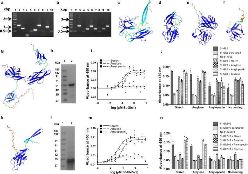
Fig. 4 Identification of starch binding protein domains. a, b Agarose gel electrophoresis of PCR amplicons indicating the presence or absence of any recombinant insert following biopanning of the metagenomic phage display library against starch. a Biopanning against starch and elution with D-glucose. Results of a single screening experiment. Lane 1: 1 kbp DNA marker. Lanes 2–9: PCR amplicons of clones St-Glc1 to St-Glc9. b Biopanning against starch and elution with starch. Results of a single screening experiment. Lane 1: 1 kbp DNA marker. Lanes 2–9: PCR amplicons of clones St-St1 to St-St9. c–f AlphaFold2 predicted structural models of St-Glc2 (c), St-Glc8 (d), St-St4 (e), and St-St5 (f). g AlphaFold2 predicted structural model of St-Glc1. h Coomassie blue stained SDS-PAGE showing purified St-Glc1 protein following Ni-NTA metal ion affinity chromatography; Lane 1: Protein molecular weight marker; Lane 2: Purified recombinant St-Glc1 protein. i Concentration dependent binding of St-Glc1 to immobilized starch, amylose, and amylopectin. Individual data points plotted after background subtraction along with regression curves. Results of a single binding experiment with two technical replicates. Source data in Supplementary Data 3. j Binding of St-Glc1 to wells containing immobilized starch, amylose, amylopectin, or buffer (blank) with or without pre-incubation with starch, amylose, amylopectin, or D-glucose. Results of a single binding experiment. Means are plotted as bars and individual data points included. Error bars represent standard deviations of three technical replicates. Source data in Supplementary Data 3. k AlphaFold2 predicted structural model of St-Glc5v2. l Coomassie blue stained SDS-PAGE showing purified St-Glc5v2 protein following Ni-NTA metal ion affinity chromatography; Lane 1: Protein molecular weight marker. Lane 2: Purified recombinant St-Glc5v2 protein. m Concentration dependent binding of St-Glc5v2 to immobilized starch, amylose, and amylopectin. Individual data points plotted after background subtraction along with regression curves. Results of a single binding experiment of two technical replicates. Source data in Supplementary Data 3. n Binding of St-Glc5v2 to wells containing immobilized starch, amylose, amylopectin, or buffer (blank) with or without pre-incubation with starch, amylose, amylopectin, or D-glucose. Results of a single binding experiment. Means are plotted as bars and individual data points included. Error bars represent standard deviations of three technical replicates. Source data in Supplementary Data 3.
Fig. 4 Identification of starch binding protein domains. a, b Agarose gel electrophoresis of PCR amplicons indicating the presence or absence of any recombinant insert following biopanning of the metagenomic phage display library against starch. a Biopanning against starch and elution with D-glucose. Results of a single screening experiment. Lane 1: 1 kbp DNA marker. Lanes 2–9: PCR amplicons of clones St-Glc1 to St-Glc9. b Biopanning against starch and elution with starch. Results of a single screening experiment. Lane 1: 1 kbp DNA marker. Lanes 2–9: PCR amplicons of clones St-St1 to St-St9. c–f AlphaFold2 predicted structural models of St-Glc2 (c), St-Glc8 (d), St-St4 (e), and St-St5 (f). g AlphaFold2 predicted structural model of St-Glc1. h Coomassie blue stained SDS-PAGE showing purified St-Glc1 protein following Ni-NTA metal ion affinity chromatography; Lane 1: Protein molecular weight marker; Lane 2: Purified recombinant St-Glc1 protein. i Concentration dependent binding of St-Glc1 to immobilized starch, amylose, and amylopectin. Individual data points plotted after background subtraction along with regression curves. Results of a single binding experiment with two technical replicates. Source data in Supplementary Data 3. j Binding of St-Glc1 to wells containing immobilized starch, amylose, amylopectin, or buffer (blank) with or without pre-incubation with starch, amylose, amylopectin, or D-glucose. Results of a single binding experiment. Means are plotted as bars and individual data points included. Error bars represent standard deviations of three technical replicates. Source data in Supplementary Data 3. k AlphaFold2 predicted structural model of St-Glc5v2. l Coomassie blue stained SDS-PAGE showing purified St-Glc5v2 protein following Ni-NTA metal ion affinity chromatography; Lane 1: Protein molecular weight marker. Lane 2: Purified recombinant St-Glc5v2 protein. m Concentration dependent binding of St-Glc5v2 to immobilized starch, amylose, and amylopectin. Individual data points plotted after background subtraction along with regression curves. Results of a single binding experiment of two technical replicates. Source data in Supplementary Data 3. n Binding of St-Glc5v2 to wells containing immobilized starch, amylose, amylopectin, or buffer (blank) with or without pre-incubation with starch, amylose, amylopectin, or D-glucose. Results of a single binding experiment. Means are plotted as bars and individual data points included. Error bars represent standard deviations of three technical replicates. Source data in Supplementary Data 3.CC BY · PubMed Central
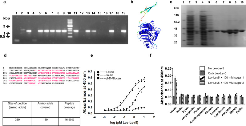
Fig. 5 Identification of a levan binding protein domain. a Agarose gel electrophoresis of PCR amplicons indicating the presence or absence of any recombinant insert following biopanning of the metagenomic phage display library against levan and elution with D-fructose or levan. Results of a single screening experiment. Lanes 1–9: PCR amplicons of clones Lev-Fru1 to Lev-Fru9. Lane 10: 1 kbp DNA marker. Lanes 11–19: PCR amplicons of clones Lev-Lev1 to Lev-Lev9. b AlphaFold2 predicted structural model of Lev-Lev5. c Coomassie blue stained SDS-PAGE showing purified Lev-Lev5 protein following Ni-NTA metal ion affinity chromatography. Lane 1: Protein molecular weight marker. Lane 2: Clarified lysate of E. coli BL21(DE3) expressing recombinant Lev-Lev5. Lane 3: Insoluble pellet fraction of E. coli BL21(DE3) expressing recombinant Lev-Lev5 following sonication. Lane 4: Flow through fraction during Ni-NTA metal ion affinity chromatography. Lane 5: 40 mM imidazole wash fraction during Ni-NTA metal ion affinity chromatography. Lanes 6–10: Purified Lev-Lev5 recombinant protein eluted with 250 mM imidazole during Ni-NTA metal ion affinity chromatography. d Sequence coverage obtained with MS/MS spectrometry of recombinant Lev-Lev5 protein followed by targeted sequence search; the peptide sequence coverage is shown in pink color. Results of a single mass spectrometry run. e Concentration dependent binding of Lev-Lev5 to immobilized levan, inulin and β-D-glucan. Results of a single binding experiment of two technical replicates. Individual values are plotted along with regression curves. Source data in Supplementary Data 3. f Binding of Lev-Lev5 to wells containing various immobilized carbohydrates with or without pre-incubation with component monosaccharide(s). The control used was wells without any immobilized carbohydrate. The polysaccharide carbohydrates with their monosaccharide units used in the assay are as follows. Levan: fructose. Inulin: D-glucose (sugar 1) and fructose (sugar 2). Xylan: xylose. Amylopectin: D-glucose. Pectin: D-galacturonic acid. Arabinogalactan: L-arabinose (sugar 1) and D-galactose (sugar 2). β-D-Glucan: D-glucose. Dextran: D-glucose. Laminarin: D-glucose. Amylose: D-glucose. Starch: D-glucose. Results of a single binding experiment of two technical replicates. Means are plotted as bars and individual data points included. Source data in Supplementary Data 3.
Fig. 5 Identification of a levan binding protein domain. a Agarose gel electrophoresis of PCR amplicons indicating the presence or absence of any recombinant insert following biopanning of the metagenomic phage display library against levan and elution with D-fructose or levan. Results of a single screening experiment. Lanes 1–9: PCR amplicons of clones Lev-Fru1 to Lev-Fru9. Lane 10: 1 kbp DNA marker. Lanes 11–19: PCR amplicons of clones Lev-Lev1 to Lev-Lev9. b AlphaFold2 predicted structural model of Lev-Lev5. c Coomassie blue stained SDS-PAGE showing purified Lev-Lev5 protein following Ni-NTA metal ion affinity chromatography. Lane 1: Protein molecular weight marker. Lane 2: Clarified lysate of E. coli BL21(DE3) expressing recombinant Lev-Lev5. Lane 3: Insoluble pellet fraction of E. coli BL21(DE3) expressing recombinant Lev-Lev5 following sonication. Lane 4: Flow through fraction during Ni-NTA metal ion affinity chromatography. Lane 5: 40 mM imidazole wash fraction during Ni-NTA metal ion affinity chromatography. Lanes 6–10: Purified Lev-Lev5 recombinant protein eluted with 250 mM imidazole during Ni-NTA metal ion affinity chromatography. d Sequence coverage obtained with MS/MS spectrometry of recombinant Lev-Lev5 protein followed by targeted sequence search; the peptide sequence coverage is shown in pink color. Results of a single mass spectrometry run. e Concentration dependent binding of Lev-Lev5 to immobilized levan, inulin and β-D-glucan. Results of a single binding experiment of two technical replicates. Individual values are plotted along with regression curves. Source data in Supplementary Data 3. f Binding of Lev-Lev5 to wells containing various immobilized carbohydrates with or without pre-incubation with component monosaccharide(s). The control used was wells without any immobilized carbohydrate. The polysaccharide carbohydrates with their monosaccharide units used in the assay are as follows. Levan: fructose. Inulin: D-glucose (sugar 1) and fructose (sugar 2). Xylan: xylose. Amylopectin: D-glucose. Pectin: D-galacturonic acid. Arabinogalactan: L-arabinose (sugar 1) and D-galactose (sugar 2). β-D-Glucan: D-glucose. Dextran: D-glucose. Laminarin: D-glucose. Amylose: D-glucose. Starch: D-glucose. Results of a single binding experiment of two technical replicates. Means are plotted as bars and individual data points included. Source data in Supplementary Data 3.CC BY · PubMed Central

Verification

Claims the paper makes

The claims and statistics of this paper haven’t been checked yet.

Run the check — free, no account needed. It reads the open-access text and checks the sources it cites.

A few free checks a day · papers already verified open instantly · results are public

Self-check

Percentages, sums and reported differences re-computed from the paper’s own numbers.

The self-check needs the paper’s full text.

Citations

Cites

Show all 1 references

Metadata

Authors
Akhtar, A., Lata, M., Sunsunwal, S., Yadav, A., LNU, K., subramanian, S., & Ramya, T. N. C.
Affiliations
  • Emory University (US)
  • Institute of Microbial Technology (IN)
  • CSIR-Institute of Microbial Technology
Corresponding author
Ramya, T.N.C.
Venue
Communications Biology
Published
2022-09-22
Volume
6
Issue
1
Pages
371-371
ISSN
2399-3642 (ISSN-L)
OpenAlex
W4362634303
Cited by
9
References
1
Citations by year
2026: 1 · 2025: 5 · 2023: 3
FWCI
0.80
Type
journal article
Language
en
Open access
Locations
  • Communications Biology (publishedVersion, cc-by)
  • Research Square (acceptedVersion, cc-by)
  • PubMed Central (publishedVersion, cc-by)
  • PubMed (publishedVersion)
  • DOAJ (DOAJ: Directory of Open Access Journals) (submittedVersion, cc-by-sa)
Funders
  • Department of Biotechnology, Ministry of Science and Technology, India
  • Council of Scientific and Industrial Research, India (BSC0119)
  • University Grants Commission
  • National Institute of General Medical Sciences (R24GM098791, P41GM103694)
  • CSIR - Institute of Microbial Technology
  • Brookhaven National Laboratory
Topics
  • Life Sciences › Biochemistry, Genetics and Molecular Biology › Molecular Biology › Gut microbiota and health
  • Life Sciences › Biochemistry, Genetics and Molecular Biology › Molecular Biology › Glycosylation and Glycoproteins Research
  • Life Sciences › Biochemistry, Genetics and Molecular Biology › Molecular Biology › Genomics and Phylogenetic Studies
Keywords
carbohydrate-binding domain, human gut microbiome, functional metagenomics, unannotated genes, polysaccharide binding, glycoconjugates, intestinal colonization, heterologous expression
MeSH terms
Gastrointestinal Microbiome, Bacteriophages, Carbohydrates, Humans, Genomics, Metagenomics

About these checks

Registry, retraction, Cites and statistics results are computed, not judged, with one exception: a printed reference that no index lists is read once by a model before it counts as a citation error. Claims are extracted by a model and kept only when their quote appears verbatim in the text; each cited source is then checked, with a model’s help, for whether it says what the claim relies on. Those verdicts can be wrong — read the claim’s quote and the cited source before relying on one.

Statistics and arithmetic are recomputed, not judged.

Confidence bands are shown only for fields calibrated on our annotated test set. None is calibrated yet, so no band is shown.

What counts as a claim ›How we measure confidence ›How we check this ›

Are you an author, or do you see a problem with this page? Request a review

How to cite

Cite CoChat for the checks on this page, and the paper for its own findings.

This page
CoChat. (2026). Citation check: New carbohydrate binding domains identified by phage display based functional metagenomic screens of human gut microbiota. https://app.cochat.ai/papers/10.21203/rs.3.rs-1898940/v1
The paper
Akhtar, A., Lata, M., Sunsunwal, S., Yadav, A., LNU, K., subramanian, S., & Ramya, T. N. C. (2022). New carbohydrate binding domains identified by phage display based functional metagenomic screens of human gut microbiota. Communications Biology. https://doi.org/10.21203/rs.3.rs-1898940/v1

Data sources

Bibliographic data: Semantic Scholar, OpenAlex, Crossref.

Contains information from the Semantic Scholar Academic Graph Datasets, made available under the ODC Attribution License (ODC-BY 1.0).

Semantic Scholar’s data is described in: Kinney, R., et al. (2023). The Semantic Scholar Open Data Platform. arXiv:2301.10140.