

The DOI matches its Crossref record; 1 of the 1 references it cites were found in a registry or an index.
Predicting Monoclonal Antibody Binding Sequences from a Sparse Sampling of All Possible Sequences
Open accessPreprint — published as 10.1038/s42003-024-06650-3 · Paper page
Abstract
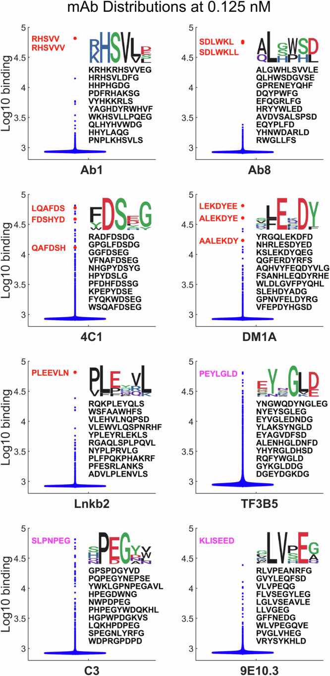
A general approach is described for creating a comprehensive and quantitative model for predicting the binding of a monoclonal antibody (mAb) to any short linear sequence of amino acids. Binding of 8 different mAbs with continuous epitopes to a common library of ~122,000 near-random peptide sequences arrayed on a silica substrate was used to train neural network models resulting in a general sequence-binding relationship for each mAb. The model was then used to predict mAb binding to 1,000,000 randomly generated sequences as well as the cognate sequence of each mAb. In every case the cognate sequence ranked in the top 250 sequences, and for 5 of the 8 mAbs it ranked in the top 10 out of one million. Practically, this approach has potential utility in selecting highly specific mAbs for therapeutics or diagnostics. Conceptually, this demonstrates that very sparse, but near random, sampling of a large sequence space is sufficient to generate comprehensive models predictive of highly specific molecular recognition.
Overview
The claims and statistics of this paper haven’t been checked yet.
Run the check — free, no account needed. It reads the open-access text and checks the sources it cites.
Integrity signals
Registry
Cites
Statistics
Figures


Verification
Claims the paper makes
The claims and statistics of this paper haven’t been checked yet.
Run the check — free, no account needed. It reads the open-access text and checks the sources it cites.
Self-check
Percentages, sums and reported differences re-computed from the paper’s own numbers.
The self-check needs the paper’s full text.
Citations
Cites
Highly accurate protein structure prediction with AlphaFold
Jumper, J., Evans, R., Pritzel, A., Green, T., Figurnov, M., Ronneberger, O., Tunyasuvunakool, K., Bates, R., Žídek, A., Potapenko, A., et al. (2021) · Nature · doi:10.1038/s41586-021-03819-2 · Cited by 43,764
Metadata
- Authors
- Woodbury, N., Bisarad, P., Kelbauskas, L., Singh, A., & Taguchi, A.
- Affiliations
- Arizona State University (US)
- Barrow Neurological Institute (US)
- iBio Inc.
- Corresponding author
- Woodbury, Neal
- Venue
- Communications Biology
- Published
- 2023-12-06
- Volume
- 7
- Issue
- 1
- Pages
- 979-979
- ISSN
- 2399-3642 (ISSN-L)
- PMC
- PMC11319732
- OpenAlex
- W4401510896
- Cited by
- 5
- References
- 1
- Citations by year
- 2026: 1 · 2025: 2 · 2024: 2
- FWCI
- 0.68
- Type
- journal article
- Language
- en
- Open access
- gold
- doi.org
- Locations
- Communications Biology (publishedVersion, cc-by)
- Research Square (acceptedVersion, cc-by)
- PubMed Central (publishedVersion, cc-by)
- PubMed (publishedVersion)
- DOAJ (DOAJ: Directory of Open Access Journals) (submittedVersion)
- Funders
- Arizona State University
- Topics
- Life Sciences › Biochemistry, Genetics and Molecular Biology › Molecular Biology › vaccines and immunoinformatics approaches
- Life Sciences › Immunology and Microbiology › Immunology › Monoclonal and Polyclonal Antibodies Research
- Physical Sciences › Chemistry › Organic Chemistry › Chemical Synthesis and Analysis
- Keywords
- monoclonal antibodies, epitope prediction, network models, binding prediction, linear epitopes
- MeSH terms
- Machine Learning, Amino Acid Sequence, Antibodies, Monoclonal, Epitopes, Binding Sites, Antibody, Computer Simulation, Humans, Protein Binding
About these checks
Registry, retraction, Cites and statistics results are computed, not judged, with one exception: a printed reference that no index lists is read once by a model before it counts as a citation error. Claims are extracted by a model and kept only when their quote appears verbatim in the text; each cited source is then checked, with a model’s help, for whether it says what the claim relies on. Those verdicts can be wrong — read the claim’s quote and the cited source before relying on one.
Statistics and arithmetic are recomputed, not judged.
Confidence bands are shown only for fields calibrated on our annotated test set. None is calibrated yet, so no band is shown.
What counts as a claim ›How we measure confidence ›How we check this ›
Are you an author, or do you see a problem with this page? Request a review
How to cite
Cite CoChat for the checks on this page, and the paper for its own findings.
- This page
- CoChat. (2026). Citation check: Predicting Monoclonal Antibody Binding Sequences from a Sparse Sampling of All Possible Sequences. https://app.cochat.ai/papers/10.21203/rs.3.rs-3560097/v1
- The paper
- Woodbury, N., Bisarad, P., Kelbauskas, L., Singh, A., & Taguchi, A. (2023). Predicting Monoclonal Antibody Binding Sequences from a Sparse Sampling of All Possible Sequences. Communications Biology. https://doi.org/10.21203/rs.3.rs-3560097/v1
Data sources
Bibliographic data: Semantic Scholar, OpenAlex, Crossref.
Contains information from the Semantic Scholar Academic Graph Datasets, made available under the ODC Attribution License (ODC-BY 1.0).
Semantic Scholar’s data is described in: Kinney, R., et al. (2023). The Semantic Scholar Open Data Platform. arXiv:2301.10140.
