

The DOI matches its Crossref record; 1 of the 1 references it cites were found in a registry or an index.
Caveolin-1 Knockout Mitigates Breast Cancer Metastasis to the Lungs via Integrin α3 Dysregulation.
Open accessPreprint — published as 10.1038/s41417-024-00821-4 · Paper page
Abstract
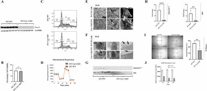
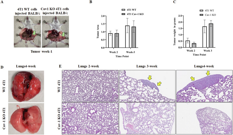
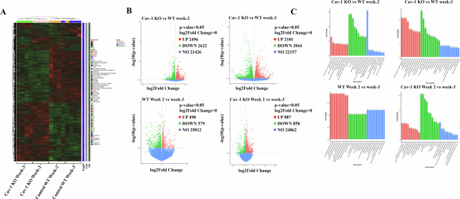
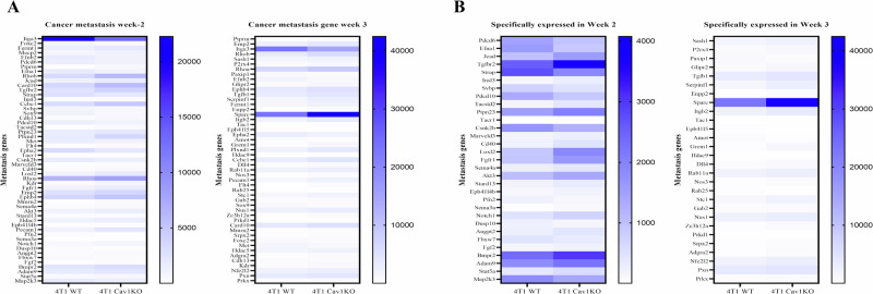
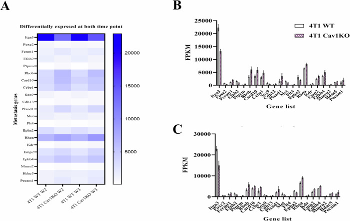
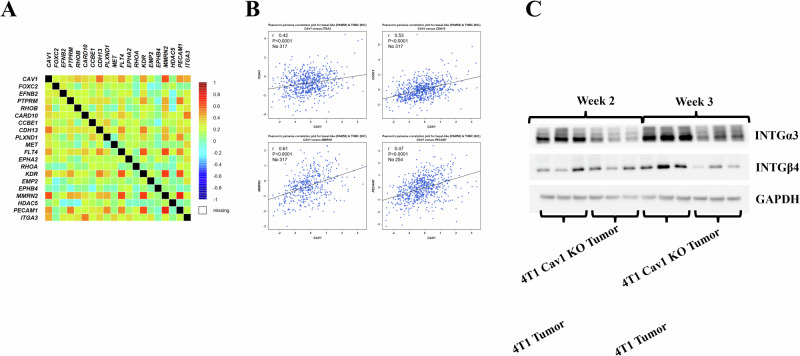
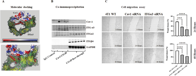
Caveolin-1 (Cav-1) is a critical lipid raft protein playing divergent roles as both a tumor suppressor and promoter. While its role in tumorigenesis, progression, and metastasis is recognized, explicit contribution of Cav-1 to the onset of lung metastasis from primary breast malignancies remained vague. We exhibit here, the first-ever evidence of Cav-1 knockout in epithelial cells leading to a significant reduction in lung metastasis in syngeneic mouse models of breast cancer. In vitro, Cav-1 knockout in 4T1 cells suppressed extracellular vesicles secretion, cellular motility, and MMP secretion vis-à-vis the controls. Complementing this, our in vivo analyses demonstrated a marked reduction in lung metastatic foci in mice injected with Cav-1 knockout 4T1 cells as compared to wild-type mice, which was further corroborated by mRNA profiling of the primary tumor. We discerned 21 epithelial cell migration genes exhibiting varied expression in tumors derived from Cav-1 knockout versus wild-type 4T1 cells. Correlation analysis and immunoblotting further divulged that Cav-1 regulates metastasis via integrin α3 (ITGα3). In silico protein docking anticipated an interaction between Cav-1 and ITGα3, substantiated by co-immunoprecipitation. Furthermore, ITGα3 knockdown corroborated its role in metastasis in a cell migration assay. Therefore, it can be inferred that Cav-1 plays a pivotal role in the pre-metastatic niche establishment and facilitates lung-specific cell migration.
Overview
The claims and statistics of this paper haven’t been checked yet.
Run the check — free, no account needed. It reads the open-access text and checks the sources it cites.
Integrity signals
Registry
Cites
Statistics
Figures
Verification
Claims the paper makes
The claims and statistics of this paper haven’t been checked yet.
Run the check — free, no account needed. It reads the open-access text and checks the sources it cites.
Self-check
Percentages, sums and reported differences re-computed from the paper’s own numbers.
The self-check needs the paper’s full text.
Citations
Cites
Highly accurate protein structure prediction with AlphaFold
Jumper, J., Evans, R., Pritzel, A., Green, T., Figurnov, M., Ronneberger, O., Tunyasuvunakool, K., Bates, R., Žídek, A., Potapenko, A., et al. (2021) · Nature · doi:10.1038/s41586-021-03819-2 · Cited by 43,764
Research works printed but not linked: 62
- Siegel RL, Miller KD, Fuchs HE, Jemal A: Cancer statistics, 2022. CA Cancer J Clin 2022, 72(1):7–33. · DOI · Paper page
- Cava E, Marzullo P, Farinelli D, Gennari A, Saggia C, Riso S, Prodam F: Breast Cancer Diet "BCD": A Review of Healthy Dietary Patterns to Prevent Breast Cancer Recurrence and Reduce Mortality. Nutrients 2022, 14(3):476. · DOI
- Schick J, Ritchie RP, Restini C: Breast Cancer Therapeutics and Biomarkers: Past, Present, and Future Approaches. Breast Cancer (Auckl) 2021, 15:1178223421995854. · DOI
- Fares J, Fares MY, Khachfe HH, Salhab HA, Fares Y: Molecular principles of metastasis: a hallmark of cancer revisited. Signal Transduct Target Ther 2020, 5(1):28. · DOI
- Dillekas H, Rogers MS, Straume O: Are 90% of deaths from cancer caused by metastases? Cancer Med 2019, 8(12):5574–5576. · DOI
- Chen W, Hoffmann AD, Liu H, Liu X: Organotropism: new insights into molecular mechanisms of breast cancer metastasis. NPJ precision oncology 2018, 2(1):1–12. · DOI
- Huang QY, Liu GF, Qian XL, Tang LB, Huang QY, Xiong LX: Long Non-Coding RNA: Dual Effects on Breast Cancer Metastasis and Clinical Applications. Cancers (Basel) 2019, 11(11). · DOI
- Luo H, Tu G, Liu Z, Liu M: Cancer-associated fibroblasts: a multifaceted driver of breast cancer progression. Cancer Lett 2015, 361(2):155–163. · DOI
- Guo Y, Ji X, Liu J, Fan D, Zhou Q, Chen C, Wang W, Wang G, Wang H, Yuan W et al: Effects of exosomes on pre-metastatic niche formation in tumors. Mol Cancer 2019, 18(1):39. · DOI
- Eric D. Young LL, Fariba Behbod, Danny R. Welch: Fundamental Relationships Between Cancer Stem Cells, the Cancer Stem Cell Niche and Metastasis, vol. 3; 2016.
- Wani NA: Abstract 1166: CXCR7-mediated signaling axis regulates breast cancer growth and metastasis by modulating tumor microenvironment. Cancer Research 2014, 74(19_Supplement):1166–1166. · DOI
- Min W: Abstract 129: AIP1 suppresses tumor metastasis by regulating tumor microenvironment and metastatic niche. Cancer Research 2014, 74(19_Supplement):129–129. · DOI
- Brena D, Huang MB, Bond V: Extracellular vesicle-mediated transport: Reprogramming a tumor microenvironment conducive with breast cancer progression and metastasis. Transl Oncol 2022, 15(1):101286. · DOI
- Qian XL, Pan YH, Huang QY, Shi YB, Huang QY, Hu ZZ, Xiong LX: Caveolin-1: a multifaceted driver of breast cancer progression and its application in clinical treatment. Onco Targets Ther 2019, 12:1539–1552. · DOI
- Ketteler J, Klein D: Caveolin-1, cancer and therapy resistance. Int J Cancer 2018, 143(9):2092–2104. · DOI
- Diaz-Valdivia NI, Calderon CC, Diaz JE, Lobos-Gonzalez L, Sepulveda H, Ortiz RJ, Martinez S, Silva V, Maldonado HJ, Silva P et al: Anti-neoplastic drugs increase caveolin-1-dependent migration, invasion and metastasis of cancer cells. Oncotarget 2017, 8(67):111943–111965. · DOI
- Karam JA, Lotan Y, Roehrborn CG, Ashfaq R, Karakiewicz PI, Shariat SF: Caveolin-1 overexpression is associated with aggressive prostate cancer recurrence. The Prostate 2007, 67(6):614–622. · DOI
- Low JY, Laiho M: Caveolae-Associated Molecules, Tumor Stroma, and Cancer Drug Resistance: Current Findings and Future Perspectives. Cancers (Basel) 2022, 14(3):589. · DOI
- Martinez-Outschoorn UE, Pavlides S, Whitaker-Menezes D, Daumer KM, Milliman JN, Chiavarina B, Migneco G, Witkiewicz AK, Martinez-Cantarin MP, Flomenberg N et al: Tumor cells induce the cancer associated fibroblast phenotype via caveolin-1 degradation: implications for breast cancer and DCIS therapy with autophagy inhibitors. Cell Cycle 2010, 9(12):2423–2433. · DOI
- Du C, Chen L, Zhang H, Wang Z, Liu W, Xie X, Xie M: Caveolin-1 limits the contribution of BKCa channel to MCF-7 breast cancer cell proliferation and invasion. Int J Mol Sci 2014, 15(11):20706–20722. · DOI
- Godina C, Chandran VI, Barbachowska M, Tryggvadottir H, Nodin B, Visse E, Borgquist S, Jirström K, Isaksson K, Bosch A: Interplay between Caveolin-1 and body and tumor size affects clinical outcomes in breast cancer. Translational Oncology 2022, 22:101464. · DOI
- Sotgia F, Martinez-Outschoorn UE, Howell A, Pestell RG, Pavlides S, Lisanti MP: Caveolin-1 and cancer metabolism in the tumor microenvironment: markers, models, and mechanisms. Annu Rev Pathol 2012, 7:423–467. · DOI
- Shi Y-B, Li J, Lai X-N, Jiang R, Zhao R-C, Xiong L-X: Multifaceted Roles of Caveolin-1 in Lung Cancer: A New Investigation Focused on Tumor Occurrence, Development and Therapy. In: Cancers. vol. 12; 2020.
- Singh DD, Lee H-J, Yadav DK: Recent Clinical Advances on Long Non-Coding RNAs in Triple-Negative Breast Cancer. In: Cells. vol. 12; 2023. · DOI
- Wang Y, Li Y, Zhong J, Li M, Zhou Y, Lin Q, Zong S, Luo W, Wang J, Wang K et al: Tumor-derived Cav-1 promotes pre-metastatic niche formation and lung metastasis in breast cancer. Theranostics 2023, 13(5):1684–1697. · DOI
- Yang H, Guan L, Li S, Jiang Y, Xiong N, Li L, Wu C, Zeng H, Liu Y: Mechanosensitive caveolin-1 activation-induced PI3K/Akt/mTOR signaling pathway promotes breast cancer motility, invadopodia formation and metastasis in vivo. Oncotarget 2016, 7(13):16227–16247. · DOI
- Godina C, Tryggvadottir H, Bosch A, Borgquist S, Belting M, Isaksson K, Jernstrom H: Caveolin-1 genotypes as predictor for locoregional recurrence and contralateral disease in breast cancer. Breast Cancer Res Treat 2023, 199(2):335–347. · DOI
- Sloan EK, Stanley KL, Anderson RL: Caveolin-1 inhibits breast cancer growth and metastasis. Oncogene 2004, 23(47):7893–7897. · DOI
- Burrows AD, Restall C, Sloan EK, Anderson RL: Abstract 1486: The contribution of stromal caveolin-1 to breast cancer metastasis. Cancer Research 2012, 72(8_Supplement):1486–1486. · DOI
- Zhang Q, Zhang S, Chen J, Xie Z: The Interplay between Integrins and Immune Cells as a Regulator in Cancer Immunology. Int J Mol Sci 2023, 24(7). · DOI
- Au - Hu X, Au - Villodre ES, Au - Woodward WA, Au - Debeb BG: Modeling Brain Metastasis Via Tail-Vein Injection of Inflammatory Breast Cancer Cells. JoVE 2021(168):e62249.
- Au - Thies KA, Au - Steck S, Au - Knoblaugh SE, Au - Sizemore ST: Pathological Analysis of Lung Metastasis Following Lateral Tail-Vein Injection of Tumor Cells. JoVE 2020(159):e61270.
- Laskowski RA, Jablonska J, Pravda L, Varekova RS, Thornton JM: PDBsum: Structural summaries of PDB entries. Protein Sci 2018, 27(1):129–134. · DOI
- Rose PW, Prlic A, Altunkaya A, Bi C, Bradley AR, Christie CH, Costanzo LD, Duarte JM, Dutta S, Feng Z et al: The RCSB protein data bank: integrative view of protein, gene and 3D structural information. Nucleic Acids Res 2017, 45(D1):D271-D281.
- Pierce BG, Wiehe K, Hwang H, Kim BH, Vreven T, Weng Z: ZDOCK server: interactive docking prediction of protein-protein complexes and symmetric multimers. Bioinformatics 2014, 30(12):1771–1773. · DOI
- Singh DP, Begum R, Kaur G, Bagam P, Kambiranda D, Singh R, Batra S: E-cig vapor condensate alters proteome and lipid profiles of membrane rafts: impact on inflammatory responses in A549 cells. Cell Biol Toxicol 2021, 37(5):773–793. · DOI
- Wu PH, Gilkes DM, Phillip JM, Narkar A, Cheng TW, Marchand J, Lee MH, Li R, Wirtz D: Single-cell morphology encodes metastatic potential. Sci Adv 2020, 6(4):eaaw6938. · DOI · Paper page
- Kleiner DE, Stetler-Stevenson WG: Matrix metalloproteinases and metastasis. Cancer Chemother Pharmacol 1999, 43 Suppl(1):S42-51.
- Lechuga S, Amin PH, Wolen AR, Ivanov AI: Adducins inhibit lung cancer cell migration through mechanisms involving regulation of cell-matrix adhesion and cadherin-11 expression. Biochimica et Biophysica Acta (BBA)-Molecular Cell Research 2019, 1866(3):395–408. · DOI
- Yu G, Wang L-G, Han Y, He Q-Y: clusterProfiler: an R package for comparing biological themes among gene clusters. Omics: a journal of integrative biology 2012, 16(5):284–287. · DOI · Paper page
- Pijuan J, Barcelo C, Moreno DF, Maiques O, Siso P, Marti RM, Macia A, Panosa A: In vitro Cell Migration, Invasion, and Adhesion Assays: From Cell Imaging to Data Analysis. Front Cell Dev Biol 2019, 7:107. · DOI
- Lee H, Carnino JM, Jin Y: Caveolin-1 regulates extracellular vesicle-miRNA packaging. Aging (Albany NY) 2019, 11(20):8733–8735. · DOI
- Rothberg KG, Heuser JE, Donzell WC, Ying YS, Glenney JR, Anderson RG: Caveolin, a protein component of caveolae membrane coats. Cell 1992, 68(4):673–682. · DOI
- Kogure A, Yoshioka Y, Ochiya T: Extracellular vesicles in cancer metastasis: Potential as therapeutic targets and materials. International Journal of Molecular Sciences 2020, 21(12):4463. · DOI
- Yamaguchi H, Takeo Y, Yoshida S, Kouchi Z, Nakamura Y, Fukami K: Lipid rafts and caveolin-1 are required for invadopodia formation and extracellular matrix degradation by human breast cancer cells. Cancer Res 2009, 69(22):8594–8602. · DOI
- Gupta VK, Sharma NS, Kesh K, Dauer P, Nomura A, Giri B, Dudeja V, Banerjee S, Bhattacharya S, Saluja A: Metastasis and chemoresistance in CD133 expressing pancreatic cancer cells are dependent on their lipid raft integrity. Cancer letters 2018, 439:101–112. · DOI
- Yu H, Shen H, Zhang Y, Zhong F, Liu Y, Qin L, Yang P: CAV1 promotes HCC cell progression and metastasis through Wnt/β-catenin pathway. PLoS One 2014, 9(9):e106451. · DOI
- Yokoi A, Yoshioka Y, Yamamoto Y, Ishikawa M, Ikeda S-i, Kato T, Kiyono T, Takeshita F, Kajiyama H, Kikkawa F et al: Malignant extracellular vesicles carrying MMP1 mRNA facilitate peritoneal dissemination in ovarian cancer. Nature Communications 2017, 8(1):14470. · DOI
- Su JL, Yang PC, Shih JY, Yang CY, Wei LH, Hsieh CY, Chou CH, Jeng YM, Wang MY, Chang KJ et al: The VEGF-C/Flt-4 axis promotes invasion and metastasis of cancer cells. Cancer Cell 2006, 9(3):209–223. · DOI
- Modi SJ, Kulkarni VM: Vascular endothelial growth factor receptor (VEGFR-2)/KDR inhibitors: medicinal chemistry perspective. Medicine in Drug Discovery 2019, 2:100009. · DOI
- Ye M, Huang T, Li J, Zhou C, Yang P, Ni C, Chen S: Role of CDH13 promoter methylation in the carcinogenesis, progression, and prognosis of colorectal cancer: A systematic meta-analysis under PRISMA guidelines. Medicine (Baltimore) 2017, 96(4):e5956. · DOI
- Andreeva AV, Kutuzov MA: Cadherin 13 in cancer. Genes Chromosomes Cancer 2010, 49(9):775–790. · DOI
- Lorenzon E, Colladel R, Andreuzzi E, Marastoni S, Todaro F, Schiappacassi M, Ligresti G, Colombatti A, Mongiat M: MULTIMERIN2 impairs tumor angiogenesis and growth by interfering with VEGF-A/VEGFR2 pathway. Oncogene 2012, 31(26):3136–3147. · DOI
- Kurozumi A, Goto Y, Matsushita R, Fukumoto I, Kato M, Nishikawa R, Sakamoto S, Enokida H, Nakagawa M, Ichikawa T: Tumor-suppressive micro RNA‐223 inhibits cancer cell migration and invasion by targeting ITGA 3/ITGB 1 signaling in prostate cancer. Cancer science 2016, 107(1):84–94. · DOI
- Idichi T, Seki N, Kurahara H, Fukuhisa H, Toda H, Shimonosono M, Yamada Y, Arai T, Kita Y, Kijima Y et al: Involvement of anti-tumor miR-124-3p and its targets in the pathogenesis of pancreatic ductal adenocarcinoma: direct regulation of ITGA3 and ITGB1 by miR-124-3p. Oncotarget 2018, 9(48):28849–28865. · DOI
- Koshizuka K, Hanazawa T, Kikkawa N, Arai T, Okato A, Kurozumi A, Kato M, Katada K, Okamoto Y, Seki N: Regulation of ITGA 3 by the anti-tumor miR‐199 family inhibits cancer cell migration and invasion in head and neck cancer. Cancer science 2017, 108(8):1681–1692. · DOI
- Munkácsy G, Santarpia L, Győrffy B: Therapeutic Potential of Tumor Metabolic Reprogramming in Triple-Negative Breast Cancer. In: International Journal of Molecular Sciences. vol. 24; 2023. · DOI
- Robb R, Kuo JC, Liu Y, Corrales-Guerrero S, Cui T, Hegazi A, Nagy G, Lee RJ, Williams TM: A novel protein-drug conjugate, SSH20, demonstrates significant efficacy in caveolin-1-expressing tumors. Mol Ther Oncolytics 2021, 22:555–564. · DOI
- Khongorzul P, Ling CJ, Khan FU, Ihsan AU, Zhang J: Antibody-Drug Conjugates: A Comprehensive Review. Mol Cancer Res 2020, 18(1):3–19. · DOI
- Pereira PMR, Mandleywala K, Monette S, Lumish M, Tully KM, Panikar SS, Cornejo M, Mauguen A, Ragupathi A, Keltee NC et al: Caveolin-1 temporal modulation enhances antibody drug efficacy in heterogeneous gastric cancer. Nat Commun 2022, 13(1):2526. · DOI
- Desgrosellier JS, Cheresh DA: Integrins in cancer: biological implications and therapeutic opportunities. Nat Rev Cancer 2010, 10(1):9–22. · DOI
- Hegazy M, Cohen-Barak E, Koetsier JL, Najor NA, Arvanitis C, Sprecher E, Green KJ, Godsel LM: Proximity Ligation Assay for Detecting Protein-Protein Interactions and Protein Modifications in Cells and Tissues in Situ. Curr Protoc Cell Biol 2020, 89(1):e115. · DOI
Metadata
- Authors
- Francis, J., Singh, D., Pathak, R., Pandit, A., Ebenezer, P., Kumar, S., Duplooy, A., White, M., Jambunathan, N., Chintalaramulu, N., & Dharmakumar, R.
- Affiliations
- Louisiana State University (US)
- Indiana University School of Medicine (US)
- Midwestern University (US)
- Corresponding author
- Francis, Joseph
- Venue
- Cancer Gene Therapy
- Published
- 2023-12-12
- Volume
- 31
- Issue
- 11
- Pages
- 1658-1668
- ISSN
- 0929-1903 (ISSN-L)
- PMC
- PMC11567888
- OpenAlex
- W4402331411
- Cited by
- 12
- References
- 1
- Citations by year
- 2026: 5 · 2025: 5 · 2024: 2
- FWCI
- 2.68
- Type
- journal article
- Language
- en
- Open access
- hybrid
- doi.org
- Locations
- Cancer Gene Therapy (publishedVersion, cc-by)
- Research Square (acceptedVersion, cc-by)
- PubMed Central (publishedVersion, cc-by)
- PubMed (publishedVersion)
- IUScholarWorks (Indiana University) (submittedVersion, cc-by)
- Funders
- National Institute of General Medical Sciences (P20GM135000)
- Topics
- Life Sciences › Biochemistry, Genetics and Molecular Biology › Cell Biology › Caveolin-1 and cellular processes
- Life Sciences › Biochemistry, Genetics and Molecular Biology › Molecular Biology › Mechanisms of cancer metastasis
- Health Sciences › Medicine › Oncology › Cancer Cells and Metastasis
- Keywords
- caveolin-1, breast cancer lung metastasis, integrin alpha3, 4T1 cells, extracellular vesicles, cell migration, matrix metalloproteinases, lipid rafts
- MeSH terms
- Animals, Breast Neoplasms, Cell Movement, Disease Models, Animal, Female, Humans, Lung Neoplasms, Mice, Inbred BALB C, Gene Expression Regulation, Neoplastic, Mice, Knockout, Integrin alpha3, Cell Line, Tumor, Caveolin 1, Mice, Gene Knockout Techniques
About these checks
Registry, retraction, Cites and statistics results are computed, not judged, with one exception: a printed reference that no index lists is read once by a model before it counts as a citation error. Claims are extracted by a model and kept only when their quote appears verbatim in the text; each cited source is then checked, with a model’s help, for whether it says what the claim relies on. Those verdicts can be wrong — read the claim’s quote and the cited source before relying on one.
Statistics and arithmetic are recomputed, not judged.
Confidence bands are shown only for fields calibrated on our annotated test set. None is calibrated yet, so no band is shown.
What counts as a claim ›How we measure confidence ›How we check this ›
Are you an author, or do you see a problem with this page? Request a review
