PreprintCited by 12Chromosomal and Genetic VariationsDNA Repair MechanismsPlant Molecular Biology Research

The DOI matches its Crossref record; 2 of the 2 references it cites were found in a registry or an index.

The novel synaptonemal complex central element SCEP3 interlinks synapsis initiation and crossover formation in Arabidopsis thaliana

Open access
Heckmann, Stefan, Feng, Chao, Lorenz, Jana, Dreissig, Steven, Schubert, Veit, Wang, Baicui, Hartmann, Franziska, Cuacos, Maria, Fernández-Jiménez, Nadia, Câmara, Amanda Souza, Himmelbach, Axel·2024·Nature Plants

Preprint — published as 10.1038/s41477-025-02030-9

Abstract

The proteinaceous synaptonemal complex (SC) structure forms between meiotic homologous chromosomes. Its central region (CR) consists of transverse filament and central element proteins, in Arabidopsis ZYP1 and SCEP1/SCEP2, respectively. We describe a novel CR protein in Arabidopsis. SCEP3 spatiotemporally overlaps with other CR components and is conserved in plants. In scep3, SC formation, crossover (CO) assurance (minimum one CO per chromosome pair), CO interference (limited closely-spaced CO) and heterochiasmy (male/female CO rate difference) vanish while genome-wide and particularly female CO increase. Compared with other CR proteins, SCEP3 is also critical for some synapsis-independent CO. SCEP3 interacts with ZYP1 but loads onto recombination intermediates independent of other CR proteins. We propose SCEP3’s loading onto recombination intermediates may stabilize and/or recruit further factors such as ZYP1 to a subset of these intermediates designated to form CO. Hence, SCEP3 interlinks SC and CO formation, being structurally likely the plant ortholog of yeast Ecm11.

Overview

The claims and statistics of this paper haven’t been checked yet.

Run the check — free, no account needed. It reads the open-access text and checks the sources it cites.

A few free checks a day · papers already verified open instantly · results are public

Integrity signals

Registry

Crossref · 10.21203/rs.3.rs-5394998/v1

Cites

2 of 2 references verified0 citation errors · 0 not checkable

Statistics

Figures

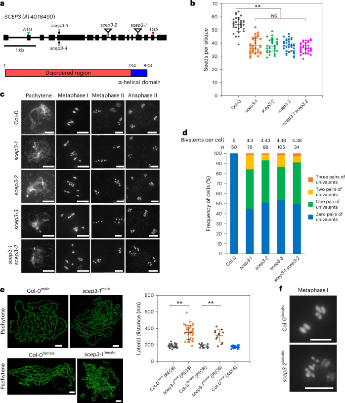
Fig. 1 Identification of SCEP3 and phenotypic analysis of scep3 mutants. a, Gene model of SCEP3 (AT4G18490; confirmed by Sanger sequencing of flower bud complementary DNA), including exons (black boxes) and introns (black lines), and a schematic depiction of the SCEP3 protein. The locations of mutant alleles are indicated: scep3-1 (initially named atc21-1 (ref. 44)) and scep3-2 (transfer DNA insertions within exon 13 and intron 8, respectively) as well as scep3-3 and scep3-4 (CRISPR–Cas9-based mutagenesis); scep3-4 is in Ler-0, while all other alleles are in the Col-0 background. b, Seeds per silique in the WT (54.33 ± 5.38, n = 40), scep3-1 (37.98 ± 5.76, n = 40), scep3-2 (37.9 ± 4.75, n = 40), scep3-3 (38.05 ± 4.44, n = 40) and scep3-1 scep3-2 (36.33 ± 3.59, n = 40). No significant differences were found among the scep3 mutants (P = 0.30). However, all scep3 mutants produced significantly fewer seeds than the WT (P < 1 × 10⁻⁷). c, Male meiotic chromosome behaviour (scale bars, 10 μm; DNA counterstained with DAPI is shown in grey) in the WT and scep3 mutants. d, Frequency of cells with zero to three pairs of univalents, including the average bivalent number per cell (n is the number of cells analysed) in the WT and scep3 mutants. e, Left, 3D-SIM analysis of REC8 immunolocalization in male and female meiocytes of Col-0 and scep3-1. Along synapsed chromosomes, the two parallel lateral elements exhibit an average distance of 188 ± 21.6 nm (range, 148–243 nm; n = 26) in WT males and 187 ± 19.4 nm (range, 162–223 nm; n = 15) in WT females. In scep3-1, within regions of alignment, the average distance increased to 352 ± 87.5 nm (range, 189–592 nm; n = 37) in males and 311 ± 74.6 nm (range, 184–423 nm; n = 12) in females, showing greater variation. Single-slice images were used to measure the distances between two aligned axes. Scale bars, 2 μm. Right, quantification of aligned axis distances (the measurement points were randomly selected) in both Col-0 and scep3-1. ASY4-labelled lateral elements exhibit an average distance of 176 ± 13.4 nm (range, 149–197 nm; n = 28) in WT males. f, Female meiotic chromosome spreads of Col-0 and scep3-2 (DAPI-stained DNA is shown in grey). The experiments were repeated three times with similar results. Scale bars, 10 μm. Distinct plants from each mutant line (or the WT) were used for seed counting and chromosome spread analysis. Significance was evaluated using one-way ANOVA with post hoc Bonferroni multiple comparison. The data are presented as mean ± s.d. **P < 0.01. NS, not significant. Source data
Fig. 1 Identification of SCEP3 and phenotypic analysis of scep3 mutants. a, Gene model of SCEP3 (AT4G18490; confirmed by Sanger sequencing of flower bud complementary DNA), including exons (black boxes) and introns (black lines), and a schematic depiction of the SCEP3 protein. The locations of mutant alleles are indicated: scep3-1 (initially named atc21-1 (ref. 44)) and scep3-2 (transfer DNA insertions within exon 13 and intron 8, respectively) as well as scep3-3 and scep3-4 (CRISPR–Cas9-based mutagenesis); scep3-4 is in Ler-0, while all other alleles are in the Col-0 background. b, Seeds per silique in the WT (54.33 ± 5.38, n = 40), scep3-1 (37.98 ± 5.76, n = 40), scep3-2 (37.9 ± 4.75, n = 40), scep3-3 (38.05 ± 4.44, n = 40) and scep3-1 scep3-2 (36.33 ± 3.59, n = 40). No significant differences were found among the scep3 mutants (P = 0.30). However, all scep3 mutants produced significantly fewer seeds than the WT (P < 1 × 10⁻⁷). c, Male meiotic chromosome behaviour (scale bars, 10 μm; DNA counterstained with DAPI is shown in grey) in the WT and scep3 mutants. d, Frequency of cells with zero to three pairs of univalents, including the average bivalent number per cell (n is the number of cells analysed) in the WT and scep3 mutants. e, Left, 3D-SIM analysis of REC8 immunolocalization in male and female meiocytes of Col-0 and scep3-1. Along synapsed chromosomes, the two parallel lateral elements exhibit an average distance of 188 ± 21.6 nm (range, 148–243 nm; n = 26) in WT males and 187 ± 19.4 nm (range, 162–223 nm; n = 15) in WT females. In scep3-1, within regions of alignment, the average distance increased to 352 ± 87.5 nm (range, 189–592 nm; n = 37) in males and 311 ± 74.6 nm (range, 184–423 nm; n = 12) in females, showing greater variation. Single-slice images were used to measure the distances between two aligned axes. Scale bars, 2 μm. Right, quantification of aligned axis distances (the measurement points were randomly selected) in both Col-0 and scep3-1. ASY4-labelled lateral elements exhibit an average distance of 176 ± 13.4 nm (range, 149–197 nm; n = 28) in WT males. f, Female meiotic chromosome spreads of Col-0 and scep3-2 (DAPI-stained DNA is shown in grey). The experiments were repeated three times with similar results. Scale bars, 10 μm. Distinct plants from each mutant line (or the WT) were used for seed counting and chromosome spread analysis. Significance was evaluated using one-way ANOVA with post hoc Bonferroni multiple comparison. The data are presented as mean ± s.d. **P < 0.01. NS, not significant. Source dataCC BY · PubMed Central
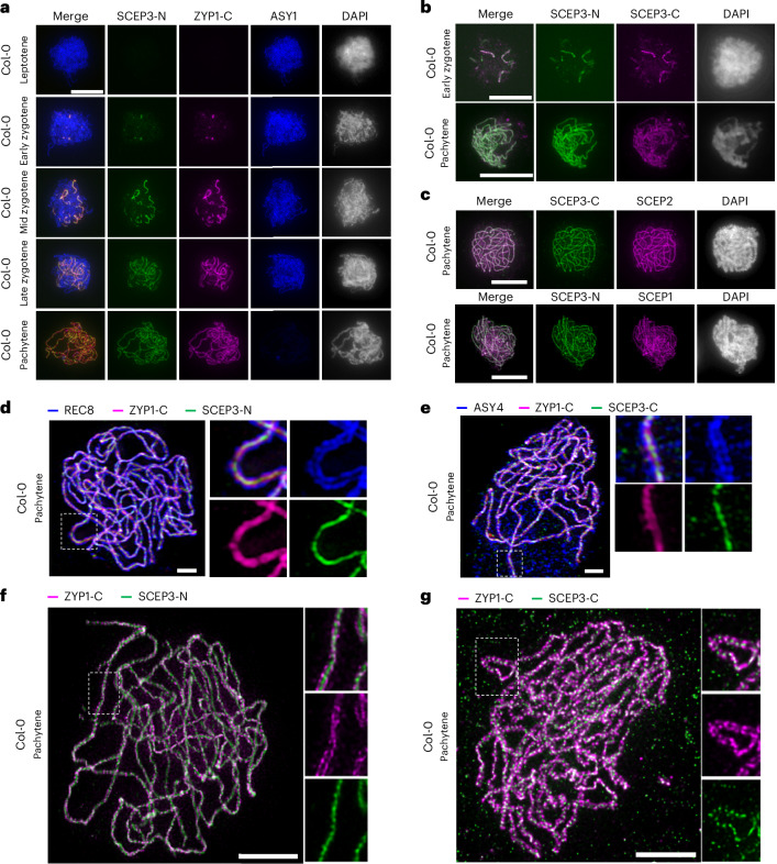
Fig. 2 Localization of SCEP3 at the CR of the SC. a–c, Immunolocalization in the WT of SCEP3-N, ZYP1-C and ASY1 during prophase I (a); SCEP3-C and SCEP3-N during zygotene and pachytene (b); and SCEP3-C with SCEP2 or SCEP3-N with SCEP1 at pachytene (c). DAPI-stained DNA is shown in grey. d,e, 3D-SIM of a pachytene nucleus in the WT immunolabelled with REC8, ZYP1-C and SCEP3-N (d) or ASY4, ZYP1-C and SCEP3-C (e). f,g, STED microscopy of a WT pachytene nucleus immunolabelled with ZYP1-C and SCEP3-N (f) or ZYP1-C and SCEP3-C (g). Scale bars, 10 μm in a–c, 2 μm in d–g. All experiments were repeated at least two times with similar results. Source data
Fig. 2 Localization of SCEP3 at the CR of the SC. a–c, Immunolocalization in the WT of SCEP3-N, ZYP1-C and ASY1 during prophase I (a); SCEP3-C and SCEP3-N during zygotene and pachytene (b); and SCEP3-C with SCEP2 or SCEP3-N with SCEP1 at pachytene (c). DAPI-stained DNA is shown in grey. d,e, 3D-SIM of a pachytene nucleus in the WT immunolabelled with REC8, ZYP1-C and SCEP3-N (d) or ASY4, ZYP1-C and SCEP3-C (e). f,g, STED microscopy of a WT pachytene nucleus immunolabelled with ZYP1-C and SCEP3-N (f) or ZYP1-C and SCEP3-C (g). Scale bars, 10 μm in a–c, 2 μm in d–g. All experiments were repeated at least two times with similar results. Source dataCC BY · PubMed Central
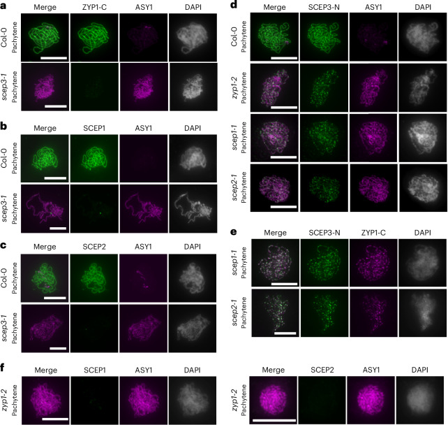
Fig. 3 SCEP3 is critical for SC assembly, and its localization is independent of other CR proteins. a–c, Immunolocalization in WT and scep3-1 pachytene nuclei of ZYP1-C and ASY1 (a), SCEP1 and ASY1 (b), and SCEP2 and ASY1 (c). d–f, Immunolocalization of SCEP3-N and ASY1 in the WT, zyp1-2, scep1-1 and scep2-1 (d); SCEP3-N and ZYP1-C in scep1-1 and scep2-1 (e); and SCEP1 or SCEP2 and ASY1 in zyp1-2 (f). DAPI-stained DNA is shown in grey. All experiments were repeated three times with similar results. Scale bars, 10 µm.
Fig. 3 SCEP3 is critical for SC assembly, and its localization is independent of other CR proteins. a–c, Immunolocalization in WT and scep3-1 pachytene nuclei of ZYP1-C and ASY1 (a), SCEP1 and ASY1 (b), and SCEP2 and ASY1 (c). d–f, Immunolocalization of SCEP3-N and ASY1 in the WT, zyp1-2, scep1-1 and scep2-1 (d); SCEP3-N and ZYP1-C in scep1-1 and scep2-1 (e); and SCEP1 or SCEP2 and ASY1 in zyp1-2 (f). DAPI-stained DNA is shown in grey. All experiments were repeated three times with similar results. Scale bars, 10 µm.CC BY · PubMed Central
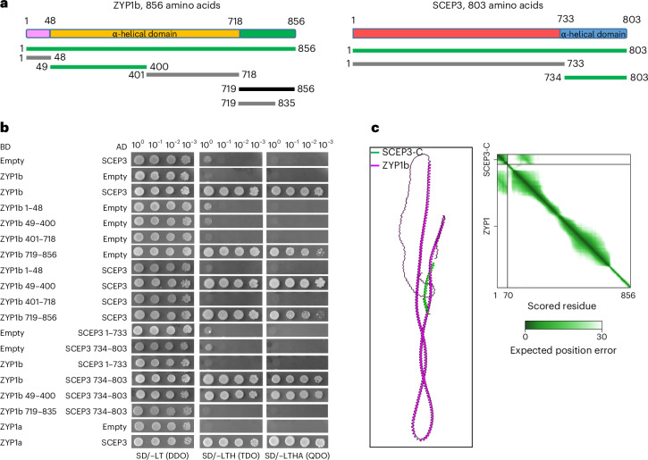
Fig. 4 SCEP3 physically interacts with ZYP1. a, Schematic depictions of ZYP1b (left) and SCEP3 (right), including fragments tested by Y2H assays. In ZYP1b, the central α-helical domain (orange) is flanked by two flexible regions (pink and green). SCEP3 consists of the C-terminal α-helical domain (blue) and the N-terminal disordered region (red). Green, grey and black lines indicate positive interaction, negative interaction and self-activation, respectively, in the Y2H experiments depicted below. b, Y2H interaction studies of ZYP1 and SCEP3. Both ZYP1a and ZYP1b interact with SCEP3 (full-length proteins). To determine the subregion within ZYP1 and SCEP3 responsible for their interaction, ZYP1 (ZYP1b was used) was divided into four fragments and SCEP3 into two. The N-terminal region of ZYP1 (amino acids 49–400) and the C-terminal region of SCEP3 (amino acids 734–803) were found responsible for the interaction between SCEP3 and ZYP1. Selection with minimal SD Base medium supplemented with DO Supplement −Leu/−Trp (SD/−LT (DDO)); DO Supplement −His/−Leu/−Trp (SD/−LT (TDO)); or DO Supplement −Ade/−His/−Leu/−Trp (SD/−LT (QDO)). c, AlphaFold3 predicts interaction between the C terminus of SCEP3 and full-length ZYP1b (~100–200 amino acids) in Arabidopsis. Predicted aligned error values are shown on the right; the interface predicted template modelling score is 0.48, and the predicted template modelling score is 0.24. Note that AlphaFold’s prediction of the global structure of ZYP1 is not consistent with cytological evidence that the N terminus and C terminus are apart, as previously described18.
Fig. 4 SCEP3 physically interacts with ZYP1. a, Schematic depictions of ZYP1b (left) and SCEP3 (right), including fragments tested by Y2H assays. In ZYP1b, the central α-helical domain (orange) is flanked by two flexible regions (pink and green). SCEP3 consists of the C-terminal α-helical domain (blue) and the N-terminal disordered region (red). Green, grey and black lines indicate positive interaction, negative interaction and self-activation, respectively, in the Y2H experiments depicted below. b, Y2H interaction studies of ZYP1 and SCEP3. Both ZYP1a and ZYP1b interact with SCEP3 (full-length proteins). To determine the subregion within ZYP1 and SCEP3 responsible for their interaction, ZYP1 (ZYP1b was used) was divided into four fragments and SCEP3 into two. The N-terminal region of ZYP1 (amino acids 49–400) and the C-terminal region of SCEP3 (amino acids 734–803) were found responsible for the interaction between SCEP3 and ZYP1. Selection with minimal SD Base medium supplemented with DO Supplement −Leu/−Trp (SD/−LT (DDO)); DO Supplement −His/−Leu/−Trp (SD/−LT (TDO)); or DO Supplement −Ade/−His/−Leu/−Trp (SD/−LT (QDO)). c, AlphaFold3 predicts interaction between the C terminus of SCEP3 and full-length ZYP1b (~100–200 amino acids) in Arabidopsis. Predicted aligned error values are shown on the right; the interface predicted template modelling score is 0.48, and the predicted template modelling score is 0.24. Note that AlphaFold’s prediction of the global structure of ZYP1 is not consistent with cytological evidence that the N terminus and C terminus are apart, as previously described18.CC BY · PubMed Central
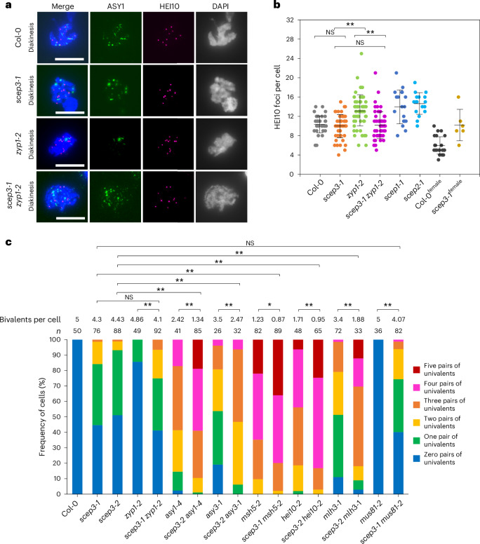
Fig. 5 SCEP3 is critical for class I and class II CO formation. a,b, Immunolocalization of ASY1 and HEI10 (a) and quantification of HEI10 foci number per diakinesis cell (b) in WT (10.34 ± 1.74, n = 41), scep3-1 (9.94 ± 2.43, n = 51), zyp1-2 (13.18 ± 2.94, n = 66), scep3-1 zyp1-2 (10.03 ± 2.43, n = 59), scep1-1 (13.94 ± 3.62, 18) and scep2-1 (14.59 ± 2.29, n = 17) male meiocytes, as well as WT (5.91 ± 1.86, n = 23) and scep3-1 (10.17 ± 3.31, n = 6) female meiocytes. Significant differences were found between scep3-1 and zyp1-2 (P < 1 × 10⁻⁷) as well as zyp1-2 and scep3-1 zyp1-2 (P < 1 × 10⁻⁷), but not between scep3-1 and the WT (P = 1) or scep3-1 and scep3-1 zyp1-2 (P = 1). DNA counterstained with DAPI is shown in merged and single-channel images in blue and grey, respectively. Scale bars, 10 μm. c, Frequency of cells with zero to five pairs of univalents including the average bivalent number per cell in a series of single or double mutants. Significant differences were found between zyp1-2 and scep3-1 zyp1-2 (P = 2.32 × 10⁻⁷), asy1-4 and scep3-2 asy1-4 (P < 1 × 10⁻⁷), scep3-2 and scep3-2 asy1-4 (P < 1 × 10⁻⁷), asy3-1 and scep3-2 asy3-1 (P = 3.26 × 10⁻⁶), scep3-2 and scep3-2 asy3-1 (P < 1 × 10⁻⁷), msh5-2 and scep3-1 msh5-2 (P = 0.016), scep3-1 and scep3-1 msh5-2 (P < 1 × 10⁻⁷), hei10-2 and scep3-2 hei10-2 (P = 4.56 × 10⁻⁷), scep3-2 and scep3-2 hei10-2 (P < 1 × 10⁻⁷), scep3-2 and scep3-2 mlh3-1 (P < 1 × 10⁻⁷), mlh3-1 and scep3-2 mlh3-1 (P < 1 × 10⁻⁷), and mus81-2 and scep3-1 mus81-2 (P < 1 × 10⁻⁷), but not between scep3-1 and scep3-1 zyp1-2 (P = 0.41) or scep3-1 and scep3-1 mus81-2 (P = 0.31). Distinct plants from each single or double mutant line (or the WT) were used for immunolocalization and chromosome spread analysis. Significance was assessed using one-way ANOVA with post hoc Bonferroni multiple comparison. The data are presented as mean ± s.d. *P < 0.05; **P < 0.01. Source data
Fig. 5 SCEP3 is critical for class I and class II CO formation. a,b, Immunolocalization of ASY1 and HEI10 (a) and quantification of HEI10 foci number per diakinesis cell (b) in WT (10.34 ± 1.74, n = 41), scep3-1 (9.94 ± 2.43, n = 51), zyp1-2 (13.18 ± 2.94, n = 66), scep3-1 zyp1-2 (10.03 ± 2.43, n = 59), scep1-1 (13.94 ± 3.62, 18) and scep2-1 (14.59 ± 2.29, n = 17) male meiocytes, as well as WT (5.91 ± 1.86, n = 23) and scep3-1 (10.17 ± 3.31, n = 6) female meiocytes. Significant differences were found between scep3-1 and zyp1-2 (P < 1 × 10⁻⁷) as well as zyp1-2 and scep3-1 zyp1-2 (P < 1 × 10⁻⁷), but not between scep3-1 and the WT (P = 1) or scep3-1 and scep3-1 zyp1-2 (P = 1). DNA counterstained with DAPI is shown in merged and single-channel images in blue and grey, respectively. Scale bars, 10 μm. c, Frequency of cells with zero to five pairs of univalents including the average bivalent number per cell in a series of single or double mutants. Significant differences were found between zyp1-2 and scep3-1 zyp1-2 (P = 2.32 × 10⁻⁷), asy1-4 and scep3-2 asy1-4 (P < 1 × 10⁻⁷), scep3-2 and scep3-2 asy1-4 (P < 1 × 10⁻⁷), asy3-1 and scep3-2 asy3-1 (P = 3.26 × 10⁻⁶), scep3-2 and scep3-2 asy3-1 (P < 1 × 10⁻⁷), msh5-2 and scep3-1 msh5-2 (P = 0.016), scep3-1 and scep3-1 msh5-2 (P < 1 × 10⁻⁷), hei10-2 and scep3-2 hei10-2 (P = 4.56 × 10⁻⁷), scep3-2 and scep3-2 hei10-2 (P < 1 × 10⁻⁷), scep3-2 and scep3-2 mlh3-1 (P < 1 × 10⁻⁷), mlh3-1 and scep3-2 mlh3-1 (P < 1 × 10⁻⁷), and mus81-2 and scep3-1 mus81-2 (P < 1 × 10⁻⁷), but not between scep3-1 and scep3-1 zyp1-2 (P = 0.41) or scep3-1 and scep3-1 mus81-2 (P = 0.31). Distinct plants from each single or double mutant line (or the WT) were used for immunolocalization and chromosome spread analysis. Significance was assessed using one-way ANOVA with post hoc Bonferroni multiple comparison. The data are presented as mean ± s.d. *P < 0.05; **P < 0.01. Source dataCC BY · PubMed Central
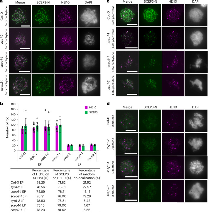
Fig. 6 SCEP3 associates with HEI10 independently of SC assembly. a, Immunolocalization of SCEP3-N and HEI10 in early pachytene nuclei of the WT, zyp1-2, scep1-1 and scep2-1. b, Quantification of SCEP3 and HEI10 foci numbers and their percentage of overlap in WT (HEI10, 83 ± 8.1; SCEP3, 94 ± 29.9; n = 7), zyp1-2 (HEI10, 88 ± 13.3; SCEP3, 95 ± 13.4; n = 10), scep1-1 (HEI10, 93 ± 24.2; SCEP3, 92 ± 23.9; n = 9) and scep2-1 (HEI10, 95 ± 16.7; SCEP3, 99 ± 22.1; n = 5) early pachytene (EP), as well as zyp1-2 (HEI10, 21 ± 3.4; SCEP3, 21 ± 0.9; n = 5), scep1-1 (HEI10, 20 ± 2.5; SCEP3, 20 ± 3.5; n = 3) and scep2-1 (HEI10, 24 ± 3.5; SCEP3, 21 ± 2.2; n = 4) late pachytene (LP). c,d, Same as a, but in late pachytene and diplotene nuclei, respectively. Distinct plants from each mutant line (or the WT) were used for immunolocalization analysis. DAPI-stained DNA is shown in grey. Scale bars, 10 µm. The data are presented as mean ± s.d. Source data
Fig. 6 SCEP3 associates with HEI10 independently of SC assembly. a, Immunolocalization of SCEP3-N and HEI10 in early pachytene nuclei of the WT, zyp1-2, scep1-1 and scep2-1. b, Quantification of SCEP3 and HEI10 foci numbers and their percentage of overlap in WT (HEI10, 83 ± 8.1; SCEP3, 94 ± 29.9; n = 7), zyp1-2 (HEI10, 88 ± 13.3; SCEP3, 95 ± 13.4; n = 10), scep1-1 (HEI10, 93 ± 24.2; SCEP3, 92 ± 23.9; n = 9) and scep2-1 (HEI10, 95 ± 16.7; SCEP3, 99 ± 22.1; n = 5) early pachytene (EP), as well as zyp1-2 (HEI10, 21 ± 3.4; SCEP3, 21 ± 0.9; n = 5), scep1-1 (HEI10, 20 ± 2.5; SCEP3, 20 ± 3.5; n = 3) and scep2-1 (HEI10, 24 ± 3.5; SCEP3, 21 ± 2.2; n = 4) late pachytene (LP). c,d, Same as a, but in late pachytene and diplotene nuclei, respectively. Distinct plants from each mutant line (or the WT) were used for immunolocalization analysis. DAPI-stained DNA is shown in grey. Scale bars, 10 µm. The data are presented as mean ± s.d. Source dataCC BY · PubMed Central
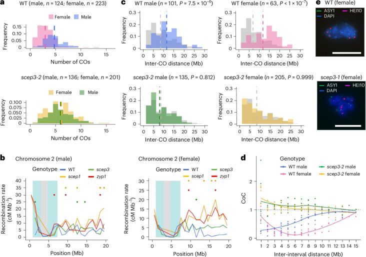
Fig. 7 CO interference and heterochiasmy vanish in scep3. a, Number of COs (the data are presented as mean ± s.e.m.) detected by sequencing of recombinant offspring of WT males (4.80 ± 0.17), WT females (3.00 ± 0.11), scep3-2 males (6.00 ± 0.24) and scep3-2 females (6.15 ± 0.23). The sample sizes used for analysis are indicated in parentheses. Significant differences were detected between WT males and females (P < 1 × 10⁻⁷), but not between scep3-2 males and females (P = 0.942) (nested ANOVA followed by Tukey’s honestly significant difference test, a two-sided test with adjustment for multiple comparisons). Compared with the WT, in scep3-2 CO numbers significantly increased in both males (P = 5.09 × 10⁻⁴) and females (P < 1 × 10⁻⁷). b, CO distribution along chromosome 2 in male and female WT, zyp1 (ref. 16), scep1-1 (ref. 18) and scep3-2. Centromere and pericentromere regions are indicated in grey and blue, respectively. CO data are presented within 1-Mb windows. Significant differences (based on χ2 tests) between the WT and scep3-2 (green dots), scep3-2 and zyp1 (red dots) and scep3-2 and scep1-1 (orange dots) are indicated. c, Distribution of inter-CO distances (only chromosomes with exactly two COs are included for analysis) in male and female WT and scep3-2. Calculated random distributions are shown in grey. The statistical significance is indicated in parentheses (nested ANOVA followed by Tukey’s honestly significant difference test, a two-sided test with adjustment for multiple comparisons). d, The coefficient of coincidence (CoC) was calculated for inter-interval distances ranging from 1 Mb to 15 Mb for each chromosome, and a LOESS curve was fitted (coloured lines). e, Immunolocalization of ASY1 and HEI10 in WT and scep3-1 female meiocytes. DAPI-stained DNA is shown in blue. Scale bars, 10 μm. Source data
Fig. 7 CO interference and heterochiasmy vanish in scep3. a, Number of COs (the data are presented as mean ± s.e.m.) detected by sequencing of recombinant offspring of WT males (4.80 ± 0.17), WT females (3.00 ± 0.11), scep3-2 males (6.00 ± 0.24) and scep3-2 females (6.15 ± 0.23). The sample sizes used for analysis are indicated in parentheses. Significant differences were detected between WT males and females (P < 1 × 10⁻⁷), but not between scep3-2 males and females (P = 0.942) (nested ANOVA followed by Tukey’s honestly significant difference test, a two-sided test with adjustment for multiple comparisons). Compared with the WT, in scep3-2 CO numbers significantly increased in both males (P = 5.09 × 10⁻⁴) and females (P < 1 × 10⁻⁷). b, CO distribution along chromosome 2 in male and female WT, zyp1 (ref. 16), scep1-1 (ref. 18) and scep3-2. Centromere and pericentromere regions are indicated in grey and blue, respectively. CO data are presented within 1-Mb windows. Significant differences (based on χ2 tests) between the WT and scep3-2 (green dots), scep3-2 and zyp1 (red dots) and scep3-2 and scep1-1 (orange dots) are indicated. c, Distribution of inter-CO distances (only chromosomes with exactly two COs are included for analysis) in male and female WT and scep3-2. Calculated random distributions are shown in grey. The statistical significance is indicated in parentheses (nested ANOVA followed by Tukey’s honestly significant difference test, a two-sided test with adjustment for multiple comparisons). d, The coefficient of coincidence (CoC) was calculated for inter-interval distances ranging from 1 Mb to 15 Mb for each chromosome, and a LOESS curve was fitted (coloured lines). e, Immunolocalization of ASY1 and HEI10 in WT and scep3-1 female meiocytes. DAPI-stained DNA is shown in blue. Scale bars, 10 μm. Source dataCC BY · PubMed Central
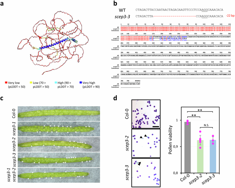
Extended Data Fig. 1 Isolation of scep3-3 and phenotypic analysis of scep3 alleles. a, SCEP3 full-length protein structure prediction by AlphaFold246. b, Isolation of scep3-3. Top: CRISPR/Cas9-mediated 22 bp deletion in exon 5 resulting in a predicted truncated protein of 201 AA; PAM sequence underlined and deleted bases as red dashes. Below: Alignment of SCEP3 WT and SCEP3-3 proteins. c, Seed abortion (indicated by asterisks) in the WT and scep3 alleles. d, Alexander staining of pollen grains (arrowheads indicate non-viable ones in blue; scale bar, 100 µm) and quantification of pollen viability in Col-0 (0.99 ± 0.01, n = 5), scep3-2 (0.92 ± 0.02, n = 5) and scep3-3 (0.93 ± 0.01, n = 3). Significance evaluated by one-way ANOVA with post-hoc Bonferroni multiple comparison (Col-0 and scep3-2, P = 1.26×10⁻⁴; Col-0 and scep3-3, P = 4.48×10⁻⁴; scep3-2 and scep3-3, P = 1). Data are presented as mean ± standard deviation (s.d.). **P < 0.01. N.S., not significant. Source data
Extended Data Fig. 1 Isolation of scep3-3 and phenotypic analysis of scep3 alleles. a, SCEP3 full-length protein structure prediction by AlphaFold246. b, Isolation of scep3-3. Top: CRISPR/Cas9-mediated 22 bp deletion in exon 5 resulting in a predicted truncated protein of 201 AA; PAM sequence underlined and deleted bases as red dashes. Below: Alignment of SCEP3 WT and SCEP3-3 proteins. c, Seed abortion (indicated by asterisks) in the WT and scep3 alleles. d, Alexander staining of pollen grains (arrowheads indicate non-viable ones in blue; scale bar, 100 µm) and quantification of pollen viability in Col-0 (0.99 ± 0.01, n = 5), scep3-2 (0.92 ± 0.02, n = 5) and scep3-3 (0.93 ± 0.01, n = 3). Significance evaluated by one-way ANOVA with post-hoc Bonferroni multiple comparison (Col-0 and scep3-2, P = 1.26×10⁻⁴; Col-0 and scep3-3, P = 4.48×10⁻⁴; scep3-2 and scep3-3, P = 1). Data are presented as mean ± standard deviation (s.d.). **P < 0.01. N.S., not significant. Source dataCC BY · PubMed Central
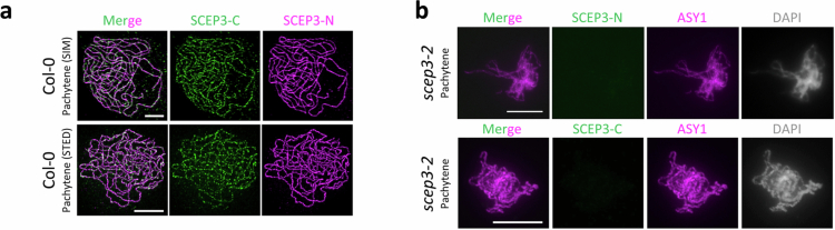
Extended Data Fig. 2 Both SCEP3-N and SCEP3-C antibodies localize to the SC and are specific for SCEP3 (signal absence in scep3). Immunolocalization in pachytene nuclei of a, SCEP3-N and SCEP3-C in Col-0 (Images acquired by 3D-SIM or STED; Scale bar, 2 μm), and of b, SCEP3-N or SCEP3-C together with ASY1 in scep3-2. DNA counterstained with DAPI in gray. The experiments were repeated three times with similar results. Scale bar, 10 μm.
Extended Data Fig. 2 Both SCEP3-N and SCEP3-C antibodies localize to the SC and are specific for SCEP3 (signal absence in scep3). Immunolocalization in pachytene nuclei of a, SCEP3-N and SCEP3-C in Col-0 (Images acquired by 3D-SIM or STED; Scale bar, 2 μm), and of b, SCEP3-N or SCEP3-C together with ASY1 in scep3-2. DNA counterstained with DAPI in gray. The experiments were repeated three times with similar results. Scale bar, 10 μm.CC BY · PubMed Central
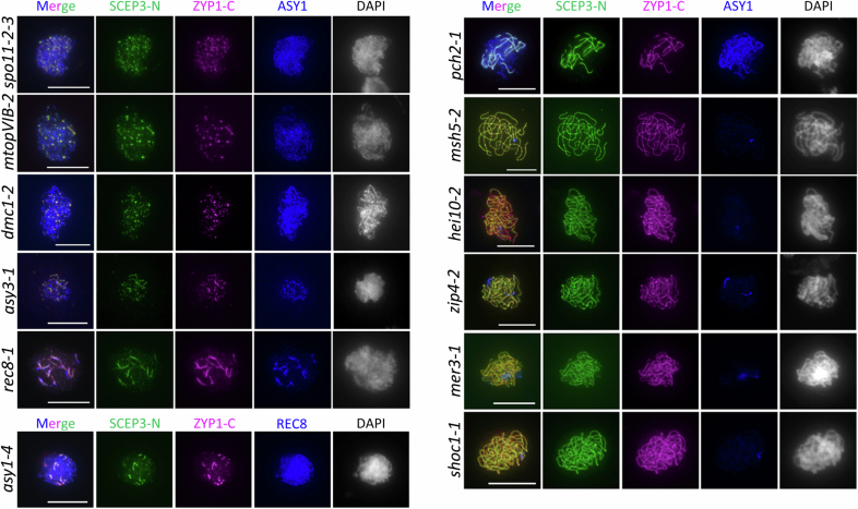
Extended Data Fig. 3 Co-localization of SCEP3 and ZYP1 in various meiotic mutants. Immunolocalization of SCEP3-N, ZYP1-C and ASY1 or REC8 in spo11-2-3, mtopVIB-2, dmc1-2, asy3-1, rec8-1, pch2-1, asy1-4, msh5-2, hei10-2, zip4-2, mer3-1, and shoc1-1 pachytene(-like) nuclei. DNA stained with DAPI in gray. All experiments were repeated at least two times with similar results. Scale bar, 10 μm.
Extended Data Fig. 3 Co-localization of SCEP3 and ZYP1 in various meiotic mutants. Immunolocalization of SCEP3-N, ZYP1-C and ASY1 or REC8 in spo11-2-3, mtopVIB-2, dmc1-2, asy3-1, rec8-1, pch2-1, asy1-4, msh5-2, hei10-2, zip4-2, mer3-1, and shoc1-1 pachytene(-like) nuclei. DNA stained with DAPI in gray. All experiments were repeated at least two times with similar results. Scale bar, 10 μm.CC BY · PubMed Central
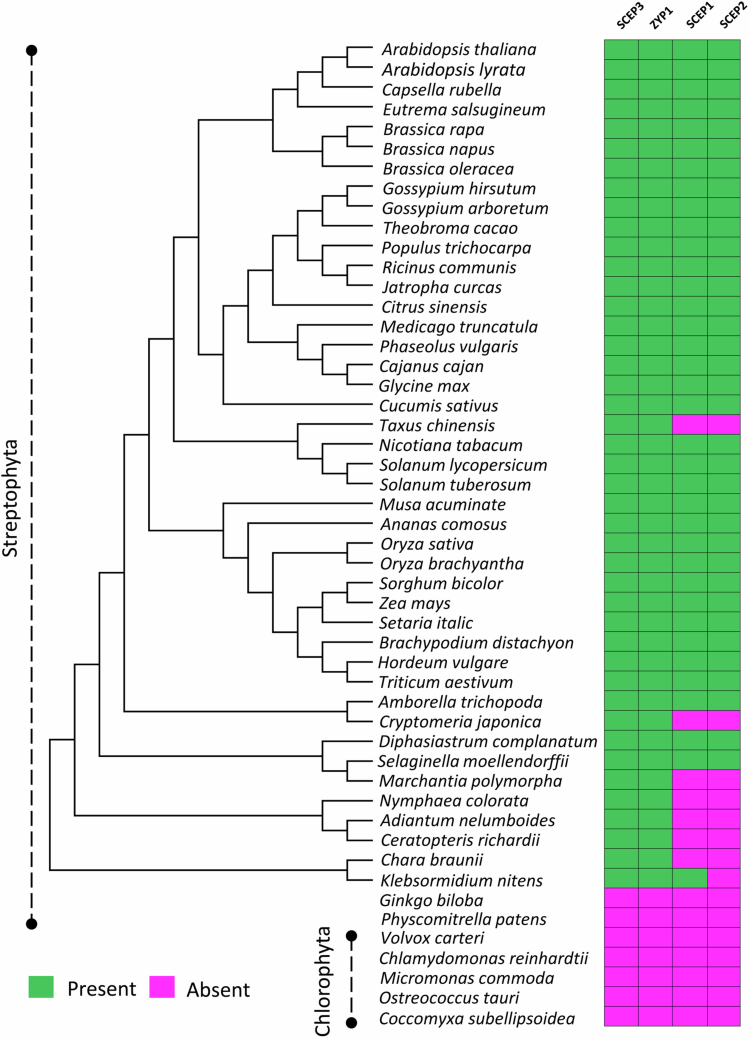
Extended Data Fig. 4 Evolutionary analysis of SCEP3 and other CR components. The evolutionary tree is constructed based on SCEP3 homologous sequences (PSI blast results using Arabidopsis SCEP3 as seed sequence) from Streptophyta species. Accessions used: A. thaliana (NP_001328810.1), A. lyrata (XP_020875481.1), Capsella rubella (XP_023635313.1), Eutrema salsugineum (XP_024005618.1), Brassica rapa (XP_009136934.2), B. napus (XP_013737233.2), B. oleracea (XP_013594171.1), Gossypium hirsutum (XP_040935483.1), G. arboretum (KAK5792206.1), Theobroma cacao (XP_017978471.1), Citrus sinensis (XP_024950445.1), Phaseolus vulgaris (XP_007153704.1), Cajanus cajan (XP_029124714.1), Medicago truncatula (XP_039689560.1), Glycine max (XP_040873294.1), Ricinus communis (XP_048229528.1), Jatropha curcas (XP_020541047.2), Populus trichocarpa (KAI5570781.1), Cucumis sativus (XP_011658243.1), Solanum lycopersicum (XP_019067843.1), S. tuberosum (KAH0717859.1), Sorghum bicolor (XP_021312987.1), Zea mays (XP_008673176.1), Setaria italic (XP_004969993.2), Oryza sativa (KAF2952333.1), O. brachyantha (XP_040380573.1), Brachypodium distachyon (KQK09796.1), Ananas comosus (XP_020098929.1), H. vulgare (XP_044977082.1), Nicotiana tabacum (XP_016437386.1), Triticum aestivum (KAF7023752.1), Musa acuminate (XP_018686223.1), Amborella trichopoda (XP_020527858.1), Taxus chinensis (KAH9309445.1), Selaginella moellendorffii (XP_024528147.1), Nymphaea colorata (XP_049931681.1), Cryptomeria japonica (XP_059065081.1), Adiantum nelumboides (MCO5557227.1), Ceratopteris richardii (KAH7301291.1), Diphasiastrum complanatum (KAJ7553908.1), Marchantia polymorpha (PTQ45356.1), Chara braunii (GBG74560.1), Klebsormidium nitens (GAQ79536.1). Some Streptophyta and Chlorophyta species without SCEP3 homolog identified are also listed. The color-coded table includes SCEP3, ZYP1, SCEP1 and SCEP2 and shows the presence (green) or absence (purple) of respective homologs in different species.
Extended Data Fig. 4 Evolutionary analysis of SCEP3 and other CR components. The evolutionary tree is constructed based on SCEP3 homologous sequences (PSI blast results using Arabidopsis SCEP3 as seed sequence) from Streptophyta species. Accessions used: A. thaliana (NP_001328810.1), A. lyrata (XP_020875481.1), Capsella rubella (XP_023635313.1), Eutrema salsugineum (XP_024005618.1), Brassica rapa (XP_009136934.2), B. napus (XP_013737233.2), B. oleracea (XP_013594171.1), Gossypium hirsutum (XP_040935483.1), G. arboretum (KAK5792206.1), Theobroma cacao (XP_017978471.1), Citrus sinensis (XP_024950445.1), Phaseolus vulgaris (XP_007153704.1), Cajanus cajan (XP_029124714.1), Medicago truncatula (XP_039689560.1), Glycine max (XP_040873294.1), Ricinus communis (XP_048229528.1), Jatropha curcas (XP_020541047.2), Populus trichocarpa (KAI5570781.1), Cucumis sativus (XP_011658243.1), Solanum lycopersicum (XP_019067843.1), S. tuberosum (KAH0717859.1), Sorghum bicolor (XP_021312987.1), Zea mays (XP_008673176.1), Setaria italic (XP_004969993.2), Oryza sativa (KAF2952333.1), O. brachyantha (XP_040380573.1), Brachypodium distachyon (KQK09796.1), Ananas comosus (XP_020098929.1), H. vulgare (XP_044977082.1), Nicotiana tabacum (XP_016437386.1), Triticum aestivum (KAF7023752.1), Musa acuminate (XP_018686223.1), Amborella trichopoda (XP_020527858.1), Taxus chinensis (KAH9309445.1), Selaginella moellendorffii (XP_024528147.1), Nymphaea colorata (XP_049931681.1), Cryptomeria japonica (XP_059065081.1), Adiantum nelumboides (MCO5557227.1), Ceratopteris richardii (KAH7301291.1), Diphasiastrum complanatum (KAJ7553908.1), Marchantia polymorpha (PTQ45356.1), Chara braunii (GBG74560.1), Klebsormidium nitens (GAQ79536.1). Some Streptophyta and Chlorophyta species without SCEP3 homolog identified are also listed. The color-coded table includes SCEP3, ZYP1, SCEP1 and SCEP2 and shows the presence (green) or absence (purple) of respective homologs in different species.CC BY · PubMed Central
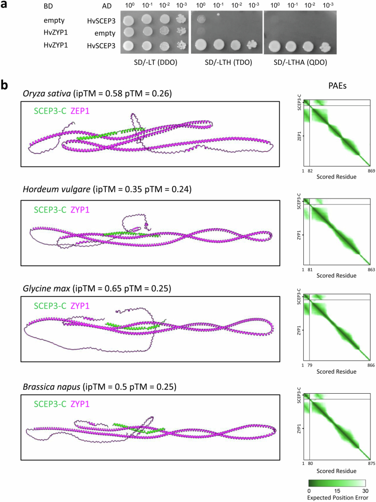
Extended Data Fig. 5 Conservation of the SCEP3 and ZYP1 interaction in plants. a, Y2H interaction of H. vulgare ZYP1 and SCEP3 (full-length proteins). Note, TDO (SD/-LTH) is a less and QDO (SD/-LTHA) a more stringent medium for selection. b, AlphaFold3 interaction prediction using the C-terminus (α-helical domain) of SCEP3 and full-length ZYP1 (ZEP1 is the rice ZYP1 homolog) from O. sativa, H. vulgare, G. max or B. napus. PAE values are shown on the right; ipTM and pTM values indicated in parentheses.
Extended Data Fig. 5 Conservation of the SCEP3 and ZYP1 interaction in plants. a, Y2H interaction of H. vulgare ZYP1 and SCEP3 (full-length proteins). Note, TDO (SD/-LTH) is a less and QDO (SD/-LTHA) a more stringent medium for selection. b, AlphaFold3 interaction prediction using the C-terminus (α-helical domain) of SCEP3 and full-length ZYP1 (ZEP1 is the rice ZYP1 homolog) from O. sativa, H. vulgare, G. max or B. napus. PAE values are shown on the right; ipTM and pTM values indicated in parentheses.CC BY · PubMed Central
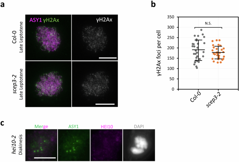
Extended Data Fig. 6 Similar γH2Ax foci numbers in scep3 when compared with the WT and absence of HEI10 immunofluorescence signals in hei10-2. a, Immunolocalization of ASY1 and γH2Ax in WT and scep3-2 meiocytes. γH2Ax signal channel is depicted on the right (gray). Scale bar, 10 μm. b, Quantification of γH2Ax foci numbers in Col-0 (187 ± 51 nm, n = 31) and scep3-2 (177 ± 31 nm, n = 30) meiocytes (late leptotene/early zygotene). Data are presented as mean ± standard deviation (s.d.). No significant difference was found between Col-0 and scep3-2 (P = 0.36, two-sided Student’s t-test). N.S., not significant. c, Immunolocalization of HEI10 and ASY1 in hei10-2. The experiment was repeated two times with similar results. Source data
Extended Data Fig. 6 Similar γH2Ax foci numbers in scep3 when compared with the WT and absence of HEI10 immunofluorescence signals in hei10-2. a, Immunolocalization of ASY1 and γH2Ax in WT and scep3-2 meiocytes. γH2Ax signal channel is depicted on the right (gray). Scale bar, 10 μm. b, Quantification of γH2Ax foci numbers in Col-0 (187 ± 51 nm, n = 31) and scep3-2 (177 ± 31 nm, n = 30) meiocytes (late leptotene/early zygotene). Data are presented as mean ± standard deviation (s.d.). No significant difference was found between Col-0 and scep3-2 (P = 0.36, two-sided Student’s t-test). N.S., not significant. c, Immunolocalization of HEI10 and ASY1 in hei10-2. The experiment was repeated two times with similar results. Source dataCC BY · PubMed Central
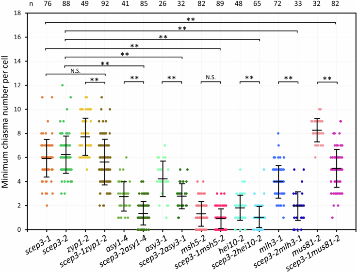
Extended Data Fig. 7 SCEP3 is required for some chiasmata in SC-deficient or ZMM mutants. Minimum chiasma number counts in single (scep3-1, scep3-2, zyp1-2, asy1-4, asy3-1, msh5-2, hei10-2, mlh3-1, mus81-2) and double (scep3-1zyp1-2, scep3-2asy1-4, scep3-2asy3-1, scep3-1msh5-2, scep3-2hei10-2, scep3-2mlh3-1, scep3-1mus81-2) mutants. Chromosome spreads were analysed for each mutant using flower buds from distinct plants. The number of cells (n) analysed is indicated at the top. Significance evaluated by one-way ANOVA with post-hoc Bonferroni multiple comparison (zyp1-2 and scep3-1zyp1-2, P < 1×10⁻⁷; zyp1-2 and scep3-1zyp1-2, P = 0.71; asy1-4 and scep3-2asy1-4, P = 1.96×10⁻⁷; scep3-2 and scep3-2asy1-4, P < 1×10⁻⁷; asy3-1 and scep3-2asy3-1, P = 6.39×10⁻⁴; scep3-2 and scep3-2asy3-1, P < 1×10⁻⁷; msh5-2 and scep3-1msh5-2, P = 0.069; scep3-1 and scep3-1msh5-2, P < 1×10⁻⁷; hei10-2 and scep3-2hei10-2, P = 4.72×10⁻³; scep3-2 and scep3-2hei10-2, P < 1×10⁻⁷; mlh3-1 and scep3-2mlh3-1, P < 1×10⁻⁷; scep3-2 and scep3-2mlh3-1, P < 1×10⁻⁷; mus81-2 and scep3-1mus81-2, P < 1×10⁻⁷; scep3-1 and scep3-1mus81-2, P = 1.51×10⁻³). Data (refer to Supplemental Table 1) are presented as mean ± standard deviation (s.d.). **P < 0.01. N.S., not significant. Source data
Extended Data Fig. 7 SCEP3 is required for some chiasmata in SC-deficient or ZMM mutants. Minimum chiasma number counts in single (scep3-1, scep3-2, zyp1-2, asy1-4, asy3-1, msh5-2, hei10-2, mlh3-1, mus81-2) and double (scep3-1zyp1-2, scep3-2asy1-4, scep3-2asy3-1, scep3-1msh5-2, scep3-2hei10-2, scep3-2mlh3-1, scep3-1mus81-2) mutants. Chromosome spreads were analysed for each mutant using flower buds from distinct plants. The number of cells (n) analysed is indicated at the top. Significance evaluated by one-way ANOVA with post-hoc Bonferroni multiple comparison (zyp1-2 and scep3-1zyp1-2, P < 1×10⁻⁷; zyp1-2 and scep3-1zyp1-2, P = 0.71; asy1-4 and scep3-2asy1-4, P = 1.96×10⁻⁷; scep3-2 and scep3-2asy1-4, P < 1×10⁻⁷; asy3-1 and scep3-2asy3-1, P = 6.39×10⁻⁴; scep3-2 and scep3-2asy3-1, P < 1×10⁻⁷; msh5-2 and scep3-1msh5-2, P = 0.069; scep3-1 and scep3-1msh5-2, P < 1×10⁻⁷; hei10-2 and scep3-2hei10-2, P = 4.72×10⁻³; scep3-2 and scep3-2hei10-2, P < 1×10⁻⁷; mlh3-1 and scep3-2mlh3-1, P < 1×10⁻⁷; scep3-2 and scep3-2mlh3-1, P < 1×10⁻⁷; mus81-2 and scep3-1mus81-2, P < 1×10⁻⁷; scep3-1 and scep3-1mus81-2, P = 1.51×10⁻³). Data (refer to Supplemental Table 1) are presented as mean ± standard deviation (s.d.). **P < 0.01. N.S., not significant. Source dataCC BY · PubMed Central
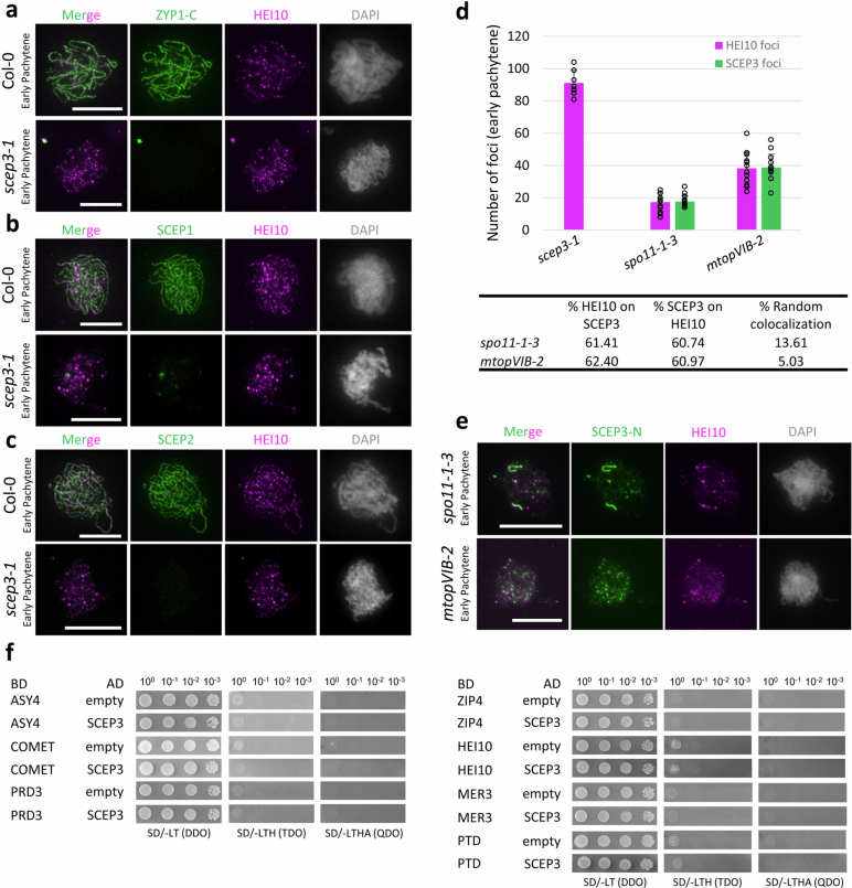
Extended Data Fig. 8 Localization of CR proteins and HEI10 in scep3, spo11-1-3 and mtopVIB-2 as well as no direct interaction of SCEP3 with selected proteins in Y2H. Immunolocalization of a, ZYP1-C and HEI10, b, SCEP1 and HEI10, and c, SCEP2 and HEI10 in early pachytene nuclei of WT and scep3-1. d, Quantification of SCEP3 and HEI10 foci numbers in scep3-1 (HEI10, 91 ± 8.1; n = 7), spo11-1-3 (HEI10, 17 ± 5.6; SCEP3, 18 ± 3.6; n = 15), and mtopVIB-2 (HEI10, 38 ± 9.7; SCEP3, 39 ± 8.1; n = 14) as well as their percentage of overlap in spo11-1-3 and mtopVIB-2. Data are presented as mean ± standard deviation (s.d.). e, Immunolocalization of SCEP3 and HEI10 in early pachytene nuclei of spo11-1-3 and mtopVIB-2. DAPI-stained DNA in gray. Scale bar, 10 µm. f, Interactions tested between SCEP3 and axis(-associated) (ASY4, COMET or PRD3) or ZMM proteins (ZIP4, HEI10, MER3 or PTD) in Y2H. TDO (SD/-LTH) is a less stringent and QDO (SD/-LTHA) is a more stringent medium used for selection. The experiments (a-c, e) were repeated at least three times with similar results. Source data
Extended Data Fig. 8 Localization of CR proteins and HEI10 in scep3, spo11-1-3 and mtopVIB-2 as well as no direct interaction of SCEP3 with selected proteins in Y2H. Immunolocalization of a, ZYP1-C and HEI10, b, SCEP1 and HEI10, and c, SCEP2 and HEI10 in early pachytene nuclei of WT and scep3-1. d, Quantification of SCEP3 and HEI10 foci numbers in scep3-1 (HEI10, 91 ± 8.1; n = 7), spo11-1-3 (HEI10, 17 ± 5.6; SCEP3, 18 ± 3.6; n = 15), and mtopVIB-2 (HEI10, 38 ± 9.7; SCEP3, 39 ± 8.1; n = 14) as well as their percentage of overlap in spo11-1-3 and mtopVIB-2. Data are presented as mean ± standard deviation (s.d.). e, Immunolocalization of SCEP3 and HEI10 in early pachytene nuclei of spo11-1-3 and mtopVIB-2. DAPI-stained DNA in gray. Scale bar, 10 µm. f, Interactions tested between SCEP3 and axis(-associated) (ASY4, COMET or PRD3) or ZMM proteins (ZIP4, HEI10, MER3 or PTD) in Y2H. TDO (SD/-LTH) is a less stringent and QDO (SD/-LTHA) is a more stringent medium used for selection. The experiments (a-c, e) were repeated at least three times with similar results. Source dataCC BY · PubMed Central
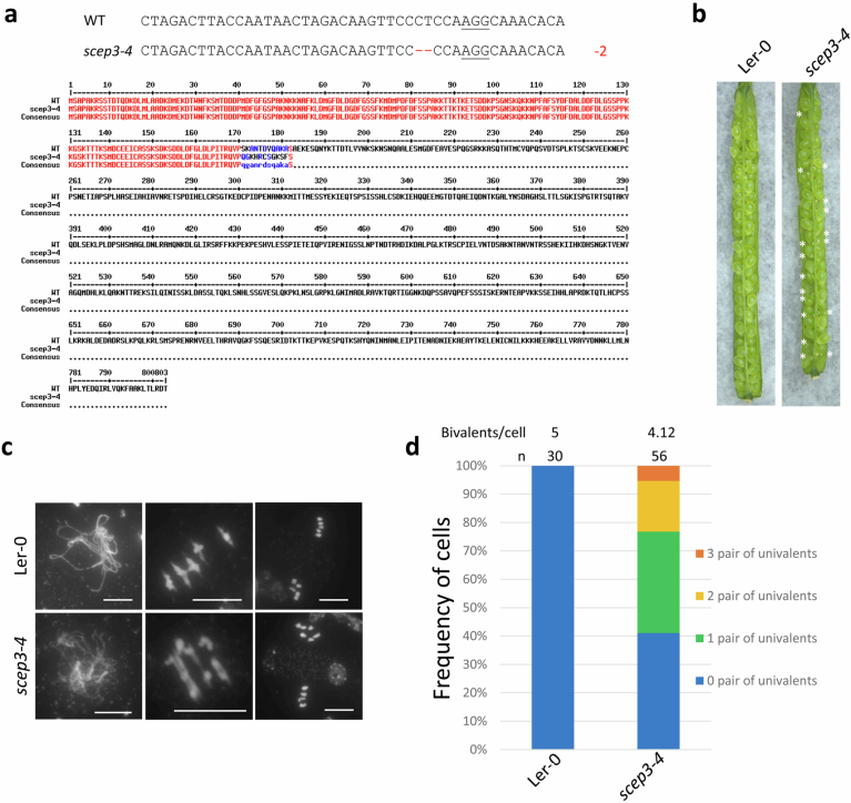
Extended Data Fig. 9 Isolation of scep3-4 in Ler-0. a, Details on the CRISPR/Cas9-mediated allele scep3-4: 2 bp deletion within exon five resulting in a predicted truncated protein of 182 AA. PAM sequence underlined and deleted bases as red dashes. Below: alignment of SCEP3 wildtype and truncated SCEP3-4 protein. b, Seed setting in siliques of Ler-0 and scep3-4. Seed abortion indicated by asterisks. c, Male meiotic chromosome spreads and d, quantification of univalent frequency in Ler-0 and scep3-4. Source data
Extended Data Fig. 9 Isolation of scep3-4 in Ler-0. a, Details on the CRISPR/Cas9-mediated allele scep3-4: 2 bp deletion within exon five resulting in a predicted truncated protein of 182 AA. PAM sequence underlined and deleted bases as red dashes. Below: alignment of SCEP3 wildtype and truncated SCEP3-4 protein. b, Seed setting in siliques of Ler-0 and scep3-4. Seed abortion indicated by asterisks. c, Male meiotic chromosome spreads and d, quantification of univalent frequency in Ler-0 and scep3-4. Source dataCC BY · PubMed Central
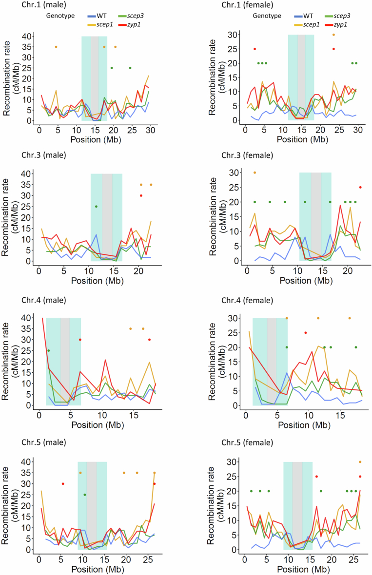
Extended Data Fig. 10 Genome-wide CO distribution in WT and scep3. CO distribution along chromosomes 1, 3, 4 and 5 in male and female of WT, zyp116, scep1-118 and scep3-2 plants (refer to Fig. 7b).
Extended Data Fig. 10 Genome-wide CO distribution in WT and scep3. CO distribution along chromosomes 1, 3, 4 and 5 in male and female of WT, zyp116, scep1-118 and scep3-2 plants (refer to Fig. 7b).CC BY · PubMed Central

Verification

Claims the paper makes

The claims and statistics of this paper haven’t been checked yet.

Run the check — free, no account needed. It reads the open-access text and checks the sources it cites.

A few free checks a day · papers already verified open instantly · results are public

Self-check

Percentages, sums and reported differences re-computed from the paper’s own numbers.

The self-check needs the paper’s full text.

Citations

Cites

Research works printed but not linked: 72

  1. Zickler D, Kleckner N. Meiosis: Dances Between Homologs. Annu. Rev. Genet. 57, 1–63 (2023). · DOI
  2. de Massy B. Initiation of meiotic recombination: how and where? Conservation and specificities among eukaryotes. Annu. Rev. Genet. 47, 563–599 (2013). · DOI
  3. Mercier R, Mezard C, Jenczewski E, Macaisne N, Grelon M. The molecular biology of meiosis in plants. Annu. Rev. Plant Biol. 66, 297–327 (2015). · DOI
  4. Börner GV, Kleckner N, Hunter N. Crossover/noncrossover differentiation, synaptonemal complex formation, and regulatory surveillance at the leptotene/zygotene transition of meiosis. Cell 117, 29–45 (2004). · DOI
  5. Snowden T, Acharya S, Butz C, Berardini M, Fishel R. hMSH4-hMSH5 recognizes Holliday Junctions and forms a meiosis-specific sliding clamp that embraces homologous chromosomes. Mol. Cell 15, 437–451 (2004). · DOI
  6. Zickler D, Kleckner N. A few of our favorite things: Pairing, the bouquet, crossover interference and evolution of meiosis. Semin. Cell Dev. Biol. 54, 135–148 (2016). · DOI
  7. Berchowitz LE, Francis KE, Bey AL, Copenhaver GP. The role of AtMUS81 in interference-insensitive crossovers in A. thaliana. PLoS Genet. 3, e132 (2007). · DOI
  8. Higgins JD, Buckling EF, Franklin FCH, Jones GH. Expression and functional analysis of AtMUS81 in Arabidopsis meiosis reveals a role in the second pathway of crossing-over. Plant J. 54, 152–162 (2008). · DOI
  9. Armstrong SJ, Caryl AP, Jones GH, Franklin FC. Asy1, a protein required for meiotic chromosome synapsis, localizes to axis-associated chromatin in Arabidopsis and Brassica. J. Cell Sci. 115, 3645–3655 (2002). · DOI
  10. Sanchez-Moran E, Santos JL, Jones GH, Franklin FC. ASY1 mediates AtDMC1-dependent interhomolog recombination during meiosis in Arabidopsis. Genes Dev. 21, 2220–2233 (2007). · DOI
  11. Ferdous M, et al. Inter-homolog crossing-over and synapsis in Arabidopsis meiosis are dependent on the chromosome axis protein AtASY3. PLoS Genet. 8, e1002507 (2012). · DOI
  12. Chambon A, et al. Identification of ASYNAPTIC4, a Component of the Meiotic Chromosome Axis. Plant Physiol. 178, 233–246 (2018). · DOI
  13. Cai X, Dong F, Edelmann RE, Makaroff CA. The Arabidopsis SYN1 cohesin protein is required for sister chromatid arm cohesion and homologous chromosome pairing. J. Cell Sci. 116, 2999–3007 (2003). · DOI
  14. Zickler D, Kleckner N. Recombination, Pairing, and Synapsis of Homologs during Meiosis. Cold Spring Harb. Perspect. Biol. 7, a016626 (2015). · DOI
  15. Capilla-Pérez L, et al. The synaptonemal complex imposes crossover interference and heterochiasmy in Arabidopsis. Proc. Natl Acad. Sci. USA 118, e2023613118 (2021).
  16. Sym M, Engebrecht J, Roeder GS. ZIP1 is a synaptonemal complex protein required for meiotic chromosome synapsis. Cell 72, 365–378 (1993). · DOI
  17. de Vries FA, et al. Mouse Sycp1 functions in synaptonemal complex assembly, meiotic recombination, and XY body formation. Genes Dev. 19, 1376–1389 (2005). · DOI
  18. Humphryes N, Leung W-K, Argunhan B, Terentyev Y, Dvorackova M, Tsubouchi H. The Ecm11-Gmc2 complex promotes synaptonemal complex formation through assembly of transverse filaments in budding yeast. PLoS Genet. 9, e1003194 (2013). · DOI
  19. Page SL, et al. Corona is required for higher-order assembly of transverse filaments into full-length synaptonemal complex in Drosophila oocytes. PLoS Genet. 4, e1000194 (2008). · DOI
  20. Collins KA, et al. Corolla is a novel protein that contributes to the architecture of the synaptonemal complex of Drosophila. Genetics 198, 219–228 (2014). · DOI
  21. Hamer G, Gell K, Kouznetsova A, Novak I, Benavente R, Höög C. Characterization of a novel meiosis-specific protein within the central element of the synaptonemal complex. J. Cell Sci. 119, 4025–4032 (2006). · DOI
  22. Bolcun-Filas E, et al. SYCE2 is required for synaptonemal complex assembly, double strand break repair, and homologous recombination. J. Cell Biol. 176, 741–747 (2007). · DOI
  23. Schramm S, et al. A novel mouse synaptonemal complex protein is essential for loading of central element proteins, recombination, and fertility. PLoS Genet. 7, e1002088 (2011). · DOI
  24. Gómez-h L, et al. C14ORF39/SIX6OS1 is a constituent of the synaptonemal complex and is essential for mouse fertility. Nat. Commun. 7, 1–16 (2016). · DOI
  25. Costa Y, et al. Two novel proteins recruited by synaptonemal complex protein 1 (SYCP1) are at the centre of meiosis. J. Cell Sci. 118, 2755–2762 (2005). · DOI
  26. Higgins JD, Sanchez-Moran E, Armstrong SJ, Jones GH, Franklin FCH. The Arabidopsis synaptonemal complex protein ZYP1 is required for chromosome synapsis and normal fidelity of crossing over. Genes Dev. 19, 2488–2500 (2005). · DOI
  27. Vrielynck N, et al. SCEP1 and SCEP2 are two new components of the synaptonemal complex central element. Nat. Plants, 9, 2016–2030 (2023). · DOI
  28. Hurlock ME, et al. Identification of novel synaptonemal complex components in C. elegans. J. Cell Biol. 219, e201910043 (2020). · DOI
  29. Lee M-S, et al. The synaptonemal complex central region modulates crossover pathways and feedback control of meiotic double-strand break formation. Nucleic Acids Res. 49, 7537–7553 (2021). · DOI
  30. Voelkel-Meiman K, Johnston C, Thappeta Y, Subramanian VV, Hochwagen A, MacQueen AJ. Separable crossover-promoting and crossover-constraining aspects of Zip1 activity during budding yeast meiosis. PLoS Genet. 11, e1005335 (2015). · DOI
  31. Voelkel-Meiman K, Cheng S-Y, Morehouse SJ, MacQueen AJ. Synaptonemal complex proteins of budding yeast define reciprocal roles in MutSγ-mediated crossover formation. Genetics 203, 1091–1103 (2016). · DOI
  32. France MG, Enderle J, Röhrig S, Puchta H, Franklin FCH, Higgins JD. ZYP1 is required for obligate cross-over formation and cross-over interference in Arabidopsis. Proc. Natl Acad. Sci. USA 118, e2021671118 (2021).
  33. Wang M, et al. The central element protein ZEP1 of the synaptonemal complex regulates the number of crossovers during meiosis in rice. Plant Cell 22, 417–430 (2010). · DOI
  34. Barakate A, et al. The synaptonemal complex protein ZYP1 is required for imposition of meiotic crossovers in barley. Plant Cell 26, 729–740 (2014). · DOI
  35. Durand S, et al. Joint control of meiotic crossover patterning by the synaptonemal complex and HEI10 dosage. Nat. Commun. 13, 5999 (2022). · DOI
  36. Fozard JA, Morgan C, Howard M. Coarsening dynamics can explain meiotic crossover patterning in both the presence and absence of the synaptonemal complex. Elife 12, e79408 (2023). · DOI
  37. Crichton JH, et al. Structural maturation of SYCP1-mediated meiotic chromosome synapsis by SYCE3. Nat. Struct. Mol. Biol. 30, 188–199 (2023). · DOI
  38. Pyatnitskaya A, Andreani J, Guérois R, De Muyt A, Borde V. The Zip4 protein directly couples meiotic crossover formation to synaptonemal complex assembly. Genes Dev. 36, 53–69 (2022). · DOI · Paper page
  39. Feng C, et al. TurboID-based proteomic profiling of meiotic chromosome axes in Arabidopsis thaliana. Nat. Plants 9, 616–630 (2023). · DOI
  40. Klepikova AV, Kasianov AS, Gerasimov ES, Logacheva MD, Penin AA. A high resolution map of the Arabidopsis thaliana developmental transcriptome based on RNA-seq profiling. Plant J. 88, 1058–1070 (2016). · DOI
  41. Mah L, El-Osta A, Karagiannis T. γH2AX: a sensitive molecular marker of DNA damage and repair. Leukemia 24, 679–686 (2010). · DOI · Paper page
  42. Su H, et al. Arabidopsis RAD51, RAD51C and XRCC3 proteins form a complex and facilitate RAD51 localization on chromosomes for meiotic recombination. PLoS Genet. 13, e1006827 (2017). · DOI
  43. Morgan C, Fozard JA, Hartley M, Henderson IR, Bomblies K, Howard M. Diffusion-mediated HEI10 coarsening can explain meiotic crossover positioning in Arabidopsis. Nat. Commun. 12, 4674 (2021). · DOI
  44. Yang C, et al. ZYP1-mediated recruitment of PCH2 to the synaptonemal complex remodels the chromosome axis leading to crossover restriction. Nucleic Acids Res. 50, 12924–12937 (2022). · DOI
  45. Mu X, Murakami H, Mohibullah N, Keeney S. Chromosome-autonomous feedback down-regulates meiotic DNA break competence upon synaptonemal complex formation. Genes Dev. 34, 1605–1618 (2020). · DOI
  46. Scholl RL, May ST, Ware DH. Seed and molecular resources for Arabidopsis. Plant Physiol. 124, 1477–1480 (2000). · DOI
  47. Higgins JD, Vignard J, Mercier R, Pugh AG, Franklin FCH, Jones GH. AtMSH5 partners AtMSH4 in the class I meiotic crossover pathway in Arabidopsis thaliana, but is not required for synapsis. Plant J. 55, 28–39 (2008). · DOI
  48. Lambing C, et al. Arabidopsis PCH2 mediates meiotic chromosome remodeling and maturation of crossovers. PLoS Genet. 11, e1005372 (2015). · DOI
  49. Pradillo M, et al. Together yes, but not coupled: new insights into the roles of RAD51 and DMC1 in plant meiotic recombination. Plant J. 69, 921–933 (2012). · DOI
  50. Watanabe K, Pacher M, Dukowic S, Schubert V, Puchta H, Schubert I. The STRUCTURAL MAINTENANCE OF CHROMOSOMES 5/6 complex promotes sister chromatid alignment and homologous recombination after DNA damage in Arabidopsis thaliana. Plant Cell 21, 2688–2699 (2009). · DOI
  51. Chelysheva L, Gendrot G, Vezon D, Doutriaux M-P, Mercier R, Grelon M. Zip4/Spo22 is required for class I CO formation but not for synapsis completion in Arabidopsis thaliana. PLoS Genet. 3, e83 (2007). · DOI
  52. Chelysheva L, et al. The Arabidopsis HEI10 is a new ZMM protein related to Zip3. PLoS Genet. 8, e1002799 (2012). · DOI
  53. Chen C, Zhang W, Timofejeva L, Gerardin Y, Ma H. The Arabidopsis ROCK-N-ROLLERS gene encodes a homolog of the yeast ATP-dependent DNA helicase MER3 and is required for normal meiotic crossover formation. Plant J. 43, 321–334 (2005). · DOI
  54. Macaisne N, et al. SHOC1, an XPF endonuclease-related protein, is essential for the formation of class I meiotic crossovers. Curr. Biol. 18, 1432–1437 (2008). · DOI
  55. Jackson N, Sanchez-Moran E, Buckling E, Armstrong SJ, Jones GH, Franklin FCH. Reduced meiotic crossovers and delayed prophase I progression in AtMLH3‐deficient Arabidopsis. EMBO J. 25, 1315–1323 (2006). · DOI
  56. Čermák T, et al. A multipurpose toolkit to enable advanced genome engineering in plants. Plant Cell 29, 1196–1217 (2017). · DOI
  57. Clough SJ, Bent AF. Floral dip: a simplified method for Agrobacterium-mediated transformation of Arabidopsis thaliana. Plant J. 16, 735–743 (1998). · DOI
  58. Mirdita M, Schütze K, Moriwaki Y, Heo L, Ovchinnikov S, Steinegger M. ColabFold: making protein folding accessible to all. Nat. Methods 19, 679–682 (2022). · DOI · Paper page
  59. Abramson J, et al. Accurate structure prediction of biomolecular interactions with AlphaFold 3. Nature, 1–3 (2024).
  60. Meng EC, et al. UCSF ChimeraX: Tools for structure building and analysis. Protein Sci. 32, e4792 (2023). · DOI
  61. Huang J, Li X, Wang C, Wang Y. Evaluation of Crossover Number, Distribution, and Interference Using Cytological Assays in Arabidopsis. Curr. Protoc. 2, e599 (2022). · DOI
  62. Fu H, et al. Interfered chromosome pairing at high temperature promotes meiotic instability in autotetraploid Arabidopsis. Plant Physiol. 188, 1210–1228 (2022). · DOI
  63. Alexander M. Differential staining of aborted and nonaborted pollen. Stain Technol. 44, 117–122 (1969). · DOI
  64. Armstrong SJ, Sanchez-Moran E, Chris F, Franklin H. Cytological analysis of Arabidopsis thaliana meiotic chromosomes. Methods Mol. Biol. 558, 131–145 (2009). · DOI
  65. Varas J, Pradillo M. Immunolabeling protocols for studying meiosis in plant mutants defective for nuclear envelope components. Methods Mol. Biol. 1840, 237–247 (2018). · DOI
  66. Cromer L, et al. Centromeric Cohesion Is Protected Twice at Meiosis, by SHUGOSHINs at Anaphase I and by PATRONUS at Interkinesis. Curr. Biol. 23, 2090–2099 (2013). · DOI
  67. Weisshart K, Fuchs J, Schubert V. Structured illumination microscopy (SIM) and photoactivated localization microscopy (PALM) to analyze the abundance and distribution of RNA polymerase II molecules on flow-sorted Arabidopsis nuclei. Bio-protoc. 6, e1725-e1725 (2016). · DOI
  68. Schindelin J, et al. Fiji: an open-source platform for biological-image analysis. Nat. Methods 9, 676–682 (2012). · DOI · Paper page
  69. Lamesch P, et al. The Arabidopsis Information Resource (TAIR): improved gene annotation and new tools. Nucleic Acids Res. 40, D1202-D1210 (2012). · DOI
  70. Li H. Aligning sequence reads, clone sequences and assembly contigs with BWA-MEM. arXiv 1303, 3997 (2013).
  71. Li H, et al. The sequence alignment/map format and SAMtools. Bioinformatics 25, 2078–2079 (2009). · DOI · Paper page
  72. Danecek P, et al. The variant call format and VCFtools. Bioinformatics 27, 2156–2158 (2011). · DOI

Metadata

Authors
Heckmann, S., Feng, C., Lorenz, J., Dreissig, S., Schubert, V., Wang, B., Hartmann, F., Cuacos, M., Fernández-Jiménez, N., Câmara, A. S., & Himmelbach, A.
Affiliations
  • Leibniz-Institut für Pflanzengenetik und Kulturpflanzenforschung (IPK) (DE)
  • Institute of Plant Genetics and Crop Plant Research
  • Universidad Complutense de Madrid
Corresponding author
Heckmann, Stefan
Venue
Nature Plants
Published
2024-11-21
Volume
11
Issue
7
Pages
1353-1366
ISSN
2055-026X (ISSN-L)
OpenAlex
W4411709756
Cited by
12
References
2
Citations by year
2026: 8 · 2025: 4
FWCI
6.48
Type
journal article
Language
en
Open access
Locations
  • Nature Plants (publishedVersion, cc-by)
  • Research Square (acceptedVersion, cc-by)
  • Europe PMC (PubMed Central) (publishedVersion, cc-by)
  • PubMed Central (publishedVersion, cc-by)
  • PubMed (publishedVersion)
  • PUBLISSO (German National Library of Medicine) (submittedVersion, cc-by)
  • Digitalen Hochschulbibliothek Sachsen-Anhalt (Universitäts- und Landesbibliothek Sachsen-Anhalt) (cc-by)
Funders
  • Deutsche Forschungsgemeinschaft (354617974, 543670370)
  • Chinese Government Scholarship (CSC202103250012)
Topics
  • Life Sciences › Biochemistry, Genetics and Molecular Biology › Genetics › Chromosomal and Genetic Variations
  • Life Sciences › Biochemistry, Genetics and Molecular Biology › Molecular Biology › DNA Repair Mechanisms
  • Life Sciences › Agricultural and Biological Sciences › Plant Science › Plant Molecular Biology Research
Keywords
synaptonemal complex, central elements, crossover formation, crossover interference, heterochiasmy, meiotic recombination, Arabidopsis thaliana, synaptonemal complex assembly, protein-protein interaction
MeSH terms
Crossing Over, Genetic, Meiosis, Synaptonemal Complex, Arabidopsis, Chromosome Pairing, Arabidopsis Proteins

About these checks

Registry, retraction, Cites and statistics results are computed, not judged, with one exception: a printed reference that no index lists is read once by a model before it counts as a citation error. Claims are extracted by a model and kept only when their quote appears verbatim in the text; each cited source is then checked, with a model’s help, for whether it says what the claim relies on. Those verdicts can be wrong — read the claim’s quote and the cited source before relying on one.

Statistics and arithmetic are recomputed, not judged.

Confidence bands are shown only for fields calibrated on our annotated test set. None is calibrated yet, so no band is shown.

What counts as a claim ›How we measure confidence ›How we check this ›

Are you an author, or do you see a problem with this page? Request a review