

The DOI matches its Crossref record; 1 of the 1 references it cites were found in a registry or an index.
Danggui-Shaoyao-San modulates sphingolipid metabolism to promote oligodendrocyte differentiation and maturation in vascular dementia rats
Open accessPreprint — published as 10.1186/s13020-025-01284-x · Paper page
Abstract
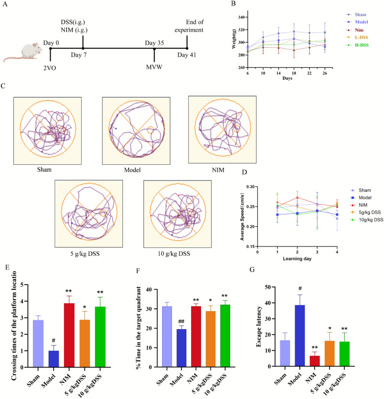
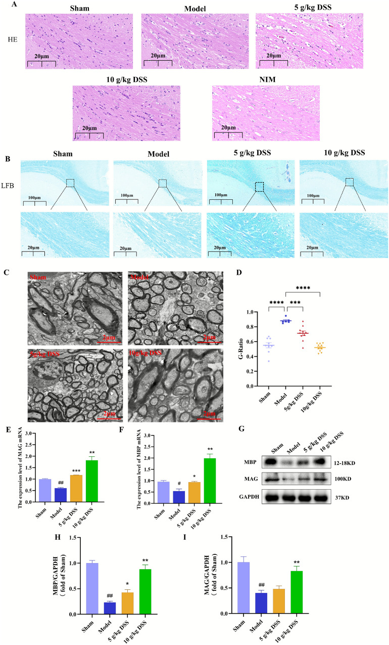
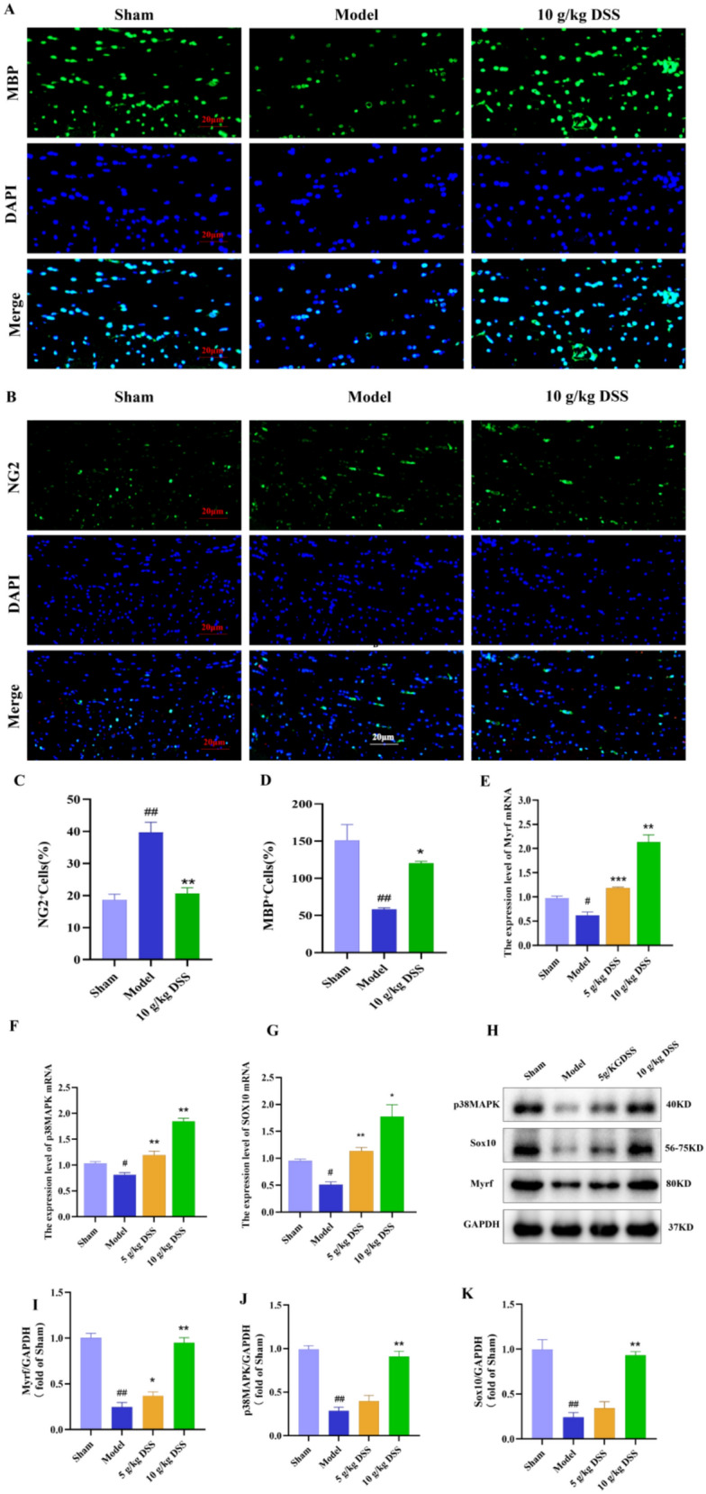
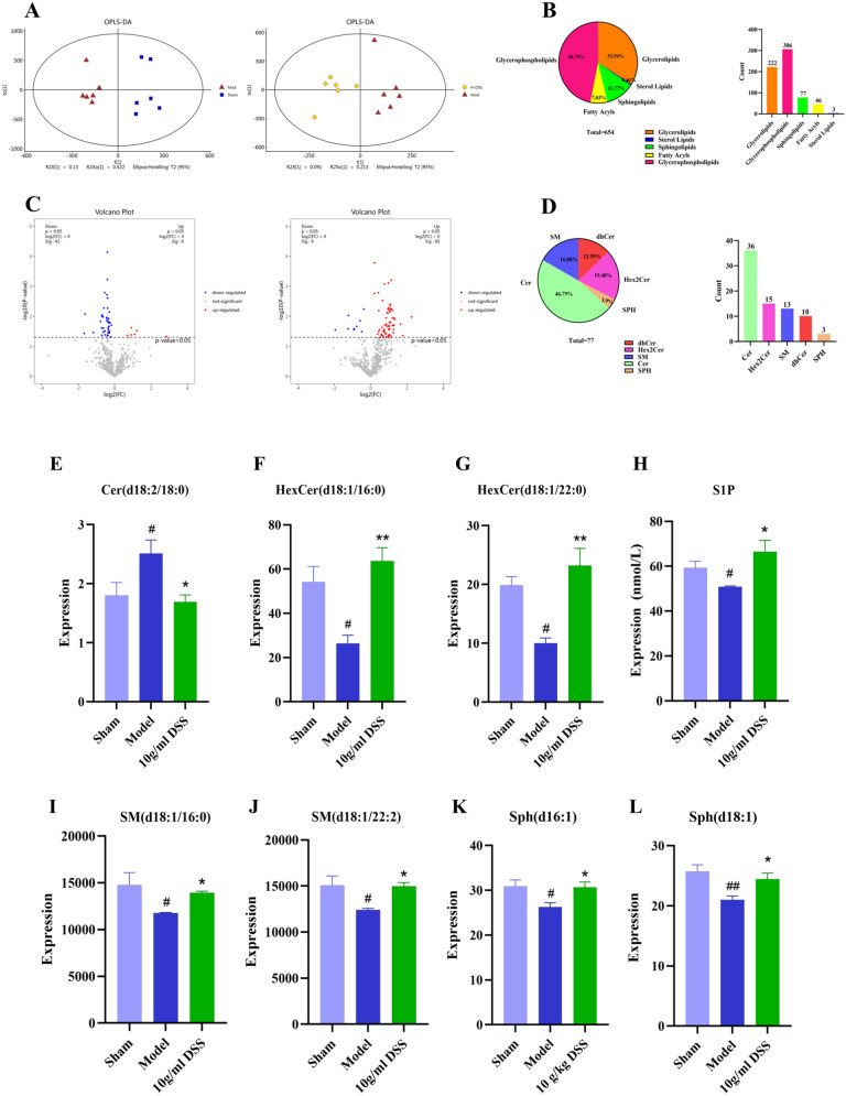
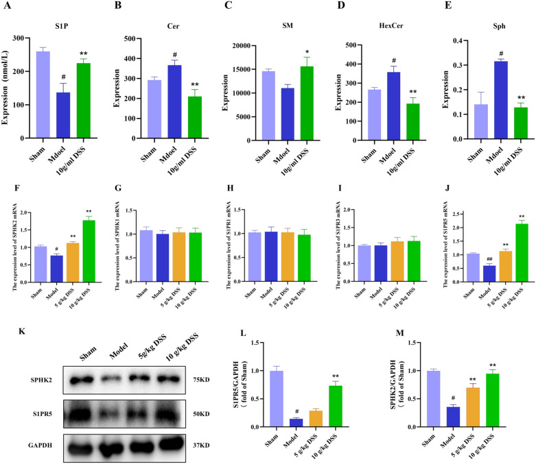
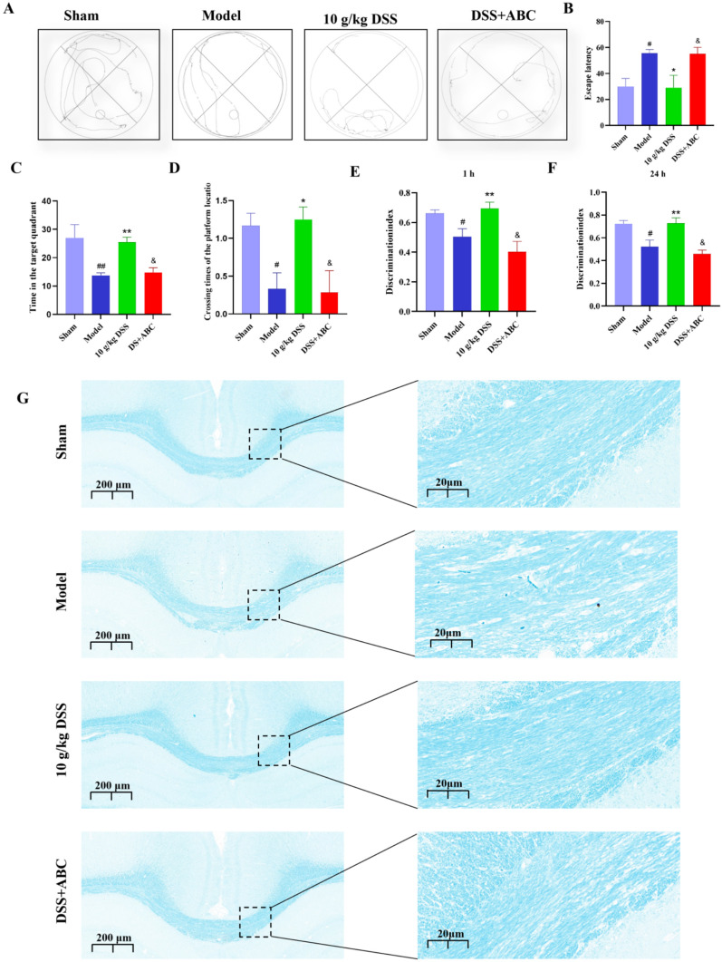
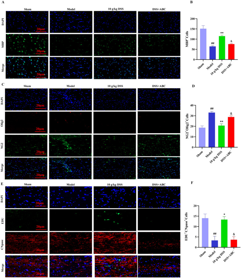
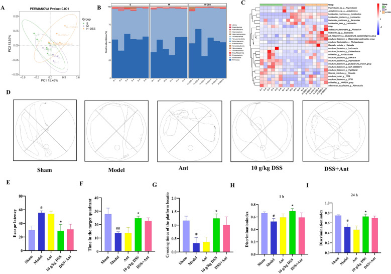
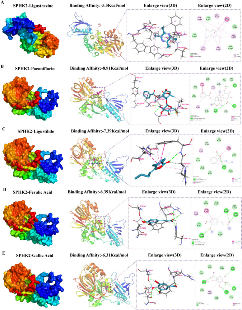
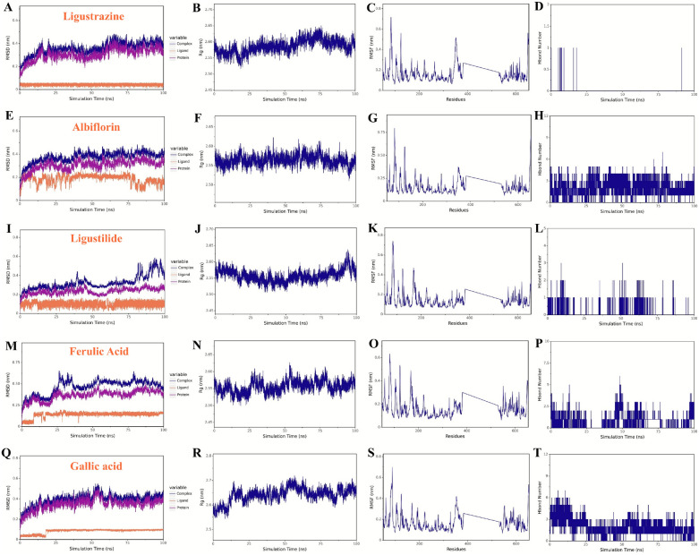
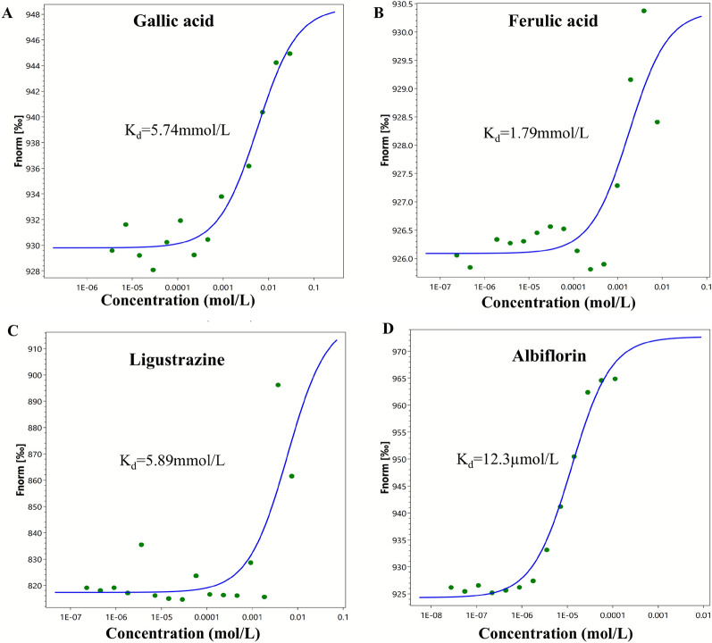
Objective Vascular dementia (VaD) is a neurodegenerative disease primarily characterized by white matter injury and myelin degeneration, and currently, there is a lack of effective treatment options. This study aims to investigate the effects of the traditional Chinese medicine formula Danggui Shaoyao San (DSS) on cognitive function and myelin repair in VaD rats and to elucidate its underlying mechanisms. Methods The VaD rat model was established using the bilateral common carotid artery ligation (2VO) method. The effects of DSS on cognitive function, myelin regeneration, sphingolipid metabolism, and SPHK2/S1P/S1PR5 pathway was conducted using behavioral tests, histological staining, Western blot, lipidomics, qPCR, immunofluorescence, LC-MS/MS, and 16S rRNA sequencing. Besides, molecular docking and molecular dynamics simulation were carried out. Results DSS treatment significantly improved learning and memory abilities in VaD rats, reduced structural damage in the hippocampus and white matter, and promoted the differentiation of oligodendrocyte precursor cells (OPCs) into mature oligodendrocytes (OLs). Lipidomics and molecular biological experiments indicated that DSS activated the SPHK2/S1P/S1PR5 pathway, ameliorated sphingolipid metabolic disorders and increased S1P levels, thereby promoting myelin repair. The specific SPHK2 inhibitor ABC294640 significantly weakened the neuroprotective effects of DSS, further confirming the central role of SPHK2/S1P/S1PR5 pathway. Antibiotic depletion experiments confirmed that the gut microbiota was not a key mediator of the therapeutic effects of DSS. Finally, molecular docking and molecular dynamics simulations indicated that the DSS components Albiflorin and Gallic acid form tighter and more stable interactions with SPHK2. Conclusion DSS improved VaD cognitive impairment by modulating sphingolipid metabolism and promote myelin regenerationa via activating the SPHK2/S1P/S1PR5/SOX10 signaling pathway. This study provides important experimental evidence for the clinical application of DSS in VaD.
Overview
The claims and statistics of this paper haven’t been checked yet.
Run the check — free, no account needed. It reads the open-access text and checks the sources it cites.
Integrity signals
Registry
Cites
Statistics
Figures
Verification
Claims the paper makes
The claims and statistics of this paper haven’t been checked yet.
Run the check — free, no account needed. It reads the open-access text and checks the sources it cites.
Self-check
Percentages, sums and reported differences re-computed from the paper’s own numbers.
The self-check needs the paper’s full text.
Citations
Cites
Varadi, M., Bertoni, D., Magana, P., Paramval, U., Pidruchna, I., Radhakrishnan, M., Tsenkov, M., Nair, S., Mirdita, M., Yeo, J., et al. (2024) · Nucleic Acids Research · doi:10.1093/nar/gkad1011 · Cited by 2,052
Research works printed but not linked: 45
- Bir SC, Khan MW, Javalkar V, Toledo EG, Kelley RE. Emerging Concepts in Vascular Dementia: A Review. J Stroke Cerebrovasc Dis. 2021;30:105864. · DOI
- Agrawal S, Schneider JA. Vascular pathology and pathogenesis of cognitive impairment and dementia in older adults. Cereb Circulation - Cognition Behav. 2022;3:100148. · DOI
- Iadecola C, Duering M, Hachinski MDV, Joutel A, Pendlebury ST, Schneider JA, et al. Vascular Cognitive Impairment and Dementia. J Am Coll Cardiol. 2019;73:3326–44. · DOI · Paper page
- Sengupta P, Ganguly J, Pal S, Ghosal M. Pattern of cognitive deficits in vascular dementia. Indian J Med Res. 2019;149:503–7. · DOI
- Macoir J. Language Impairment in Vascular Dementia: A Clinical Review. J Geriatr Psychiatry Neurol. 2024;37:87–95. · DOI
- Kuang H, Zhou Z-F, Zhu Y-G, Wan Z-K, Yang M-W, Hong F-F, et al. Pharmacological Treatment of Vascular Dementia: A Molecular Mechanism Perspective. Aging Dis. 2021;12:308–26. · DOI
- Gc JPGHALMP. D. Regional contribution of vascular dysfunction in white matter dementia: clinical and neuropathological insights. Frontiers in neurology [Internet]. 2023 [cited 2025 May 22];14. Available from: https://pubmed.ncbi.nlm.nih.gov/37396778/
- Khelfaoui H, Ibaceta-Gonzalez C, Angulo MC. Functional myelin in cognition and neurodevelopmental disorders. Cell Mol Life Sci. 2024;81:181. · DOI
- Zhang H, Yang Y, Zhang J, Huang L, Niu Y, Chen H, et al. Oligodendrocytes Play a Critical Role in White Matter Damage of Vascular Dementia. Neuroscience. 2024;538:1–10. · DOI
- Shimizu S, Ishino Y, Tohyama M, Miyata S. NDE1 positively regulates oligodendrocyte morphological differentiation. Sci Rep. 2018;8:7644. · DOI
- Kalafatakis I, Karagogeos D. Oligodendrocytes and Microglia: Key Players in Myelin Development, Damage and Repair. Biomolecules. 2021;11:1058. · DOI
- Chen D, Huang Y, Shi Z, Li J, Zhang Y, Wang K, et al. Demyelinating processes in aging and stroke in the central nervous system and the prospect of treatment strategy. CNS Neurosci Ther. 2020;26:1219–29. · DOI
- Garcia-Martin G, Alcover-Sanchez B, Wandosell F, Cubelos B. Pathways Involved in Remyelination after Cerebral Ischemia. Curr Neuropharmacol. 2022;20:751–65. · DOI
- Zveik O, Rechtman A, Ganz T, Vaknin-Dembinsky A. The interplay of inflammation and remyelination: rethinking MS treatment with a focus on oligodendrocyte progenitor cells. Mol Neurodegener. 2024;19:53. · DOI
- Binish F, Xiao J. Deciphering the role of sphingosine 1-phosphate in central nervous system myelination and repair. J Neurochem. 2025;169:e16228. · DOI
- Moro K, Ichikawa H, Koyama Y, Abe S, Uchida H, Naruse K, et al. Oral Administration of Glucosylceramide Suppresses Tumor Growth by Affecting the Ceramide/Sphingosine-1-Phosphate Balance in Breast Cancer Tissue. World J Oncol. 2023;14:430–7. · DOI
- Parveen F, Bender D, Law S-H, Mishra VK, Chen C-C, Ke L-Y. Role of Ceramidases in Sphingolipid Metabolism and Human Diseases. Cells. 2019;8:1573. · DOI
- Czubowicz K, Jęśko H, Wencel P, Lukiw WJ, Strosznajder RP. The Role of Ceramide and Sphingosine-1-Phosphate in Alzheimer’s Disease and Other Neurodegenerative Disorders. Mol Neurobiol. 2019;56:5436–55. · DOI · Paper page
- Tringali C, Giussani P. Ceramide and Sphingosine-1-Phosphate in Neurodegenerative Disorders and Their Potential Involvement in Therapy. Int J Mol Sci. 2022;23:7806. · DOI
- Sassoli C, Pierucci F, Zecchi-Orlandini S, Meacci E. Sphingosine 1-Phosphate (S1P)/ S1P Receptor Signaling and Mechanotransduction: Implications for Intrinsic Tissue Repair/Regeneration. Int J Mol Sci. 2019;20:5545. · DOI
- Ningning Y, Ying X, Xiang L, Yue S, Zhongda W, Ruoyu J, et al. Danggui-Shaoyao San alleviates cognitive impairment via enhancing HIF-1α/EPO axis in vascular dementia rats. J Ethnopharmacol. 2024;331:118306. · DOI
- Cheng X, Dai Y, Shang B, Zhang S, Lin L, Wu Q, et al. Danggui Shaoyao San and disassembled prescription: neuroprotective effects via AMPK/mTOR-mediated autophagy in mice. BMC Complement Med Ther. 2024;24:298. · DOI
- Kim Y, Cho S-H. Danggui-Shaoyao-San for dementia. Med (Baltim). 2020;99:e18507. · DOI
- Liu X, Yao L, Ye X, Qin Y, Chen S, Jiang Q, et al. Danggui-Shaoyao-San (DSS) ameliorating cognitive impairment in ischemia–reperfusion vascular dementia mice through miR-124 regulating PI3K/Akt signaling pathway. Brain Res. 2024;1845:149135. · DOI
- He J, Jin Y, He C, Li Z, Yu W, Zhou J, et al. Danggui Shaoyao San: comprehensive modulation of the microbiota-gut-brain axis for attenuating Alzheimer’s disease-related pathology. Front Pharmacol. 2024;14:1338804. · DOI
- Denecke J, Dewenter A, Lee J, Franzmeier N, Valentim C, Kopczak A, et al. Reduced myelin contributes to cognitive impairment in patients with monogenic small vessel disease. Alzheimers Dement. 2025;21:e70127. · DOI
- Liu C, Yin H, Chen X, Ban W, Zhu G, Wang J. Electroacupuncture promotes oligodendrocyte differentiation and myelin repair in a rat model of vascular dementia: Investigation of the mechanism from NF-κB-mediated inflammation. Neuroscience. 2025;572:21–34. · DOI
- Gong K, Zhou S, Xiao L, Xu M, Zhou Y, Lu K, et al. Danggui Shaoyao San ameliorates Alzheimer’s disease by regulating lipid metabolism and inhibiting neuronal ferroptosis through the AMPK/Sp1/ACSL4 signaling pathway. Front Pharmacol. 2025;16:1588375. · DOI
- Lorenzini L, Fernandez M, Baldassarro VA, Bighinati A, Giuliani A, Calzà L, et al. White Matter and Neuroprotection in Alzheimer’s Dementia. Molecules. 2020;25:503. · DOI
- Li Y, Wan LP, Song N-N, Ding Y-Q, Zhao S, Niu J et al. RNF220-mediated K63-linked polyubiquitination stabilizes Olig proteins during oligodendroglial development and myelination. Sci Adv 10:eadk3931. · DOI
- Chen X, Chen L, Lin G, Wang Z, Kodali MC, Li M, et al. White matter damage as a consequence of vascular dysfunction in a spontaneous mouse model of chronic mild chronic hypoperfusion with eNOS deficiency. Mol Psychiatry. 2022;27:4754–69. · DOI
- Walker KA, Duggan MR, Gong Z, Dark HE, Laporte JP, Faulkner ME, et al. MRI and fluid biomarkers reveal determinants of myelin and axonal loss with aging. Ann Clin Transl Neurol. 2023;10:397–407. · DOI
- Pappas I, Lohman T, Dutt S, Kapoor A, Engstrom AC, Alitin JPM et al. Cerebral hypoperfusion, brain structural integrity, and cognitive impairment in older APOE4 carriers. GeroScience [Internet]. 2025 [cited 2025 May 20]; Available from: https://doi.org/10.1007/s11357-025-01642-5 · DOI
- Luo Q, Li F, Liu X, Yan T, Yang L, Zhu W, et al. Puerarin mitigates cognitive decline and white matter injury via CD36-Mediated microglial phagocytosis in chronic cerebral hypoperfusion. Phytomedicine. 2025;138:156396. · DOI
- Davis DL, Mahawar U, Pope VS, Allegood J, Sato-Bigbee C, Wattenberg BW. Dynamics of sphingolipids and the serine palmitoyltransferase complex in rat oligodendrocytes during myelination. J Lipid Res. 2020;61:505–22. · DOI
- Giussani P, Prinetti A, Tringali C. The role of Sphingolipids in myelination and myelin stability and their involvement in childhood and adult demyelinating disorders. J Neurochem. 2021;156:403–14. · DOI · Paper page
- Simon MV, Basu SK, Qaladize B, Grambergs R, Rotstein NP, Mandal N. Sphingolipids as critical players in retinal physiology and pathology. J Lipid Res. 2021;62:100037. · DOI
- Qiu S, Palavicini JP, Wang J, Gonzalez NS, He S, Dustin E, et al. Adult-onset CNS myelin sulfatide deficiency is sufficient to cause Alzheimer’s disease-like neuroinflammation and cognitive impairment. Mol Neurodegener. 2021;16:64. · DOI
- Xu X, Han Y, Zhu T, Fan F, Wang X, Liu Y, et al. The role of SphK/S1P/S1PR signaling pathway in bone metabolism. Biomed Pharmacother. 2023;169:115838. · DOI
- Pérez-Jeldres T, Alvarez-Lobos M, Rivera-Nieves J. Targeting Sphingosine-1-Phosphate Signaling in Immune-Mediated Diseases: Beyond Multiple Sclerosis. Drugs. 2021;81:985–1002. · DOI · Paper page
- Ji X, Chen Z, Wang Q, Li B, Wei Y, Li Y, et al. Sphingolipid metabolism controls mammalian heart regeneration. Cell Metabol. 2024;36:839–e8568. · DOI
- Lucaciu A, Brunkhorst R, Pfeilschifter JM, Pfeilschifter W, Subburayalu J. The S1P–S1PR Axis in Neurological Disorders—Insights into Current and Future Therapeutic Perspectives. Cells. 2020;9:1515. · DOI
- Bai X, Ya R, Tang X, Cai M. Role and interaction of bacterial sphingolipids in human health. Front Microbiol. 2023;14:1289819. · DOI
- Le HH, Lee M-T, Besler KR, Johnson EL. Host hepatic metabolism is modulated by gut microbiota–derived sphingolipids. Cell Host Microbe. 2022;30:798–e8087. · DOI
- Brown EM, Ke X, Hitchcock D, Jeanfavre S, Avila-Pacheco J, Nakata T, et al. Bacteroides-derived sphingolipids are critical for maintaining intestinal homeostasis and symbiosis. Cell Host Microbe. 2019;25:668–e6807. · DOI · Paper page
Metadata
- Authors
- Su, Y., Zhong, Y., Yuan, N., Li, X., Xu, Y., Yang, H., Huang, M., Zhang, Y., & Cheng, X.
- Affiliations
- Nanjing University of Chinese Medicine (CN)
- Nanjing University of Chinese Medicine
- Corresponding author
- Cheng, Xiaolan
- Venue
- Chinese Medicine
- Published
- 2025-08-12
- Volume
- 21
- Issue
- 1
- Pages
- 11-11
- ISSN
- 1749-8546 (ISSN-L)
- PMC
- PMC12777056
- OpenAlex
- W7119109165
- Cited by
- 0
- References
- 1
- FWCI
- 0.00
- Type
- journal article
- Language
- en
- Open access
- gold
- doi.org
- Locations
- Chinese Medicine (publishedVersion, cc-by)
- Research Square (acceptedVersion, cc-by)
- PubMed (publishedVersion)
- PubMed Central (publishedVersion, cc-by)
- DOAJ (DOAJ: Directory of Open Access Journals) (submittedVersion, cc-by-sa)
- Funders
- National Natural Science Foundation of China (82274218)
- Topics
- Life Sciences › Neuroscience › Neurology › Neurological Disease Mechanisms and Treatments
- Life Sciences › Biochemistry, Genetics and Molecular Biology › Molecular Biology › Sphingolipid Metabolism and Signaling
- Life Sciences › Neuroscience › Developmental Neuroscience › Neurogenesis and neuroplasticity mechanisms
- Keywords
- Danggui Shaoyao San, vascular dementia, sphingolipid metabolism, oligodendrocyte differentiation, remyelination, sphingosine-1-phosphate, oligodendrocyte precursor cells, cognitive impairment, albiflorin, gallic acid, traditional Chinese medicine formula, bilateral common carotid artery occlusion, gut microbiota
About these checks
Registry, retraction, Cites and statistics results are computed, not judged, with one exception: a printed reference that no index lists is read once by a model before it counts as a citation error. Claims are extracted by a model and kept only when their quote appears verbatim in the text; each cited source is then checked, with a model’s help, for whether it says what the claim relies on. Those verdicts can be wrong — read the claim’s quote and the cited source before relying on one.
Statistics and arithmetic are recomputed, not judged.
Confidence bands are shown only for fields calibrated on our annotated test set. None is calibrated yet, so no band is shown.
What counts as a claim ›How we measure confidence ›How we check this ›
Are you an author, or do you see a problem with this page? Request a review
How to cite
Cite CoChat for the checks on this page, and the paper for its own findings.
- This page
- CoChat. (2026). Citation check: Danggui-Shaoyao-San modulates sphingolipid metabolism to promote oligodendrocyte differentiation and maturation in vascular dementia rats. https://app.cochat.ai/papers/10.21203/rs.3.rs-7241365/v1
- The paper
- Su, Y., Zhong, Y., Yuan, N., Li, X., Xu, Y., Yang, H., Huang, M., Zhang, Y., & Cheng, X. (2025). Danggui-Shaoyao-San modulates sphingolipid metabolism to promote oligodendrocyte differentiation and maturation in vascular dementia rats. Chinese Medicine. https://doi.org/10.21203/rs.3.rs-7241365/v1
Data sources
Bibliographic data: Semantic Scholar, OpenAlex, Crossref.
Contains information from the Semantic Scholar Academic Graph Datasets, made available under the ODC Attribution License (ODC-BY 1.0).
Semantic Scholar’s data is described in: Kinney, R., et al. (2023). The Semantic Scholar Open Data Platform. arXiv:2301.10140.
