

The DOI matches its Crossref record; 2 of the 2 references it cites were found in a registry or an index.
Molecular dynamics simulation analysis of structural dynamic cross correlation induced by odorant hydrogen-bonding in mouse eugenol ol- factory receptor
Open accessPublished version of a preprint
Abstract
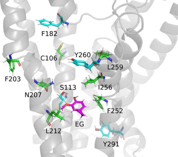
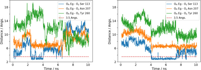
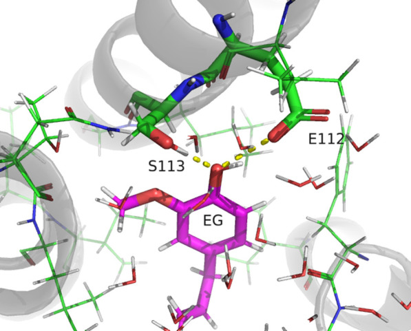
Structural fluctuations and dynamic cross-correlations in the mouse eugenol olfactory receptor (Olfr73) were studied by molecular dynamics (MD) simulation to characterize the dynamic response of the protein upon ligand binding. The initial structure was generated by the artificial intelligence tool AlphaFold2 due to the current lack of experimental data. We focused on the hydrogen (H) bond of the odorant eugenol to Ser113, Asn207, and Tyr260 of the receptor protein, the importance of which has been suggested by previous experimental studies. The H-bond was not observed in docking simulations, but in subsequent MD simulations the H-bond to Ser113 was formed in 2-4 ns. The lifetime of the H-bond was in the range of 1-20 ns. On the trajectory with the most stable (20 ns) H-bond, the structural fluctuation of the α-carbon atoms of the receptor main chain was studied by calculating the root mean square fluctuations, the dynamic cross-correlation map, and the time-dependent dynamic cross-correlation. The analysis suggested a correlation transfer pathway Ser113 → Phe182 → (Leu259 or Tyr260) → Tyr291 induced by the ligand binding with a time scale of 4-6 ns.
Overview
The claims and statistics of this paper haven’t been checked yet.
Run the check — free, no account needed. It reads the open-access text and checks the sources it cites.
Integrity signals
Registry
Cites
Statistics
Figures
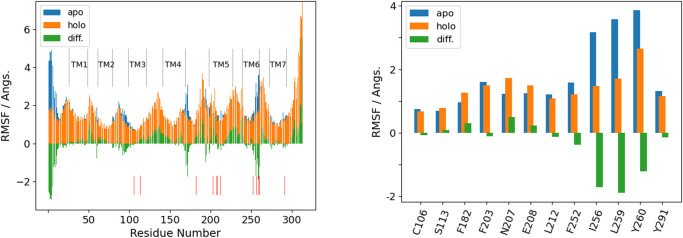
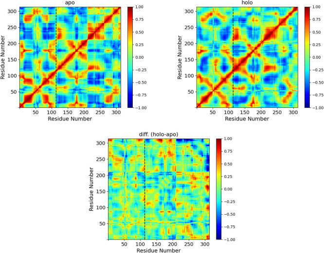
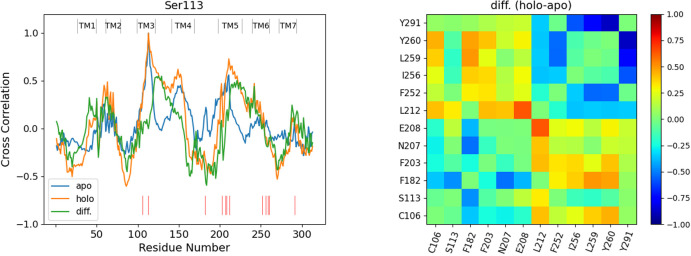
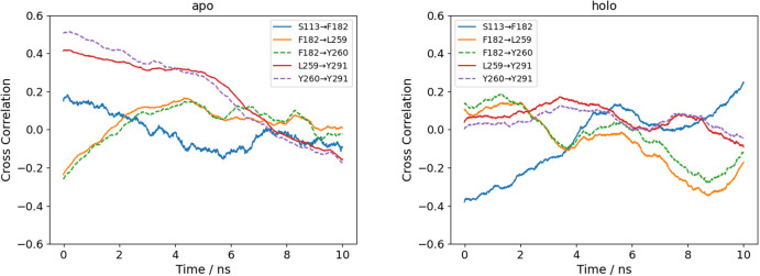
Verification
Claims the paper makes
The claims and statistics of this paper haven’t been checked yet.
Run the check — free, no account needed. It reads the open-access text and checks the sources it cites.
Self-check
Percentages, sums and reported differences re-computed from the paper’s own numbers.
The self-check needs the paper’s full text.
Citations
Cites
Highly accurate protein structure prediction with AlphaFold
Jumper, J., Evans, R., Pritzel, A., Green, T., Figurnov, M., Ronneberger, O., Tunyasuvunakool, K., Bates, R., Žídek, A., Potapenko, A., et al. (2021) · Nature · doi:10.1038/s41586-021-03819-2 · Cited by 43,764
A structural biology community assessment of AlphaFold2 applications
Akdel, M., Pires, D. E. V., Pardo, E. P., Jänes, J., Zalevsky, A. O., Mészáros, B., Bryant, P., Good, L. L., Laskowski, R. A., Pozzati, G., et al. (2022) · Nature Structural & Molecular Biology · doi:10.1038/s41594-022-00849-w · Cited by 675
Research works printed but not linked: 43
- [1] Buck, L., Axel, R. A novel multigene family may encode odorant receptors: A molecular basis for odor recognition. Cell 65, 175–187 (1991). https://doi.org/10.1016/0092-8674(91)90418-x · DOI · Paper page
- [2] Kraft, P., Bajgrowicz, J. A., Denis, C., Fráter, G. Odds and trends: Recent developments in the chemistry of odorants. Angew. Chem. Int. Ed. Engl. 39, 2980–3010 (2000). https://doi.org/10.1002/1521-3773(20000901)39:17<2980::AID-ANIE2980>3.0.CO;2-%23 · DOI
- [3] Firestein, S. How the olfactory system makes sense of scents. Nature 413, 211–218 (2001). https://doi.org/10.1038/35093026 · DOI
- [4] Zozulya, S., Echeverri, F., Nguyen, T. The human olfactory receptor repertoire. Genome Biol. 2, (2001). https://doi.org/10.1186/gb-2001-2-6-research0018 · DOI
- [5] Zhang, X., Firestein, S. The olfactory receptor gene superfamily of the mouse. Nat. Neurosci. 5, 124–133 (2002). https://doi.org/10.1038/nn800 · DOI
- [6] Serizawa, S., Miyamichi, K., Nakatani, H., Suzuki, M., Saito, M., Yoshihara, Y., et al. Negative feedback fegulation ensures the one receptor-one olfactory neuron rule in mouse. Science 302, 2088–2094 (2003). https://doi.org/10.1126/science.1089122 · DOI
- [7] Malnic, B., Godfrey, P. A., Buck, L. B. The human olfactory receptor gene family. Proc. Natl. Acad. Sci. U.S.A. 101, 2584–2589 (2004). https://doi.org/10.1073/pnas.0307882100 · DOI
- [8] Reed, R. R. After the holy grail. Cell 116, 329–336 (2004). https://doi.org/10.1016/s0092-8674(04)00047-9 · DOI
- [9] Sell, C. S. Chemistry and the Sense of Smell (Wiley, New Jersey, 2014). · DOI
- [10] Yang, D., Zhou, Q., Labroska, V., Qin, S., Darbalaei, S., Wu, Y., et al. G protein-coupled receptors: structure- and function-based drug discovery. Signal Transduct. Target. Ther. 6, 7 (2021). https://doi.org/10.1038/s41392-020-00435-w · DOI
- [11] DiPizio, A., Niv, M. Y. Computational studies of smell and taste receptors. Isr. J. Chem. 54, 1205–1218 (2014). https://doi.org/10.1002/ijch.201400027 · DOI
- [12] Doszczak, L., Kraft, P., Weber, H.-P., Bertermann, R., Triller, A., Hatt, H., et al. Prediction of perception: Probing the hOR17-4 olfactory receptor model with silicon analogues of bourgeonal and lilial. Angew. Chem. Int. Ed. Engl. 46, 3367–3371 (2007). https://doi.org/10.1002/anie.200605002 · DOI
- [13] Gelis, L., Wolf, S., Hatt, H., Neuhaus, E. M., Gerwert, K. Prediction of a ligand-binding niche within a human olfactory receptor by combining site-directed mutagenesis with dynamic homology modeling. Angew. Chem. Int. Ed. Engl. 51, 1274–1278 (2011). https://doi.org/10.1002/anie.201103980 · DOI
- [14] Floriano, W. B., Vaidehi, N., Goddard, W. A., Singer, M. S., Shepherd, G. M. Molecular mechanisms underlying differential odor responses of a mouse olfactory receptor. Proc. Natl. Acad. Sci. U.S.A. 97, 10712–10716 (2000). https://doi.org/10.1073/pnas.97.20.10712 · DOI
- [15] Floriano, W. B. Making sense of olfaction through predictions of the 3-D structure and function of olfactory receptors. Chem. Senses 29, 269–290 (2004). https://doi.org/10.1093/chemse/bjh030 · DOI
- [16] Katada, S., Hirokawa, T., Oka, Y., Suwa, M., Touhara, K. Structural basis for a broad but selective ligand spectrum of a mouse olfactory receptor: Mapping the odorant-binding site. J. Neurosci. 25, 1806–1815 (2005). https://doi.org/10.1523/jneurosci.4723-04.2005 · DOI
- [17] Baud, O., Etter, S., Spreafico, M., Bordoli, L., Schwede, T., Vogel, H., et al. The mouse eugenol odorant receptor: Structural and functional plasticity of a broadly tuned odorant binding pocket. Biochemistry 50, 843–853 (2010). https://doi.org/10.1021/bi1017396 · DOI
- [18] Baud, O., Yuan, S., Veya, L., Filipek, S., Vogel, H., Pick, H. Exchanging ligand-binding specificity between a pair of mouse olfactory receptor paralogs reveals odorant recognition principles. Sci. Rep. 5, 14948 (2015). https://doi.org/10.1038/srep14948 · DOI
- [19] Yuan, S., Dahoun, T., Brugarolas, M., Pick, H., Filipek, S., Vogel, H. Computational modeling of the olfactory receptor Olfr73 suggests a molecular basis for low potency of olfactory receptor-activating compounds. Commun. Biol. 2, 141 (2019). https://doi.org/10.1038/s42003-019-0384-8 · DOI
- [20] Pándy-Szekeres, G., Caroli, J., Mamyrbekov, A., Kermani, A. A., Keserü, G. M., Kooistra, A. J., et al. GPCRdb in 2023: state-specific structure models using AlphaFold2 and new ligand resources. Nucleic Acids Res. 51, D395–D402 (2022). https://doi.org/10.1093/nar/gkac1013 · DOI
- [21] Ballesteros, J. A., Weinstein, H. Integrated methods for the construction of three-dimensional models and computational probing of structure-function relations in G protein-coupled receptors. Methods Neurosci. 25, 366–428 (1995). https://doi.org/10.1016/S1043-9471(05)80049-7 · DOI
- [24] Yang, Z., Zeng, X., Zhao, Y., Chen, R. AlphaFold2 and its applications in the fields of biology and medicine. Signal Transduct. Target. Ther. 8, 115 (2023). https://doi.org/10.1038/s41392-023-01381-z · DOI
- [25] McCammon, J. A. Protein dynamics. Rep. Prog. Phys. 47, 1 (1984). https://doi.org/10.1088/0034-4885/47/1/001 · DOI
- [26] da Penha Valente, R. P., de Souza, R. C., de Medeiros Muniz, G., Ferreira, J. E. V., de Miranda, R. M., e Lima, A. H. L., et al. Using accelerated molecular dynamics simulation to elucidate the effects of the T198F mutation on the molecular flexibility of the West Nile virus envelope protein. Sci. Rep. 10, 9625 (2020). https://doi.org/10.1038/s41598-020-66344-8 · DOI
- [27] Parida, P. K., Paul, D., Chakravorty, D. The natural way forward: Molecular dynamics simulation analysis of phytochemicals from Indian medicinal plants as potential inhibitors of SARS-CoV-2 targets. Phytother. Res. 34, 3420–3433 (2020). https://doi.org/10.1002/ptr.6868 · DOI
- [28] Wang, Z., Hu, B., An, Y., Wang, J. Exploring the resistance mechanisms of distal D835V mutation in FLT3 to inhibitors. Oxid. Med. Cell. Longev. 2022, 3720026 (2022). https://doi.org/10.1155/2022/3720026 · DOI
- [29] Dash, R., Munni, Y. A., Mitra, S., Choi, H. J., Jahan, S. I., Chowdhury, A., et al. Dynamic insights into the effects of nonsynonymous polymorphisms (nsSNPs) on loss of TREM2 function. Sci. Rep. 12, 9378 (2022). https://doi.org/10.1038/s41598-022-13120-5 · DOI
- [30] Mirdita, M., Schütze, K., Moriwaki, Y., Heo, L., Ovchinnikov, S., Steinegger, M. ColabFold: making protein folding accessible to all. Nat. Methods 19, 679–682 (2022). https://doi.org/10.1038/s41592-022-01488-1 · DOI · Paper page
- [31] Fukunishi, Y., Mikami, Y., Nakamura, H. The filling potential method: A method for estimating the free energy surface for protein-ligand docking. J. Phys. Chem. B 107, 13201–13210 (2003). https://doi.org/10.1021/jp035478e · DOI
- [32] Berendsen, H. J. C., Postma, J. P. M., van Gunsteren, W. F., DiNola, A., Haak, J. R. Molecular dynamics with coupling to an external bath. J. Chem. Phys. 81, 3684–3690 (1984). https://doi.org/10.1063/1.448118 · DOI
- [33] Evans, D. J., Hoover, W. G., Failor, B. H., Moran, B., Ladd, A. J. C. Nonequilibrium molecular dynamics via Gauss’s principle of least constraint. Phys. Rev. A 28, 1016–1021 (1983). https://doi.org/10.1103/physreva.28.1016 · DOI
- [34] Nosé, S. Constant temperature molecular dynamics methods. Prog. Theor. Phys. Suppl. 103, 1–46 (1991). https://doi.org/10.1143/ptps.103.1 · DOI
- [35] Ryckaert, J.-P., Ciccotti, G., Berendsen, H. J. Numerical integration of the Cartesian equations of motion of a system with constraints: molecular dynamics of n-alkanes. J. Comput. Phys. 23, 327–341 (1977). https://doi.org/10.1016/0021-9991(77)90098-5 · DOI
- [36] Kasahara, K., Ma, B., Goto, K., Dasgupta, B., Higo, J., Fukuda, I., et al. myPresto/omegagene: a GPU-accelerated molecular dynamics simulator tailored for enhanced conformational sampling methods with a non-Ewald electrostatic scheme. Biophys. Physicobiol. 13, 209–216 (2016). https://doi.org/10.2142/biophysico.13.0_209 · DOI
- [37] Fukuda, I., Yonezawa, Y., Nakamura, H. Molecular dynamics scheme for precise estimation of electrostatic interaction via zero-dipole summation principle. J. Chem. Phys. 134, 164107 (2011). https://doi.org/10.1063/1.3582791 · DOI
- [38] Mashimo, T., Fukunishi, Y., Kamiya, N., Takano, Y., Fukuda, I., Nakamura, H. Molecular dynamics simulations accelerated by GPU for biological macromolecules with a non-Ewald scheme for electrostatic interactions. J. Chem. Theory Comput. 9, 5599–5609 (2013). https://doi.org/10.1021/ct400342e · DOI
- [39] Wang, J., Cieplak, P., Kollman, P. A. How well does a restrained electrostatic potential (RESP) model perform in calculating conformational energies of organic and biological molecules? J. Comput. Chem. 21, 1049–1074 (2000). https://doi.org/10.1002/1096-987x(200009)21:12<1049::AID-JCC3>3.0.CO;2-F · DOI
- [40] Dickson, C. J., Madej, B. D., Skjevik, Å. A., Betz, R. M., Teigen, K., Gould, I. R., et al. Lipid14: The Amber lipid force field. J. Chem. Theory Comput. 10, 865–879 (2014). https://doi.org/10.1021/ct4010307 · DOI
- [41] Fukunishi, Y., Mikami, Y., Nakamura, H. Similarities among receptor pockets and among compounds: Analysis and application to in silico ligand screening. J. Mol. Graph. Model 24, 34–45 (2005). https://doi.org/10.1016/j.jmgm.2005.04.004 · DOI
- [42] Schmidt, M. W., Baldridge, K. K., Boatz, J. A., Elbert, S. T., Gordon, M. S., Jensen, J. H., et al. General atomic and molecular electronic structure system. J. Comput. Chem. 14, 1347–1363 (1993). https://doi.org/10.1002/jcc.540141112 · DOI
- [44] Nguyen, T. A., Puhl, H. L., Hines, K., Liput, D. J., Vogel, S. S. Binary-FRET reveals transient excited-state structure associated with activity-dependent CaMKII - NR2B binding and adaptation. Nat. Commun. 13, 6335 (2022). https://doi.org/10.1038/s41467-022-33795-8 · DOI
- [45] Yamashita, S., Mizuno, M., Mizutani, Y. High suitability of tryptophan residues as a spectroscopic thermometer for local temperature in proteins under nonequilibrium conditions. J. Chem. Phys. 156, 075101 (2022). https://doi.org/10.1063/5.0079797 · DOI
- [46] Terazima, M. Spectrally silent protein reaction dynamics revealed by time-resolved thermodynamics and diffusion techniques. Acc. Chem. Res. 54, 2238–2248 (2021). https://doi.org/10.1021/acs.accounts.1c00113 · DOI
Metadata
- Authors
- Okamoto, C., & Ando, K.
- Affiliations
- Tokyo Woman's Christian University (JP)
- Corresponding author
- Ando, Koji
- ORCID
- Ando, Koji
- Venue
- Biophysics and Physicobiology
- Publisher
- Biophysical Society of Japan
- Published
- 2024
- Volume
- 21
- Issue
- 1
- Pages
- n/a
- ISSN
- 2189-4779 (ISSN-L)
- PMC
- PMC11128758
- OpenAlex
- W4390939122
- Cited by
- 19
- References
- 2
- Citations by year
- 2026: 7 · 2025: 11 · 2024: 1
- FWCI
- 2.59
- Type
- journal article
- Language
- en
- Open access
- gold
- doi.org
- Locations
- Biophysics and Physicobiology (publishedVersion)
- bioRxiv (Cold Spring Harbor Laboratory) (acceptedVersion, cc-by-nc-nd)
- PubMed Central (publishedVersion, cc-by-nc-sa)
- PubMed (publishedVersion)
- arXiv (Cornell University) (submittedVersion, cc-by-nc-nd)
- DOAJ (DOAJ: Directory of Open Access Journals) (submittedVersion)
- arXiv (Cornell University)
- Funders
- Japan Society for the Promotion of Science (19K22173)
- Topics
- Life Sciences › Biochemistry, Genetics and Molecular Biology › Molecular Biology › Protein Structure and Dynamics
- Life Sciences › Biochemistry, Genetics and Molecular Biology › Molecular Biology › Receptor Mechanisms and Signaling
- Life Sciences › Neuroscience › Sensory Systems › Olfactory and Sensory Function Studies
- Keywords
- eugenol, odorant binding, hydrogen bonding, structural fluctuations, ligand binding, RMSF
About these checks
Registry, retraction, Cites and statistics results are computed, not judged, with one exception: a printed reference that no index lists is read once by a model before it counts as a citation error. Claims are extracted by a model and kept only when their quote appears verbatim in the text; each cited source is then checked, with a model’s help, for whether it says what the claim relies on. Those verdicts can be wrong — read the claim’s quote and the cited source before relying on one.
Statistics and arithmetic are recomputed, not judged.
Confidence bands are shown only for fields calibrated on our annotated test set. None is calibrated yet, so no band is shown.
What counts as a claim ›How we measure confidence ›How we check this ›
Are you an author, or do you see a problem with this page? Request a review
