

The DOI matches its Crossref record; 57 of the 57 references it cites were found in a registry or an index.
Biodistribution, Clearance And Morphological Alterations Of Intravenously Administered Iron Oxide Nanoparticles In Male Wistar Rats
Open accessAbstract
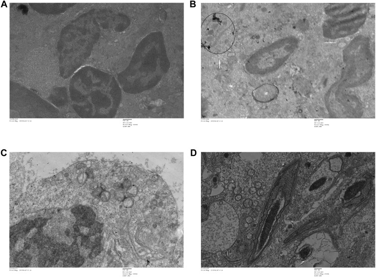
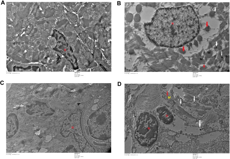
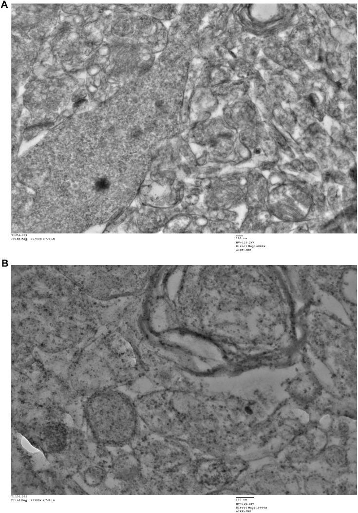
INTRODUCTION: Nanoparticles are used worldwide because of their unique properties, with large-scale application in various fields, such as medicine, cosmetics and industries. In view of their widespread use, the potential adverse effects of nanoparticles have become a significant cause for concern, in terms of not only human health and safety but also the environment. The present investigation focused on establishing the bioaccumulation patterns and ultrastructural changes induced by retained iron oxide nanoparticles (IONPs) in various target organs of rats. METHODS: Twenty-four male Wistar rats were randomly divided into four groups. Experimental animals were intravenously administered different doses of IONPs (7.5 mg/kg, 15 mg/kg and 30 mg/kg) once in a week for 4 weeks. Urine and feces samples were collected on a daily basis to assess nanoparticle clearance and analyzed via atomic absorption spectroscopy (AAS). At the end of the experiment, rats were euthanized and different organs, including spleen, liver, kidney, lung, heart, testis and brain, were dissected. Bioaccumulation of iron in organs and ultrastructural changes induced by IONPs were determined. RESULTS: The maximal concentration of iron was detected in spleen and minimal concentration in the brain. The level of iron accumulation in organs was as follows: spleen>blood>liver>kidney>lung>heart>testis>brain. The excretion profile in urine revealed maximum excretion on the day following administration that was maintained until day 28, whereas the iron content in feces remained high during the first three days after injection. A similar pattern was observed throughout the duration of the experiment. Ultrastructural alterations were detected in spleen, kidney, lung, heart, testis, brain and liver, indicative of cellular damage induced by accumulating nanoparticles in these organs. CONCLUSION: Intravenous administration of IONPs results in ultrastructural changes and dose-dependent bioaccumulation in different organs of rats.
Overview
The claims and statistics of this paper haven’t been checked yet.
Run the check — free, no account needed. It reads the open-access text and checks the sources it cites.
Integrity signals
Registry
Cites
Statistics
Figures
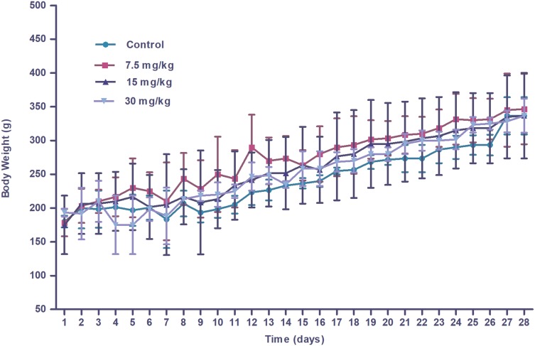
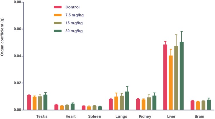
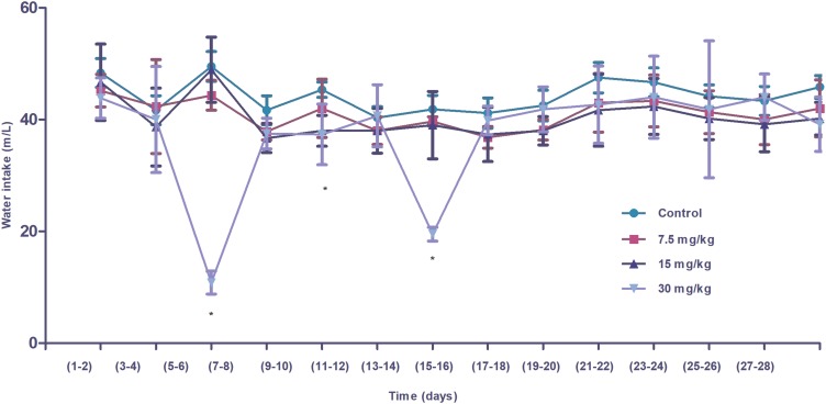
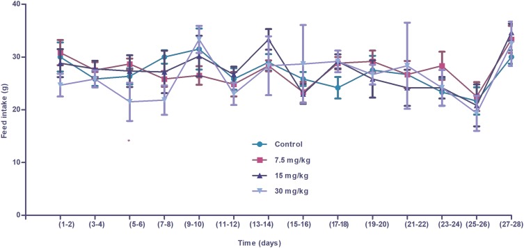
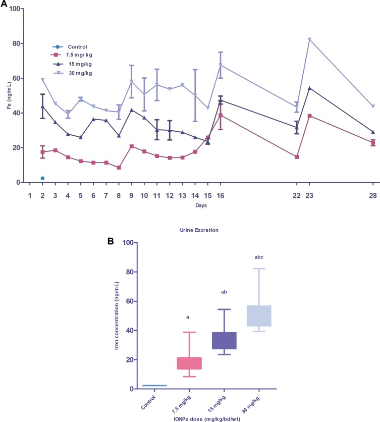
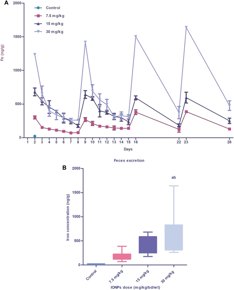
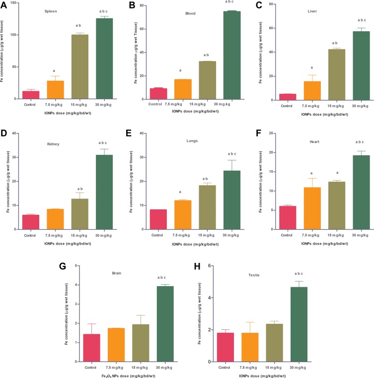
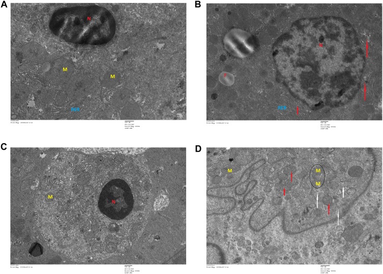
Verification
Claims the paper makes
The claims and statistics of this paper haven’t been checked yet.
Run the check — free, no account needed. It reads the open-access text and checks the sources it cites.
Claims other papers make about this one
Each is checked against this paper’s own text: does it say what the citing paper says it does?
The cited source says what the claim attributes to it.
In the citing paper
“Their accumulation pattern in organs of rodent models was revealed as follows: spleen > blood > liver > kidney > lungs > heart > testis > brain.”
From The blood–brain barrier: Structure, regulation and drug delivery (2023) · cites this paper as [328]
Self-check
Percentages, sums and reported differences re-computed from the paper’s own numbers.
The self-check needs the paper’s full text.
Citations
Cites
Yaşlı Kadınlarda Üreme Sağlığı
Kokanalı, D., & Üstün, Y. E. (2019) · DergiPark (Istanbul University) · Cited by 169,621
Oberdörster, G., Maynard, A., Donaldson, K., Castranova, V., Fitzpatrick, J., Ausman, K., Carter, J., Karn, B., Kreyling, W., Lai, D., et al. (2005) · Particle and Fibre Toxicology · doi:10.1186/1743-8977-2-8 · Cited by 2,134
Neuberger, T., Schöpf, B., Hofmann, H., Hofmann, M., & von Rechenberg, B. (2005) · Journal of Magnetism and Magnetic Materials · doi:10.1016/j.jmmm.2005.01.064 · Cited by 1,727
Particle size-dependent organ distribution of gold nanoparticles after intravenous administration
de Jong, W. H., Hagens, W. I., Krystek, P., Burger, M. C., Sips, A. J. A. M., & Geertsma, R. E. (2008) · Biomaterials · doi:10.1016/j.biomaterials.2007.12.037 · Cited by 1,620
Pharmacokinetics and Biodistribution of Nanoparticles
Li, S., & Huang, L. (2008) · Molecular Pharmaceutics · doi:10.1021/mp800049w · Cited by 1,603
Åkerman, M. E., Chan, W. C. W., Laakkonen, P. M., Bhatia, S. N., & Ruoslahti, E. I. (2002) · Proceedings of the National Academy of Sciences · doi:10.1073/pnas.152463399 · Cited by 1,436
Wijnhoven, S., Peijnenburg, W. J. G. M., Herberts, C. A., Hagens, W. I., Oomen, A. G., Heugens, E. H. W., Roszek, B., Bisschops, J., Gosens, I., van de Meent, D., et al. (2009) · Nanotoxicology · doi:10.1080/17435390902725914 · Cited by 1,321
Wang, J., Zhou, G.-D., Chen, C., YU, H., WANG, T., Ma, Y., JIA, G., Gao, Y., Li, B., & SUN, J. (2006) · Toxicology Letters · doi:10.1016/j.toxlet.2006.12.001 · Cited by 1,145
Noninvasive Imaging of Quantum Dots in Mice
Ballou, B. T., Lagerholm, B. C., Ernst, L. A., Bruchez, M. P., & Waggoner, A. S. (2003) · Bioconjugate Chemistry · doi:10.1021/bc034153y · Cited by 1,141
Sonavane, G. S., Tomoda, K., & Makino, K. (2008) · Colloids and Surfaces B Biointerfaces · doi:10.1016/j.colsurfb.2008.07.004 · Cited by 1,105
Acute toxicological effects of copper nanoparticles in vivo
Chen, Z., Meng, H., Xing, G., Chen, C., Zhao, Y., Jia, G., Wang, T., Yuan, H. Y., Ye, C., Zhao, F., et al. (2005) · Toxicology Letters · doi:10.1016/j.toxlet.2005.10.003 · Cited by 967
Kim, Y. S., Kim, J. S., Cho, H. S., Rha, D.-S., Kim, J. M., Park, J. D., Choi, B., Lim, R., Chang, H. K., Chung, Y. H., et al. (2008) · Inhalation Toxicology · doi:10.1080/08958370701874663 · Cited by 878
Biodistribution, Clearance, and Biocompatibility of Iron Oxide Magnetic Nanoparticles in Rats
Jain, T. K., Reddy, M. K., Morales, M. A., Leslie‐Pelecky, D. L., & Labhasetwar, V. D. (2008) · Molecular Pharmaceutics · doi:10.1021/mp7001285 · Cited by 709
Warheit, D. B., Webb, T. R., Reed, K. L., Frerichs, S., & Sayes, C. M. (2006) · Toxicology · doi:10.1016/j.tox.2006.11.002 · Cited by 642
Subchronic oral toxicity of silver nanoparticles
Kim, Y. S., Song, M. Y., Park, J. D., Song, K. S., Ryu, H. R., Chung, Y. H., Chang, H. K., Lee, J. H., Oh, K. H., Kelman, B. J., et al. (2010) · Particle and Fibre Toxicology · doi:10.1186/1743-8977-7-20 · Cited by 589
Kumar, R., Roy, I., Ohulchanskyy, T. Y., Vathy, L. A., Bergey, E. J., Sajjad, M., & Prasad, P. N. (2010) · ACS Nano · doi:10.1021/nn901146y · Cited by 558
Nanotoxicology and in vitro studies: The need of the hour
Arora, S., Rajwade, J. M., & Paknikar, K. M. (2011) · Toxicology and Applied Pharmacology · doi:10.1016/j.taap.2011.11.010 · Cited by 553
Fluorescent Silica Nanoparticles with Efficient Urinary Excretion for Nanomedicine
Burns, A., Vider, J., Ow, H., Herz, E., Medina, O. P., Baumgart, M., Larson, S. M., Wiesner, U. B., & Bradbury, M. S. (2008) · Nano Letters · doi:10.1021/nl803405h · Cited by 487
What do we (need to) know about the kinetic properties of nanoparticles in the body?
Hagens, W. I., Oomen, A. G., de Jong, W. H., Cassee, F. R., & Sips, A. J. A. M. (2007) · Regulatory Toxicology and Pharmacology · doi:10.1016/j.yrtph.2007.07.006 · Cited by 463
Schipper, M. L., Iyer, G., Koh, A. L., Cheng, Z., Ebenstein, Y., Aharoni, A., Keren, S., Bentolila, L. A., Li, J., Rao, J., et al. (2008) · Small · doi:10.1002/smll.200800003 · Cited by 451
Cytotoxicity and oxidative stress induced by different metallic nanoparticles on human kidney cells
Pujalté, I., Passagne, I., Brouillaud, B., Tréguer, M., Durand, E., Ohayon-Courtès, C., & L'Azou, B. (2011) · Particle and Fibre Toxicology · doi:10.1186/1743-8977-8-10 · Cited by 398
Powers, K. W., Palazuelos, M., Moudgil, B. M., & Roberts, S. M. (2007) · Nanotoxicology · doi:10.1080/17435390701314902 · Cited by 387
Surface charge-mediated rapid hepatobiliary excretion of mesoporous silica nanoparticles
Souris, J. S., Lee, C., Cheng, S., Chen, C.-T., Yang, C., Ho, J. A., Mou, C., & Lo, L. (2010) · Biomaterials · doi:10.1016/j.biomaterials.2010.03.048 · Cited by 328
In vivo studies of fullerene-based materials using endohedral metallofullerene radiotracers
Cagle, D. W., Kennel, S. J., Mirzadeh, S., Alford, J. M., & Wilson, L. J. (1999) · Proceedings of the National Academy of Sciences · doi:10.1073/pnas.96.9.5182 · Cited by 303
Dose-dependent in-vivo toxicity assessment of silver nanoparticle in Wistar rats
Tiwari, D. K., Jin, T., & Behari, J. (2010) · Toxicology Mechanisms and Methods · doi:10.3109/15376516.2010.529184 · Cited by 280
Comparative study of pulmonary responses to nano- and submicron-sized ferric oxide in rats
Zhu, M.-T., Feng, W.-Y., Wang, B., Wang, T.-C., Gu, Y.-Q., Wang, M., Wang, Y., Ouyang, H., Zhao, Y.-L., & Chai, Z.-F. (2008) · Toxicology · doi:10.1016/j.tox.2008.02.011 · Cited by 276
Red blood cell membrane camouflaged magnetic nanoclusters for imaging-guided photothermal therapy
Ren, X., Zheng, R., Fang, X., Wang, X., Zhang, X., Yang, W., & Sha, X. (2016) · Biomaterials · doi:10.1016/j.biomaterials.2016.03.026 · Cited by 259
Evaluation of iron oxide nanoparticle biocompatibility
Gavard, J., Hanini, Schmitt, Kacem, Chau, Ammar, S., & Gavard, J. (2011) · International Journal of Nanomedicine · doi:10.2147/ijn.s17574 · Cited by 246
Time‐dependent biodistribution and excretion of silver nanoparticles in male Wistar rats
Dziendzikowska, K., Gromadzka-Ostrowska, J., Lankoff, A., Oczkowski, M., Krawczyńska, A., Chwastowska, J., Sadowska‐Bratek, M., Chajduk, E., Wojewódzka, M., Dušinská, M., et al. (2012) · Journal of Applied Toxicology · doi:10.1002/jat.2758 · Cited by 238
Time and space resolved uptake study of silicananoparticles by human cells
Shapero, K. S., Fenaroli, F., Lynch, I., Cottell, D. C., Salvati, A. L., & Dawson, K. A. (2010) · Molecular BioSystems · doi:10.1039/c0mb00109k · Cited by 229
In VivoClearance and Toxicity of Monodisperse Iron Oxide Nanocrystals
Gu, L., Fang, R. H., Sailor, M. J., & Park, J. (2012) · ACS Nano · doi:10.1021/nn300456z · Cited by 216
Brain Iron Toxicity: Differential Responses of Astrocytes, Neurons, and Endothelial Cells
Lockman, J. A., Lockman, P. R., Geldenhuys, W. J., Allen, D. D., & der Schyf, C. J. V. (2007) · Neurochemical Research · doi:10.1007/s11064-007-9290-4 · Cited by 192
Kateb, B., Chiu, K., Black, K. L., Yamamoto, V. N., Khalsa, B., Ljubimova, J. Y., Ding, H., Patil, R., Portilla‐Arias, J., Modo, M., et al. (2010) · NeuroImage · doi:10.1016/j.neuroimage.2010.01.105 · Cited by 192
Receptor-mediated transcytosis of transferrin through blood-brain barrier endothelial cells
Descamps, L., Dehouck, M., Torpier, G., & Cecchelli, R. (1996) · American Journal of Physiology-Heart and Circulatory Physiology · doi:10.1152/ajpheart.1996.270.4.h1149 · Cited by 175
Pharmacokinetic parameters and tissue distribution of magnetic Fe3O4 nanoparticles in mice
Chen, B. (2010) · International Journal of Nanomedicine · doi:10.2147/ijn.s13662 · Cited by 174
Calculation of LD50 values from the method of Miller and Tainter, 1944.
Randhawa, M. A. (2009) · PubMed · Cited by 168
Wang, B., Feng, W. Y., Wang, M., Shi, J., Zhang, F., Ouyang, H. W., Zhao, Y. L., Chai, Z., Huang, Y., Xie, Y., et al. (2007) · Biological Trace Element Research · doi:10.1007/s12011-007-0028-6 · Cited by 161
Kim, W., Kim, J., Park, J. D., Ryu, H. Y., & Yu, I. J. (2009) · Journal of Toxicology and Environmental Health · doi:10.1080/15287390903212287 · Cited by 156
Brain Iron Toxicity: Differential Responses of Astrocytes, Neurons, and Endothelial Cells
Gaasch, J. A., Lockman, P. R., Geldenhuys, W. J., Allen, D. D., & der Schyf, C. J. V. (2007) · Neurochemical Research · doi:10.1007/s11064-007-9488-5 · Cited by 149
Acute Toxicity of Ferric Oxide and Zinc Oxide Nanoparticles in Rats
Wang, L., Wang, L., Ding, W., & Zhang, F. (2010) · Journal of Nanoscience and Nanotechnology · doi:10.1166/jnn.2010.2483 · Cited by 133
Hussein, R., & Sarhan, O. M. (2014) · International Journal of Nanomedicine · doi:10.2147/ijn.s56729 · Cited by 121
Thakur, M. K., Gupta, H., Singh, D., Mohanty, I. R., Maheswari, U., Vanage, G. R., & Joshi, D. (2014) · Journal of Nanobiotechnology · doi:10.1186/s12951-014-0042-8 · Cited by 119
Iron oxide nanoparticles induced cytotoxicity, oxidative stress and DNA damage in lymphocytes
Gaharwar, U. S., Meena, R., & Rajamani, P. (2017) · Journal of Applied Toxicology · doi:10.1002/jat.3485 · Cited by 112
Self-citationSingh, S. P., Rahman, M. F., Murty, U. S. N., Mahboob, M., & Grover, P. (2012) · Toxicology and Applied Pharmacology · doi:10.1016/j.taap.2012.10.016 · Cited by 110
Meng, Z., Wei, F., Ma, W., Yu, N., Wei, P., Wang, Z., Tang, Y., Chen, Z., Wang, H.-P., & Zhu, M. (2016) · Advanced Functional Materials · doi:10.1002/adfm.201603776 · Cited by 103
Wu, F., Zhang, M., Lu, H., Liang, D., Huang, Y., Xia, Y., Hu, Y., Hu, S., Wang, J., Yi, X., et al. (2018) · ACS Applied Materials & Interfaces · doi:10.1021/acsami.8b07213 · Cited by 95
Meena, R., Kumari, K., & Paulraj, R. (2014) · Applied Biochemistry and Biotechnology · doi:10.1007/s12010-014-1299-y · Cited by 87
Self-citationToxicity and immunological activity of silver nanoparticles
Daniel, S. C. G. K., Vairaperumal, T., Sironmani, T. A., & Pitchumani, K. (2010) · Applied Clay Science · doi:10.1016/j.clay.2010.03.001 · Cited by 84
Patri, A. K., Umbreit, T. H., Zheng, J., Nagashima, K., Goering, P. L., Francke‐Carroll, S., Gordon, E. A., Weaver, J. L., Miller, T. J., Sadrieh, N. K., et al. (2009) · Journal of Applied Toxicology · doi:10.1002/jat.1454 · Cited by 80
Biochemical alterations induced by acute oral doses of iron oxide nanoparticles in Wistar rats
Kumari, M., Rajak, S., Singh, S. P., Murty, U. S. N., Mahboob, M., Grover, P., & Rahman, M. F. (2012) · Drug and Chemical Toxicology · doi:10.3109/01480545.2012.720988 · Cited by 72
Iversen, N., Frische, S., Thomsen, K. L., Laustsen, C., Pedersen, M., Hansen, P., Bie, P., Fresnais, J., Berret, J., Baatrup, E., et al. (2012) · Toxicology and Applied Pharmacology · doi:10.1016/j.taap.2012.10.014 · Cited by 64
Expression of ferroportin1, hephaestin and ceruloplasmin in rat heart
Qian, Z., Chang, Y. Z., Leung, G., Du, J. R., Zhu, L., Wang, Q. L., Niu, L., Xu, Y. J., Yang, L., Ho, K. P., et al. (2007) · Biochimica et Biophysica Acta (BBA) - Molecular Basis of Disease · doi:10.1016/j.bbadis.2007.02.006 · Cited by 55
Evaluating the role of silver nanoparticles on acrosomal reaction and spermatogenic cells in rat.
Miresmaeili, S. M., Halvaei, I., Fesahat, F., Fallah, A., Nikoonahad, N., & Taherinejad, M. (2013) · Europe PMC (PubMed Central) · Cited by 47
Biodistribution, kinetics, and biological fate of SPION microbubbles in the rat
Hassan, M., Barrefelt, Å., Saghafian, M., Kuiper, R., Ye, F., Egri, G., Klickermann, M., Brismar, T. B., Aspelin, P., Muhammed, M. A., et al. (2013) · International Journal of Nanomedicine · doi:10.2147/ijn.s49948 · Cited by 45
Park, E., Kim, H., Kim, Y., & Choi, K. (2010) · Toxicological & Environmental Chemistry Reviews · doi:10.1080/02772248.2010.495191 · Cited by 38
Feng, L., He, X. B., Xiao, H., Li, Z., Li, F., Liu, N., Chai, Z., Zhao, Y., & Zhang, Z. (2007) · Biological Trace Element Research · doi:10.1007/bf02698086 · Cited by 33
[Nanomedicine: application of nanotechnology in medicine. Opportunities in neuropsychiatry].
Szebeni, J. (2011) · PubMed · Cited by 12
Cited by
Sorted by citations
The blood–brain barrier: Structure, regulation and drug delivery
Wu, D., Chen, Q., Chen, X., Han, F., Chen, Z., & Wang, Y. (2023) · Signal Transduction and Targeted Therapy · doi:10.1038/s41392-023-01481-w · Cited by 1,906
“Their accumulation pattern in organs of rodent models was revealed as follows: spleen > blood > liver > kidney > lungs > heart > testis > brain.(328) Covalent conjugation of targeting ligands, and/or polymeric materials such as PEG chains, carboxymethyl cellulose, dextran could distinctively facilitate the transport o…”
BackgroundKumar, M., Kulkarni, P., Liu, S., Chemuturi, N. V., & Shah, D. K. (2023) · Advanced Drug Delivery Reviews · doi:10.1016/j.addr.2023.114708 · Cited by 272
Yusefi, M., Shameli, K., Yee, O. S., Teow, S., Hedayatnasab, Z., Jahangirian, H., Webster, T. J., & Kuča, K. (2021) · International Journal of Nanomedicine · doi:10.2147/ijn.s284134 · Cited by 207
Vilas‐Boas, V., Carvalho, F. D., & Espiña, B. (2020) · Molecules · doi:10.3390/molecules25122874 · Cited by 153
Medina‐Reyes, E. I., Rodríguez‐Ibarra, C., Déciga‐Alcaraz, A., Díaz‐Urbina, D., Chirino, Y. I., & Pedraza‐Chaverrí, J. (2020) · Food and Chemical Toxicology · doi:10.1016/j.fct.2020.111814 · Cited by 116
Metadata
- Authors
- Gaharwar, U. S., Meena, R., & Rajamani, P.
- Affiliations
- Jawaharlal Nehru University (IN)
- Corresponding author
- Gaharwar, Usha Singh
- Meena, Ramovatar
- Rajamani, Paulraj
- Venue
- International Journal of Nanomedicine
- Publisher
- Informa UK Limited
- Published
- 2019-12
- Volume
- Volume 14
- Pages
- 9677-9692
- ISSN
- 1176-9114 (ISSN-L)
- 1178-2013
- PMC
- PMC6902883
- OpenAlex
- W2993868899
- Cited by
- 75
- References
- 57
- Citations by year
- 2026: 5 · 2025: 14 · 2024: 7 · 2023: 10 · 2022: 15 · 2021: 14 · 2020: 10
- FWCI
- 3.52
- Type
- journal article
- Language
- en
- Open access
- gold
- doi.org
- Licence
- Locations
- International Journal of Nanomedicine (publishedVersion, cc-by-nc)
- PubMed Central (publishedVersion, cc-by-nc)
- PubMed (publishedVersion)
- DOAJ (DOAJ: Directory of Open Access Journals) (submittedVersion, cc-by-sa)
- Funders
- Jawaharlal Nehru University
- Advanced Instrumentation Research Facility
- Topics
- Physical Sciences › Materials Science › Materials Chemistry › Nanoparticles: synthesis and applications
- Health Sciences › Medicine › Hematology › Iron Metabolism and Disorders
- Life Sciences › Immunology and Microbiology › Immunology › Immunotoxicology and immune responses
- Keywords
- iron oxide nanoparticles, tissue distribution, nanoparticle clearance, bioaccumulation, ultrastructural changes, intravenous administration, dose-dependent toxicity, nanoparticle excretion, spleen, kidney, liver, lung, testis, brain, heart
- MeSH terms
- Animals, Brain, Ferric Compounds, Heart, HeLa Cells, Humans, Iron, Kidney, Liver, Lung, Male, Spleen, Testis, Tissue Distribution, Rats, Wistar, Metal Nanoparticles, Administration, Intravenous
About these checks
Registry, retraction, Cites and statistics results are computed, not judged, with one exception: a printed reference that no index lists is read once by a model before it counts as a citation error. Claims are extracted by a model and kept only when their quote appears verbatim in the text; each cited source is then checked, with a model’s help, for whether it says what the claim relies on. Those verdicts can be wrong — read the claim’s quote and the cited source before relying on one.
Statistics and arithmetic are recomputed, not judged.
Confidence bands are shown only for fields calibrated on our annotated test set. None is calibrated yet, so no band is shown.
What counts as a claim ›How we measure confidence ›How we check this ›
Are you an author, or do you see a problem with this page? Request a review
