

The DOI matches its Crossref record; 6 of the 6 references it cites were found in a registry or an index.
CGI-58/ABHD5-Derived Signaling Lipids Regulate Systemic Inflammation and Insulin Action
Abstract
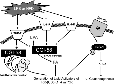
Mutations of comparative gene identification 58 (CGI-58) in humans cause Chanarin-Dorfman syndrome, a rare autosomal recessive disease in which excess triacylglycerol (TAG) accumulates in multiple tissues. CGI-58 recently has been ascribed two distinct biochemical activities, including coactivation of adipose triglyceride lipase and acylation of lysophosphatidic acid (LPA). It is noteworthy that both the substrate (LPA) and the product (phosphatidic acid) of the LPA acyltransferase reaction are well-known signaling lipids. Therefore, we hypothesized that CGI-58 is involved in generating lipid mediators that regulate TAG metabolism and insulin sensitivity. Here, we show that CGI-58 is required for the generation of signaling lipids in response to inflammatory stimuli and that lipid second messengers generated by CGI-58 play a critical role in maintaining the balance between inflammation and insulin action. Furthermore, we show that CGI-58 is necessary for maximal TH1 cytokine signaling in the liver. This novel role for CGI-58 in cytokine signaling may explain why diminished CGI-58 expression causes severe hepatic lipid accumulation yet paradoxically improves hepatic insulin action. Collectively, these findings establish that CGI-58 provides a novel source of signaling lipids. These findings contribute insight into the basic mechanisms linking TH1 cytokine signaling to nutrient metabolism.
Overview
The claims and statistics of this paper haven’t been checked yet.
Run the check — free, no account needed. It reads the open-access text and checks the sources it cites.
Integrity signals
Registry
Cites
Statistics
Figures
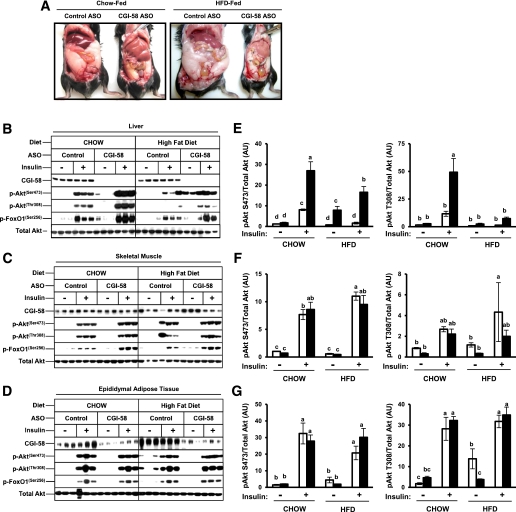
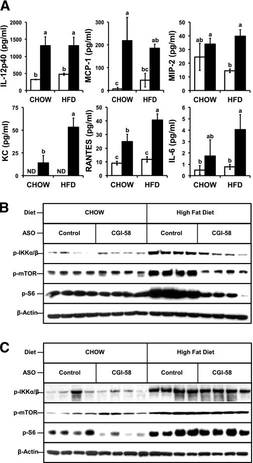
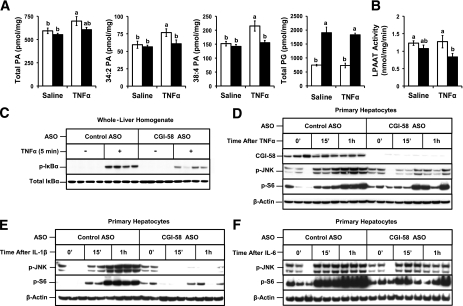
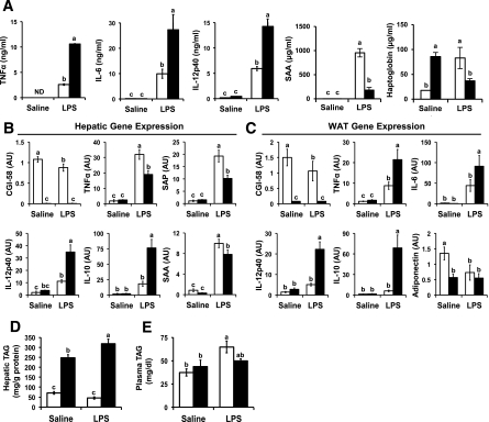
Verification
Claims the paper makes
The claims and statistics of this paper haven’t been checked yet.
Run the check — free, no account needed. It reads the open-access text and checks the sources it cites.
Claims other papers make about this one
Each is checked against this paper’s own text: does it say what the citing paper says it does?
The cited source says what the claim attributes to it.
In the citing paper
“In addition to these critical effectors, several members of the AD-specific miR-132 predicted targetome that we identified, e.g. TRIM44, NUCKS1, NFE2L2, LYN, ZEB2, SPRED1, ABHD5, RASA1, and NAP1L1 [143, 147–154], have known roles in inflammation and/or microglial cell states/function.”
From microRNA-132 attenuates inflammation in induced pluripotent stem cell-derived microglia from Alzheimer’s disease patients (2026) · cites this paper as [152]
Self-check
Percentages, sums and reported differences re-computed from the paper’s own numbers.
The self-check needs the paper’s full text.
Citations
Cites
A RAPID METHOD OF TOTAL LIPID EXTRACTION AND PURIFICATION
Bligh, E. G., & Dyer, W. J. (1959) · Canadian Journal of Biochemistry and Physiology · doi:10.1139/o59-099 · Cited by 54,751
Yuan, M., Konstantopoulos, N., Lee, J., Hansen, L., Li, Z.-W., Karin, M., & Shoelson, S. E. (2001) · Science · doi:10.1126/science.1061620 · Cited by 1,884
IKK-β links inflammation to obesity-induced insulin resistance
Arkan, M. C., Hevener, A. L., Greten, F. R., Maeda, S., Li, Z.-W., Long, J. M., Wynshaw-Boris, A., Poli, G., Olefsky, J., & Karin, M. (2005) · Nature Medicine · doi:10.1038/nm1185 · Cited by 1,773
Absence of S6K1 protects against age- and diet-induced obesity while enhancing insulin sensitivity
Um, S. H., Frigerio, F., Watanabe, M., Picard, F., Joaquin, M., Sticker, M., Fumagalli, S., Allegrini, P. R., Kozma, S. C., Auwerx, J., et al. (2004) · Nature · doi:10.1038/nature02866 · Cited by 1,677
Self-citationPhosphatidic Acid-Mediated Mitogenic Activation of mTOR Signaling
Fang, Y., Vilella-Bach, M., Bachmann, R., Flanigan, A., & Chen, J. (2001) · Science · doi:10.1126/science.1066015 · Cited by 1,055
A calculated response: control of inflammation by the innate immune system
Barton, G. M. (2008) · Journal of Clinical Investigation · doi:10.1172/jci34431 · Cited by 532
Research works printed but not linked: 44
- Lass, 2006, Adipose triglyceride lipase-mediated lipolysis of cellular fat stores is activated by CGI-58 and defective in Chanarin-Dorfman Syndrome, Cell Metab, 3, 309, 10.1016/j.cmet.2006.03.005 · DOI
- Brown, 2010, CGI-58 knockdown in mice causes hepatic steatosis but prevents diet-induced obesity and glucose intolerance, J Lipid Res, 51, 3306, 10.1194/jlr.M010256 · DOI
- Montero-Moran, 2010, CGI-58/ABHD5 is a coenzyme A-dependent lysophosphatidic acid acyltransferase, J Lipid Res, 51, 709, 10.1194/jlr.M001917 · DOI
- Ghosh, 2008, CGI-58, the causative gene for Chanarin-Dorfman syndrome, mediates acylation of lysophosphatidic acid, J Biol Chem, 283, 24525, 10.1074/jbc.M801783200 · DOI
- Lefèvre, 2001, Mutations in CGI-58, the gene encoding a new protein of the esterase/lipase/thioesterase subfamily, in Chanarin-Dorfman syndrome, Am J Hum Genet, 69, 1002, 10.1086/324121 · DOI
- Radner, 2010, Growth retardation, impaired triacylglycerol catabolism, hepatic steatosis, and lethal skin barrier defect in mice lacking comparative gene identification-58 (CGI-58), J Biol Chem, 285, 7300, 10.1074/jbc.M109.081877 · DOI
- Wymann, 2008, Lipid signalling in disease, Nat Rev Mol Cell Biol, 9, 162, 10.1038/nrm2335 · DOI
- Hla, 2001, Lysophospholipids: receptor revelations, Science, 294, 1875, 10.1126/science.1065323 · DOI
- Igal, 1996, Acylglycerol recycling from triacylglycerol to phospholipid, not lipase activity, is defective in neutral lipid storage disease fibroblasts, J Biol Chem, 271, 16644, 10.1074/jbc.271.28.16644 · DOI
- Williams, 1991, Neutral lipid storage disease: a possible functional defect in phospholipid-linked triacylglycerol metabolism, Biochim Biophys Acta, 1096, 162, 10.1016/0925-4439(91)90055-E · DOI
- Crooke, 2005, An apolipoprotein B antisense oligonucleotide lowers LDL cholesterol in hyperlipidemic mice without causing hepatic steatosis, J Lipid Res, 46, 872, 10.1194/jlr.M400492-JLR200 · DOI
- Brown, 2004, Conjugated linoleic acid induces human adipocyte delipidation: autocrine/paracrine regulation of MEK/ERK signaling by adipocytokines, J Biol Chem, 279, 26735, 10.1074/jbc.M401766200 · DOI
- Brown, 2008, Inhibition of stearoyl-coenzyme A desaturase 1 dissociates insulin resistance and obesity from atherosclerosis, Circulation, 118, 1467, 10.1161/CIRCULATIONAHA.108.793182 · DOI
- Brown, 2008, Targeted depletion of hepatic ACAT2-driven cholesterol esterification reveals a non-biliary route for fecal neutral sterol loss, J Biol Chem, 283, 10522, 10.1074/jbc.M707659200 · DOI
- Ivanova, 2007, Glycerophospholipid identification and quantitation by electrospray ionization mass spectrometry, Methods Enzymol, 432, 21, 10.1016/S0076-6879(07)32002-8 · DOI
- Chung, 2010, Targeted deletion of hepatocyte ABCA1 leads to very low density lipoprotein triglyceride overproduction and low density lipoprotein hypercatabolism, J Biol Chem, 285, 12197, 10.1074/jbc.M109.096933 · DOI · Paper page
- Draper, 2010, ATP-binding cassette transporter G1 deficiency dysregulates host defense in the lung, Am J Respir Crit Care Med, 182, 404, 10.1164/rccm.200910-1580OC · DOI
- Lowry, 1951, Protein measurement with the Folin phenol reagent, J Biol Chem, 193, 265, 10.1016/S0021-9258(19)52451-6 · DOI
- Callender, 2007, Quantification of diacylglycerol species from cellular extracts by electrospray ionization mass spectrometry using a linear regression algorithm, Anal Chem, 79, 263, 10.1021/ac061083q · DOI
- Erion, 2010, Diacylglycerol-mediated insulin resistance, Nat Med, 16, 400, 10.1038/nm0410-400 · DOI
- Hotamisligil, 1996, IRS-1-mediated inhibition of insulin receptor tyrosine kinase activity in TNF-alpha- and obesity-induced insulin resistance, Science, 271, 665, 10.1126/science.271.5249.665 · DOI · Paper page
- Fingar, 2004, Target of rapamycin (TOR): an integrator of nutrient and growth factor signals and coordinator of cell growth and cell cycle progression, Oncogene, 23, 3151, 10.1038/sj.onc.1207542 · DOI
- Hirosumi, 2002, A central role for JNK in obesity and insulin resistance, Nature, 420, 333, 10.1038/nature01137 · DOI · Paper page
- Tong, 1999, Involvement of lipid mediators on cytokine signaling and induction of secretory phospholipase A2 in immortalized astrocytes (DITNC), J Mol Neurosci, 12, 89, 10.1007/BF02736923 · DOI
- Bursten, 1991, Interleukin-1 rapidly stimulates lysophosphatidate acyltransferase and phosphatidate phosphohydrolase activities in human mesangial cells, J Biol Chem, 266, 20732, 10.1016/S0021-9258(18)54770-0 · DOI
- Bursten, 1992, Lipid A activation of glomerular mesangial cells: mimicry of the bioactive lipid, phosphatidic acid, Am J Physiol, 262, C328, 10.1152/ajpcell.1992.262.2.C328 · DOI
- Rice, 1994, Protection from endotoxic shock in mice by pharmacologic inhibition of phosphatidic acid, Proc Natl Acad Sci USA, 91, 3857, 10.1073/pnas.91.9.3857 · DOI
- Abraham, 1995, Phosphatidic acid signaling mediates lung cytokine expression and lung inflammatory injury after hemorrhage in mice, J Exp Med, 181, 569, 10.1084/jem.181.2.569 · DOI
- Hardardóttir, 1994, Effects of endotoxin and cytokines on lipid metabolism, Curr Opin Lipidol, 5, 207, 10.1097/00041433-199405030-00008 · DOI
- Shindou, 2009, Recent progress on acyl CoA: lysophospholipid acyltransferase research, J Lipid Res, 50, S46, 10.1194/jlr.R800035-JLR200 · DOI
- Nathan, 2002, Points of control in inflammation, Nature, 420, 846, 10.1038/nature01320 · DOI · Paper page
- Williams, 1988, Neutral lipid storage disease with ichthyosis: lipid content and metabolism of fibroblasts, J Inherit Metab Dis, 11, 131, 10.1007/BF01799862 · DOI
- Hilaire, 1993, Cytoplasmic triacylglycerols and cholesteryl esters are degraded in two separate catabolic pools in cultured human fibroblasts, FEBS Lett, 328, 230, 10.1016/0014-5793(93)80933-L · DOI
- Hilaire, 1994, Cellular uptake and catabolism of high-density-lipoprotein triacylglycerols in human cultured fibroblasts: degradation block in neutral lipid storage disease, Biochem J, 297, 467, 10.1042/bj2970467 · DOI
- Hilaire, 1995, The turnover of cytoplasmic triacylglycerols in human fibroblasts involves two separate acyl chain length-dependent degradation pathways, J Biol Chem, 270, 27027, 10.1074/jbc.270.45.27027 · DOI
- Brown, 2007, CGI-58 facilitates the mobilization of cytoplasmic triglyceride for lipoprotein secretion in hepatoma cells, J Lipid Res, 48, 2295, 10.1194/jlr.M700279-JLR200 · DOI
- Subramanian, 2004, Perilipin A mediates the reversible binding of CGI-58 to lipid droplets in 3T3-L1 adipocytes, J Biol Chem, 279, 42062, 10.1074/jbc.M407462200 · DOI
- Yamaguchi, 2004, CGI-58 interacts with perilipin and is localized to lipid droplets: possible involvement of CGI-58 mislocalization in Chanarin-Dorfman syndrome, J Biol Chem, 279, 30490, 10.1074/jbc.M403920200 · DOI
- Jenkins, 2005, Phospholipase D: a lipid centric review, Cell Mol Life Sci, 62, 2305, 10.1007/s00018-005-5195-z · DOI
- Andresen, 2002, The role of phosphatidic acid in the regulation of the Ras/MEK/Erk signaling cascade, FEBS Lett, 531, 65, 10.1016/S0014-5793(02)03483-X · DOI
- Exton, 1997, Phospholipase D: enzymology, mechanisms of regulation, and function, Physiol Rev, 77, 303, 10.1152/physrev.1997.77.2.303 · DOI
- Topham, 2009, Mammalian diacylglycerol kinases: molecular interactions and biological functions of selected isoforms, 416
- Bleich, 1996, Lisofylline, an inhibitor of unsaturated phosphatidic acid generation, ameliorates interleukin-1 beta-induced dysfunction in cultured rat islets, Endocrinology, 137, 4871, 10.1210/endo.137.11.8895359 · DOI
- Guidot, 1997, Modulating phosphatidic acid metabolism decreases oxidative injury in rat lungs, Am J Physiol, 273, L957
Cited by
Sorted by citations
Penning, A., Snoeck, S., Ormaechea, O. R., Ayyildiz, D., Polzer, O., Buitrago-Arango, M., Capobianco, R., de Winter, F., Balusu, S., Verhaagen, J., et al. (2026) · Acta Neuropathologica Communications · doi:10.1186/s40478-026-02228-8 · Cited by 6
Metadata
- Authors
- Lord, C. C., Betters, J. L., Ivanova, P. T., Milne, S. B., Myers, D. S., Madenspacher, J., Thomas, G., Chung, S., Liu, M., Davis, M. A., Lee, R. G., Crooke, R. M., Graham, M. J., Parks, J. S., Brasaemle, D. L., Fessler, M. B., Brown, H. A., & Brown, J. M.
- Affiliations
- Department of Pathology, Section on Lipid Sciences, Wake Forest University School of Medicine, Winston-Salem, North Carolina
- Department of Pharmacology, Vanderbilt University School of Medicine, Nashville, Tennessee
- Laboratory of Respiratory Biology, National Institute of Environmental Health Sciences, Research Triangle Park, North Carolina
- Cardiovascular Group, Antisense Drug Discovery, Isis Pharmaceuticals, Carlsbad, California
- Department of Nutritional Sciences, Rutgers, The State University of New Jersey, New Brunswick, New Jersey
- Venue
- Diabetes
- Publisher
- American Diabetes Association
- Published
- 2012-01-17
- Volume
- 61
- Issue
- 2
- Pages
- 355-363
- ISSN
- 0012-1797
- 1939-327X
- PMC
- PMC3266405
- Cited by
- 45
- References
- 6
- Language
- en
About these checks
Registry, retraction, Cites and statistics results are computed, not judged, with one exception: a printed reference that no index lists is read once by a model before it counts as a citation error. Claims are extracted by a model and kept only when their quote appears verbatim in the text; each cited source is then checked, with a model’s help, for whether it says what the claim relies on. Those verdicts can be wrong — read the claim’s quote and the cited source before relying on one.
Statistics and arithmetic are recomputed, not judged.
Confidence bands are shown only for fields calibrated on our annotated test set. None is calibrated yet, so no band is shown.
What counts as a claim ›How we measure confidence ›How we check this ›
Are you an author, or do you see a problem with this page? Request a review
