

The DOI matches its Crossref record; 240 of the 240 references it cites were found in a registry or an index.
Exploitation of Hetero- and Phototrophic Metabolic Modules for Redox-Intensive Whole-Cell Biocatalysis
Open accessAbstract
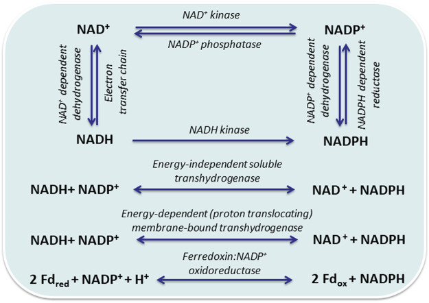
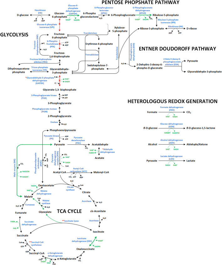
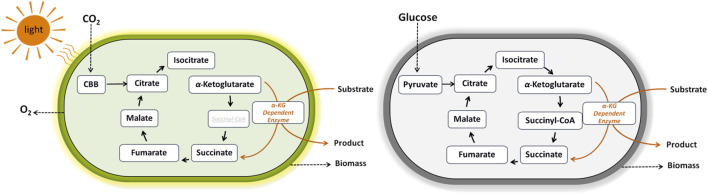
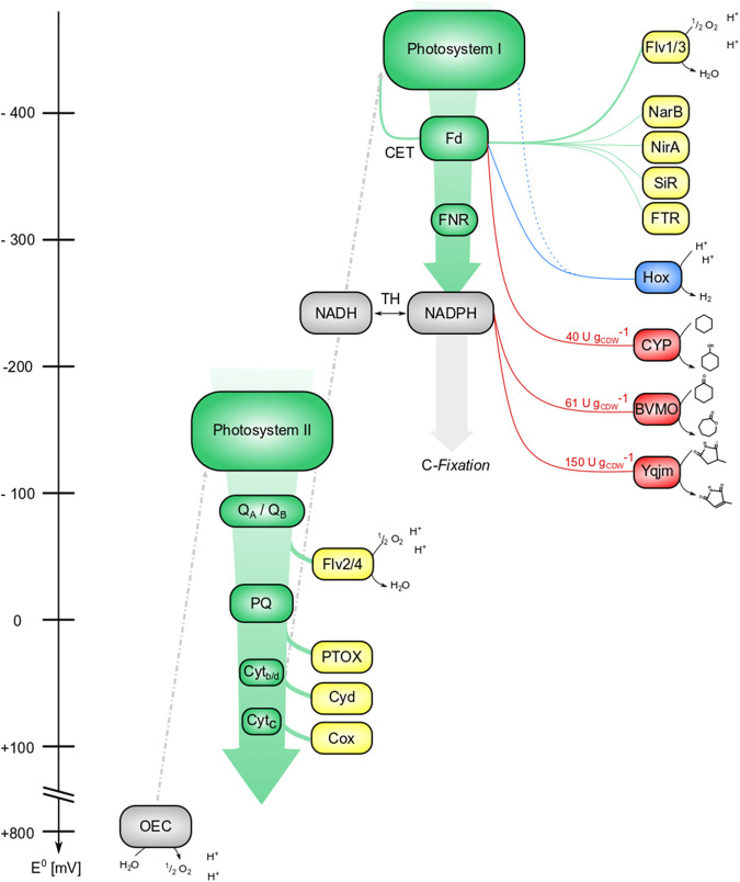
The successful realization of a sustainable manufacturing bioprocess and the maximization of its production potential and capacity are the main concerns of a bioprocess engineer. A main step towards this endeavor is the development of an efficient biocatalyst. Isolated enzyme(s), microbial cells, or (immobilized) formulations thereof can serve as biocatalysts. Living cells feature, beside active enzymes, metabolic modules that can be exploited to support energy-dependent and multi-step enzyme-catalyzed reactions. Metabolism can sustainably supply necessary cofactors or cosubstrates at the expense of readily available and cheap resources, rendering external addition of costly cosubstrates unnecessary. However, for the development of an efficient whole-cell biocatalyst, in depth comprehension of metabolic modules and their interconnection with cell growth, maintenance, and product formation is indispensable. In order to maximize the flux through biosynthetic reactions and pathways to an industrially relevant product and respective key performance indices (i.e., titer, yield, and productivity), existing metabolic modules can be redesigned and/or novel artificial ones established. This review focuses on whole-cell bioconversions that are coupled to heterotrophic or phototrophic metabolism and discusses metabolic engineering efforts aiming at 1) increasing regeneration and supply of redox equivalents, such as NAD(P/H), 2) blocking competing fluxes, and 3) increasing the availability of metabolites serving as (co)substrates of desired biosynthetic routes.
Overview
The claims and statistics of this paper haven’t been checked yet.
Run the check — free, no account needed. It reads the open-access text and checks the sources it cites.
Integrity signals
Registry
Cites
Statistics
Figures
Verification
Claims the paper makes
The claims and statistics of this paper haven’t been checked yet.
Run the check — free, no account needed. It reads the open-access text and checks the sources it cites.
Claims other papers make about this one
Each is checked against this paper’s own text: does it say what the citing paper says it does?
The source backs part of the claim, not all of it — or backs it for a different population, condition or comparison.
In the citing paper
“Nonetheless, competition with native metabolic processes frequently restricts the efficiency of heterologous pathway activity.”
From Cyanobacteria as a Photosynthetic Chassis for Metabolic Pathway Engineering with Heterologous Gene Expression (2026) · cites this paper as [143]
Experimental — accuracy not yet measuredThe cited source says what the claim attributes to it.
In the citing paper
“Since cyanobacterial ATP and NADPH production is tightly linked to PET, pathway efficiency can be heavily affected by factors such as light intensity, carbon availability, and cellular redox balance [143].”
From Cyanobacteria as a Photosynthetic Chassis for Metabolic Pathway Engineering with Heterologous Gene Expression (2026) · cites this paper as [143]
Self-check
Percentages, sums and reported differences re-computed from the paper’s own numbers.
The self-check needs the paper’s full text.
Citations
Cites
Redesigning photosynthesis to sustainably meet global food and bioenergy demand
Ort, D. R., Merchant, S., Alric, J., Barkan, A., Blankenship, R. E., Bock, R., Croce, R., Hanson, M. R., Hibberd, J. M., Long, S. P., et al. (2015) · Proceedings of the National Academy of Sciences · doi:10.1073/pnas.1424031112 · Cited by 1,018
The oxidative pentose phosphate pathway: structure and organisation
Kruger, N. J., & von Schaewen, A. (2003) · Current Opinion in Plant Biology · doi:10.1016/s1369-5266(03)00039-6 · Cited by 879
Redesign of the coenzyme specificity of a dehydrogenase by protein engineering
Scrutton, N. S., Berry, A., & Perham, R. N. (1990) · Nature · doi:10.1038/343038a0 · Cited by 728
Sauer, U., Canonaco, F., Heri, S., Perrenoud, A., & Fischer, E. (2004) · Journal of Biological Chemistry · doi:10.1074/jbc.m311657200 · Cited by 619
Fuelling the future: microbial engineering for the production of sustainable biofuels
Liao, J. C., Mi, L., Pontrelli, S., & Luo, S. (2016) · Nature Reviews Microbiology · doi:10.1038/nrmicro.2016.32 · Cited by 604
Sauer, U., & Eikmanns, B. J. (2004) · FEMS Microbiology Reviews · doi:10.1016/j.femsre.2004.11.002 · Cited by 538
Lipid production in Yarrowia lipolytica is maximized by engineering cytosolic redox metabolism
Qiao, K., Wasylenko, T. M., Zhou, K., Xu, P., & Stephanopoulos, G. N. (2017) · Nature Biotechnology · doi:10.1038/nbt.3763 · Cited by 468
Flux balance analysis of primary metabolism in Chlamydomonas reinhardtii
Boyle, N. R., & Morgan, J. A. (2009) · BMC Systems Biology · doi:10.1186/1752-0509-3-4 · Cited by 466
Whole-cell biocatalysts by design
Lin, B., & Tao, Y. (2017) · Microbial Cell Factories · doi:10.1186/s12934-017-0724-7 · Cited by 424
Systems and Photosystems: Cellular Limits of Autotrophic Productivity in Cyanobacteria
Burnap, R. L. (2015) · Frontiers in Bioengineering and Biotechnology · doi:10.3389/fbioe.2015.00001 · Cited by 422
Cited by
Sorted by citations
Cyanobacteria as whole-cell factories: Current status and future prospectives
Toepel, J., Karande, R., Klähn, S., & Bühler, B. (2023) · Current Opinion in Biotechnology · doi:10.1016/j.copbio.2023.102892 · Cited by 52
Self-citationGrowth-coupled enzyme engineering through manipulation of redox cofactor regeneration
Nielsen, J. R., Weusthuis, R. A., & Huang, W. E. (2023) · Biotechnology Advances · doi:10.1016/j.biotechadv.2023.108102 · Cited by 48
Schröder, G. C., Smit, M. S., & Opperman, D. J. (2022) · Current Opinion in Green and Sustainable Chemistry · doi:10.1016/j.cogsc.2022.100734 · Cited by 39
Light‐driven redox biocatalysis on gram‐scale in Synechocystis sp. PCC 6803 via an in vivo cascade
Tüllinghoff, A., Djaya‐Mbissam, H., Toepel, J., & Bühler, B. (2023) · Plant Biotechnology Journal · doi:10.1111/pbi.14113 · Cited by 18
Self-citationZhang, M., Tian, Y., Xu, D., Li, W., Cheng, S., & Song, H. (2025) · Coordination Chemistry Reviews · doi:10.1016/j.ccr.2025.216774 · Cited by 14
Metadata
- Authors
- Theodosiou, E., Tüllinghoff, A., Toepel, J., & Bühler, B.
- Affiliations
- Centre for Research and Technology Hellas (GR)
- Helmholtz Centre for Environmental Research (DE)
- Corresponding author
- Bühler, Bruno
- Venue
- Frontiers in Bioengineering and Biotechnology
- Publisher
- Frontiers Media SA
- Published
- 2022-04-13
- Volume
- 10
- Pages
- 855715-855715
- ISSN
- 2296-4185 (ISSN-L)
- PMC
- PMC9043136
- OpenAlex
- W4224125904
- Semantic Scholar
- e00eaee1b714c8d312839e3acb7eed6543ad4153
- Cited by
- 17
- References
- 239
- Citations by year
- 2026: 6 · 2025: 4 · 2024: 1 · 2023: 5 · 2022: 1
- FWCI
- 0.28
- Type
- journal article
- Language
- en
- Open access
- gold
- doi.org
- Licence
- Locations
- Frontiers in Bioengineering and Biotechnology (publishedVersion, cc-by)
- PubMed Central (publishedVersion, cc-by)
- PubMed (publishedVersion)
- DOAJ (DOAJ: Directory of Open Access Journals) (submittedVersion, cc-by-sa)
- Funders
- Helmholtz-Zentrum für Umweltforschung
- European Regional Development Fund
- Topics
- Life Sciences › Biochemistry, Genetics and Molecular Biology › Molecular Biology › Microbial Metabolic Engineering and Bioproduction
- Life Sciences › Biochemistry, Genetics and Molecular Biology › Biotechnology › Biochemical and biochemical processes
- Life Sciences › Biochemistry, Genetics and Molecular Biology › Molecular Biology › Enzyme Catalysis and Immobilization
- Keywords
- whole-cell biocatalysis, metabolic modules, heterotrophic metabolism, redox metabolism, NAD(P)H regeneration, metabolic engineering, flux control, bioprocess intensification
About these checks
Registry, retraction, Cites and statistics results are computed, not judged, with one exception: a printed reference that no index lists is read once by a model before it counts as a citation error. Claims are extracted by a model and kept only when their quote appears verbatim in the text; each cited source is then checked, with a model’s help, for whether it says what the claim relies on. Those verdicts can be wrong — read the claim’s quote and the cited source before relying on one.
Statistics and arithmetic are recomputed, not judged.
Confidence bands are shown only for fields calibrated on our annotated test set. None is calibrated yet, so no band is shown.
What counts as a claim ›How we measure confidence ›How we check this ›
Are you an author, or do you see a problem with this page? Request a review
