

The DOI matches its Crossref record; 1 of the 1 references it cites were found in a registry or an index.
MnO2 coated multi-layer nanoplatform for enhanced sonodynamic therapy and MR imaging of breast cancer
Open accessAbstract
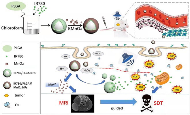
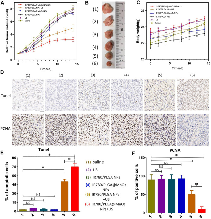
Sonodynamic therapy (SDT) is a promising new anti-tumor therapy that inhibits tumor growth by ultrasound activation of sonosensitizers to produce reactive oxygen species (ROS). However, the problems of hypoxia in the microenvironment within solid tumors and the effectiveness of SDT will decrease due to the little accumulation of sonosensitizers at the tumor site, as well as tumor cell tolerance, have limited the development of SDT. To overcome these problems, a core-shell structured nanoparticle (IR780/PLGA@MnO 2 NPs) loaded with IR780 and manganese dioxide (MnO 2 ) was developed as a nanocarrier to transport the sonosensitizer IR780 and the generated oxygen into the tumor tissue. The MnO 2 shell layer of IR780/PLGA@MnO 2 NPs can prevent the premature release of IR780 in the blood and also it can react with acidic and high H 2 O 2 , the generated oxygen can relieve tumor tissue hypoxia, and the generated Mn can enhance magnetic resonance imaging (MRI) signal intensity by acting as a contrast agent for MRI. More importantly, the released IR780 can produce ROS to kill tumor cells under ultrasound excitation. This PH-responsive and H 2 O 2 -triggered SDT based on the IR780/PLGA@MnO 2 NPs is an effective platform to inhibit tumor growth with negligible systemic toxicity. This work develops a multifunctional therapeutic integrated nanoplatform for breast cancer treatment, which is expected to be used in the clinic.
Overview
The claims and statistics of this paper haven’t been checked yet.
Run the check — free, no account needed. It reads the open-access text and checks the sources it cites.
Integrity signals
Registry
Cites
Statistics
Figures
Verification
Claims the paper makes
The claims and statistics of this paper haven’t been checked yet.
Run the check — free, no account needed. It reads the open-access text and checks the sources it cites.
Claims other papers make about this one
Each is checked against this paper’s own text: does it say what the citing paper says it does?
The source backs part of the claim, not all of it — or backs it for a different population, condition or comparison.
In the citing paper
“Furthermore, hypoxia and high glutathione (GSH) concentrations in the tumor microenvironment (TME) greatly limit the efficacy of SDT [24–26].”
From Nanosensitizer-assisted sonodynamic therapy for breast cancer (2025) · cites this paper as [24]
Experimental — accuracy not yet measured
Self-check
Percentages, sums and reported differences re-computed from the paper’s own numbers.
The self-check needs the paper’s full text.
Citations
Cites
Multifunctional sonosensitizers in sonodynamic cancer therapy
Son, S., Kim, J. H., Wang, X., Zhang, C., Yoon, S. A., Shin, J., Sharma, A., Lee, M. H., Cheng, L., Wu, J., et al. (2020) · Chemical Society Reviews · doi:10.1039/c9cs00648f · Cited by 1,150
Research works printed but not linked: 34
- Ahmadi, 2020, Recent advances in ultrasound-triggered drug delivery through lipid-based nanomaterials, Drug Discov. Today, 25, 2182, 10.1016/j.drudis.2020.09.026 · DOI
- Chen, 2020, Mitochondria-targeting oxygen-sufficient perfluorocarbon nanoparticles for imaging-guided tumor phototherapy, Int. J. Nanomedicine, 15, 8641, 10.2147/IJN.S281649 · DOI
- Chu, 2017, Tumor microenvironment-triggered supramolecular system as an in situ nanotheranostic generator for cancer phototherapy, Adv. Mat., 29, 1605928, 10.1002/adma.201605928 · DOI
- Costley, 2015, Treating cancer with sonodynamic therapy: A review, Int. J. Hyperth., 31, 107, 10.3109/02656736.2014.992484 · DOI
- Ding, 2020, Manganese oxide nanomaterials: Synthesis, properties, and theranostic applications, Adv. Mat., 32, e1905823, 10.1002/adma.201905823 · DOI
- Dong, 2021, Inorganic chemoreactive nanosonosensitzers with unique physiochemical properties and structural features for versatile sonodynamic nanotherapies, Biomed. Mat., 16, 032006, 10.1088/1748-605X/abef58 · DOI
- Fu, 2019, Targeted magnetic resonance imaging and modulation of hypoxia with multifunctional hyaluronic acid-MnO2 nanoparticles in glioma, Adv. Healthc. Mat., 8, e1900047, 10.1002/adhm.201900047 · DOI
- Gordijo, 2015, Design of hybrid MnO2-polymer-lipid nanoparticles with tunable oxygen generation rates and tumor accumulation for cancer treatment, Adv. Funct. Mat., 25, 1858, 10.1002/adfm.201404511 · DOI
- Hockel, 2001, Tumor hypoxia: Definitions and current clinical, biologic, and molecular aspects, JNCI J. Natl. Cancer Inst., 93, 266, 10.1093/jnci/93.4.266 · DOI · Paper page
- Huang, 2020, Oxygen-sufficient nanoplatform for chemo-sonodynamic therapy of hypoxic tumors, Front. Chem., 8, 358, 10.3389/fchem.2020.00358 · DOI
- Huang, 2017, Metalloporphyrin-encapsulated biodegradable nanosystems for highly efficient magnetic resonance imaging-guided sonodynamic cancer therapy, J. Am. Chem. Soc., 139, 1275, 10.1021/jacs.6b11846 · DOI
- Hussain, 2014, Molecular imaging for cancer diagnosis and surgery, Adv. Drug Deliv. Rev., 66, 90, 10.1016/j.addr.2013.09.007 · DOI
- Liu, 2021, Enzymatically synthesised MnO2 nanoparticles for efficient near-infrared photothermal therapy and dual-responsive magnetic resonance imaging, Nanoscale, 13, 11093, 10.1039/d1nr02400k · DOI
- Liu, 2018, Theranostic 2D ultrathin MnO2 nanosheets with fast responsibility to endogenous tumor microenvironment and exogenous NIR irradiation, Biomaterials, 155, 54, 10.1016/j.biomaterials.2017.11.015 · DOI
- Ma, 2017, MnO2Gatekeeper: An intelligent and O2-evolving shell for preventing premature release of high cargo payload core, overcoming tumor hypoxia, and acidic H2O2-sensitive MRI, Adv. Funct. Mat., 27, 1604258, 10.1002/adfm.201604258 · DOI
- Niu, 2017, Near-infrared induced phase-shifted ICG/Fe3O4 loaded PLGA nanoparticles for photothermal tumor ablation, Sci. Rep., 7, 5490, 10.1038/s41598-017-06122-1 · DOI
- Pei, 2020, Platelet-Mimicking drug delivery nanoparticles for enhanced chemo-photothermal therapy of breast cancer , Int. J. Nanomedicine, 15, 10151, 10.2147/IJN.S285952 · DOI
- Qian, 2016, Micro/nanoparticle-augmented sonodynamic therapy (SDT): Breaking the depth shallow of photoactivation, Adv. Mat., 28, 8097, 10.1002/adma.201602012 · DOI
- Rosenthal, 2004, Sonodynamic therapy--a review of the synergistic effects of drugs and ultrasound, Ultrason. Sonochem., 11, 349, 10.1016/j.ultsonch.2004.03.004 · DOI
- Song, 2016, Perfluorocarbon-loaded hollow Bi2Se3 nanoparticles for timely supply of oxygen under near-infrared light to enhance the radiotherapy of cancer, Adv. Mat., 28, 2716, 10.1002/adma.201504617 · DOI
- Song, 2016, Ultrasound triggered tumor oxygenation with oxygen-shuttle nanoperfluorocarbon to overcome hypoxia-associated resistance in cancer therapies, Nano Lett., 16, 6145, 10.1021/acs.nanolett.6b02365 · DOI
- Sun, 2021, MnO2 nanoflowers as a multifunctional nano-platform for enhanced photothermal/photodynamic therapy and MR imaging, Biomater. Sci., 9, 3662, 10.1039/d1bm00033k · DOI
- Wan, 2016, Recent advances of sonodynamic therapy in cancer treatment, Cancer Biol. Med., 13, 325, 10.20892/j.issn.2095-3941.2016.0068 · DOI
- Wang, 2018, Triple-modal imaging-guided chemo-photothermal synergistic therapy for breast cancer with magnetically targeted phase-shifted nanoparticles, ACS Appl. Mat. Interfaces, 10, 42102, 10.1021/acsami.8b16323 · DOI
- Wang, 2020, Newly developed strategies for improving sonodynamic therapy, Mat. Horiz., 7, 2028, 10.1039/d0mh00613k · DOI
- Wells, 2006, Ultrasound imaging, Phys. Med. Biol., 51, R83, 10.1088/0031-9155/51/13/R06 · DOI
- Wood, 2015, A review of low-intensity ultrasound for cancer therapy, Ultrasound Med. Biol., 41, 905, 10.1016/j.ultrasmedbio.2014.11.019 · DOI
- Wu, 2005, Feasibility of US-guided high-intensity focused ultrasound treatment in patients with advanced pancreatic cancer: Initial experience, Radiology, 236, 1034, 10.1148/radiol.2362041105 · DOI
- Xu, 2020, Near-Infrared responsive phase-shifted nanoparticles for magnetically targeted MR/US imaging and photothermal therapy of cancer, Front. Bioeng. Biotechnol., 8, 599107, 10.3389/fbioe.2020.599107 · DOI
- Yang, 2019, Biomimetic hybrid nanozymes with self-supplied H(+) and accelerated O2 generation for enhanced starvation and photodynamic therapy against hypoxic tumors, Nano Lett., 19, 4334, 10.1021/acs.nanolett.9b00934 · DOI
- Zhang, 2019, Mitochondria-targeted and ultrasound-activated nanodroplets for enhanced deep-penetration sonodynamic cancer therapy, ACS Appl. Mat. Interfaces, 11, 9355, 10.1021/acsami.8b21968 · DOI
- Zhang, 2020, Hollow magnetic nanosystem-boosting synergistic effect between magnetic hyperthermia and sonodynamic therapy via modulating reactive oxygen species and heat shock proteins, Chem. Eng. J., 390, 124521, 10.1016/j.cej.2020.124521 · DOI
- Zhang, 2020, Nanostructured manganese dioxide for anticancer applications: Preparation, diagnosis, and therapy, Nanoscale, 12, 17982, 10.1039/d0nr04067c · DOI
- Zhu, 2018, Nanoenzyme-augmented cancer sonodynamic therapy by catalytic tumor oxygenation, ACS Nano, 12, 3780, 10.1021/acsnano.8b00999 · DOI
Cited by
Sorted by citations
Nanosensitizer-assisted sonodynamic therapy for breast cancer
Yu, J., Hu, J.-R., Tian, Y., Lei, Y.-M., Hu, H.-M., Lei, B.-S., Zhang, G., Sun, Y., & Ye, H.-R. (2025) · Journal of Nanobiotechnology · doi:10.1186/s12951-025-03311-3 · Cited by 43
Metadata
- Authors
- Xu, Y., Tan, W., Chen, M., Chen, S., Tang, K., Liao, H., & Niu, C.
- Affiliations
- Central South University (CN)
- Corresponding author
- Niu, Chengcheng
- Venue
- Frontiers in Bioengineering and Biotechnology
- Publisher
- Frontiers Media SA
- Published
- 2022-10-19
- Volume
- 10
- Pages
- 955127-955127
- ISSN
- 2296-4185 (ISSN-L)
- PMC
- PMC9627152
- OpenAlex
- W4306842424
- Cited by
- 24
- References
- 1
- Citations by year
- 2026: 11 · 2025: 5 · 2024: 5 · 2023: 3
- FWCI
- 0.94
- Type
- journal article
- Language
- en
- Open access
- gold
- doi.org
- Licence
- Locations
- Frontiers in Bioengineering and Biotechnology (publishedVersion, cc-by)
- PubMed Central (publishedVersion, cc-by)
- PubMed (publishedVersion)
- DOAJ (DOAJ: Directory of Open Access Journals) (submittedVersion, cc-by-sa)
- Funders
- National Natural Science Foundation of China
- Science and Technology Program of Hunan Province
- Topics
- Physical Sciences › Engineering › Biomedical Engineering › Nanoplatforms for cancer theranostics
- Life Sciences › Biochemistry, Genetics and Molecular Biology › Cancer Research › Photodynamic Therapy Research Studies
- Physical Sciences › Physics and Astronomy › Radiation › Radiation Therapy and Dosimetry
- Keywords
- sonodynamic therapy, breast cancer, IR780, manganese dioxide, PLGA nanoparticles, core-shell nanoparticles, tumor hypoxia, reactive oxygen species, pH-responsive drug release, MRI contrast agents
About these checks
Registry, retraction, Cites and statistics results are computed, not judged, with one exception: a printed reference that no index lists is read once by a model before it counts as a citation error. Claims are extracted by a model and kept only when their quote appears verbatim in the text; each cited source is then checked, with a model’s help, for whether it says what the claim relies on. Those verdicts can be wrong — read the claim’s quote and the cited source before relying on one.
Statistics and arithmetic are recomputed, not judged.
Confidence bands are shown only for fields calibrated on our annotated test set. None is calibrated yet, so no band is shown.
What counts as a claim ›How we measure confidence ›How we check this ›
Are you an author, or do you see a problem with this page? Request a review
How to cite
Cite CoChat for the checks on this page, and the paper for its own findings.
- This page
- CoChat. (2026). Citation check: MnO2 coated multi-layer nanoplatform for enhanced sonodynamic therapy and MR imaging of breast cancer. https://app.cochat.ai/papers/10.3389/fbioe.2022.955127
- The paper
- Xu, Y., Tan, W., Chen, M., Chen, S., Tang, K., Liao, H., & Niu, C. (2022). MnO2 coated multi-layer nanoplatform for enhanced sonodynamic therapy and MR imaging of breast cancer. Frontiers in Bioengineering and Biotechnology. https://doi.org/10.3389/fbioe.2022.955127
