

The DOI matches its Crossref record; 42 of the 42 references it cites were found in a registry or an index.
Carboxymethyl chitosan nanoparticle-modulated cationic hydrogels doped with copper ions for combating bacteria and facilitating wound healing
Open accessAbstract
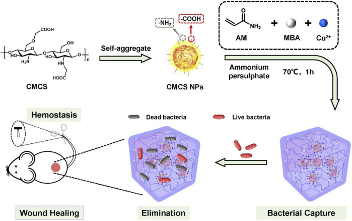
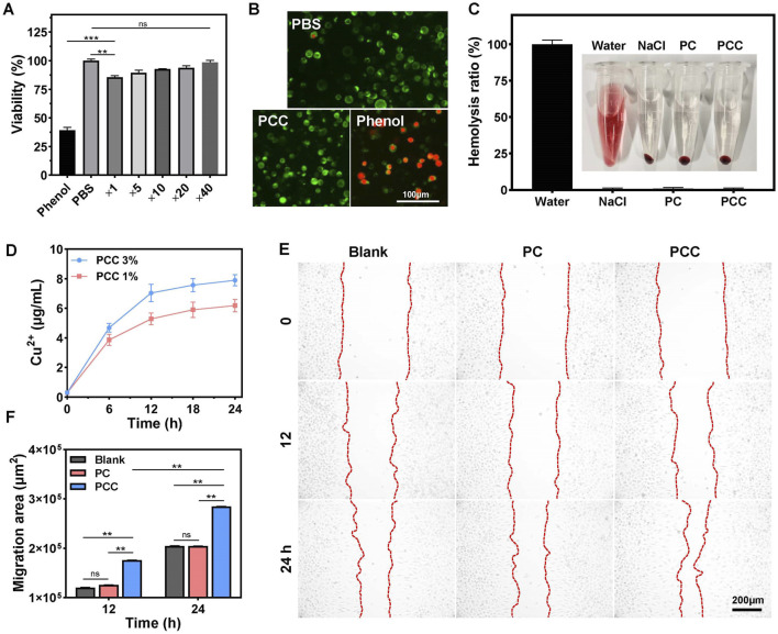
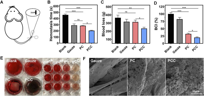
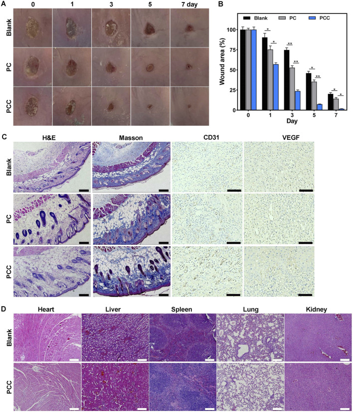
The simultaneous administration of antibacterial treatment and acceleration of tissue regeneration are crucial for the effective healing of infected wounds. In this work, we developed a facile hydrogel (PCC hydrogel) through coordination and hydrogen interactions by polymerizing acrylamide monomers in the presence of carboxymethyl chitosan nanoparticles and copper ions. The prepared PCC hydrogel demonstrated effective bacterial capture from wound exudation and exhibited a potent bactericidal activity against methicillin-resistant Staphylococcus aureus (MRSA) and Pseudomonas aeruginosa . Furthermore, slow release of copper ions from the hydrogel facilitated wound healing by promoting cell migration, collagen deposition and angiogenesis. Additionally, the PCC hydrogel possessed excellent biocompatibility and hemostatic properties. The practical effectiveness of PCC hydrogel in addressing bacterial infections and facilitating wound healing was verified using a mouse model of MRSA-induced wound infections. Overall, this work presents a simple yet efficient multifunctional hydrogel platform that integrates antibacterial activity, promotion of wound healing, and hemostasis for managing bacteria-associated wounds.
Overview
The claims and statistics of this paper haven’t been checked yet.
Run the check — free, no account needed. It reads the open-access text and checks the sources it cites.
Integrity signals
Registry
Cites
Statistics
Figures
Verification
Claims the paper makes
The claims and statistics of this paper haven’t been checked yet.
Run the check — free, no account needed. It reads the open-access text and checks the sources it cites.
Claims other papers make about this one
Each is checked against this paper’s own text: does it say what the citing paper says it does?
The source backs part of the claim, not all of it — or backs it for a different population, condition or comparison.
In the citing paper
“One of the notorious effects noticed in some of these studies was the significant increase in VEGF and the number of blood vessels in the copper-treated wounds as compared to the controls [72,73,75,77,78,84,97,103].”
From The Journey of Copper-Impregnated Dressings in Wound Healing: From a Medical Hypothesis to Clinical Practice (2025) · cites this paper as [97]
Experimental — accuracy not yet measuredThe cited source says what the claim attributes to it.
In the citing paper
“Additionally, copper-containing biomaterials, mostly wound dressings, have shown significant potential in promoting the healing of infected wounds [68,87,91,92,93,94,95,96,97,98], including those infected with MRSA [99] and Escherichia coli biofilm [100].”
From The Journey of Copper-Impregnated Dressings in Wound Healing: From a Medical Hypothesis to Clinical Practice (2025) · cites this paper as [97]
Self-check
Percentages, sums and reported differences re-computed from the paper’s own numbers.
The self-check needs the paper’s full text.
Citations
Cites
Liang, Y., Zhao, X., Hu, T., Chen, B., Yin, Z., Ma, P. X., & Guo, B. (2019) · Small · doi:10.1002/smll.201900046 · Cited by 1,294
Zhao, X., Guo, B., Wu, H., Liang, Y., & Ma, P. X. (2018) · Nature Communications · doi:10.1038/s41467-018-04998-9 · Cited by 1,173
Highly Stretchable, Adhesive, Biocompatible, and Antibacterial Hydrogel Dressings for Wound Healing
Yang, Z., Huang, R., Zheng, B., Guo, W., Li, C., He, W., Wei, Y., Du, Y., Wang, H., Wu, D., et al. (2021) · Advanced Science · doi:10.1002/advs.202003627 · Cited by 614
Bioinspired mechanically active adhesive dressings to accelerate wound closure
Blacklow, S. O., Li, J., Freedman, B. R., Zeidi, M., Chen, C., & Mooney, D. J. (2019) · Science Advances · doi:10.1126/sciadv.aaw3963 · Cited by 555
Godiya, C. B., Cheng, X., Li, D., Chen, Z., & Lü, X. (2018) · Journal of Hazardous Materials · doi:10.1016/j.jhazmat.2018.09.076 · Cited by 477
A Cooperative Copper Metal–Organic Framework‐Hydrogel System Improves Wound Healing in Diabetes
Xiao, J., Chen, S., Yi, J., Zhang, H. F., & Ameer, G. A. (2016) · Advanced Functional Materials · doi:10.1002/adfm.201604872 · Cited by 445
Huang, W., Ying, R., Wang, W., Guo, Y., He, Y., Mo, X., Xue, C., & Mao, X. (2020) · Advanced Functional Materials · doi:10.1002/adfm.202000644 · Cited by 427
Xiao, J., Zhu, Y., Huddleston, S. E., Li, P., Xiao, B., Farha, O. K., & Ameer, G. A. (2018) · ACS Nano · doi:10.1021/acsnano.7b01850 · Cited by 401
Microenvironment‐Based Diabetic Foot Ulcer Nanomedicine
Huang, F., Lu, X., Yang, Y., Yang, Y., Li, Y., Kuai, L., Li, B., Dong, H., & Shi, J. (2022) · Advanced Science · doi:10.1002/advs.202203308 · Cited by 388
Self-citationYu, Y., Li, P., Zhu, C., Ning, N., Zhang, S., & Vancso, G. J. (2019) · Advanced Functional Materials · doi:10.1002/adfm.201904402 · Cited by 363
Cited by
Davoodian, D., Rashkhar, S. K., & Es‐haghi, A. (2025) · Materials Advances · doi:10.1039/d4ma01220h · Cited by 35
Zaffar, S., Saha, S., Agrawal, T., Gupta, S., Gulati, P., Paul, D., Verma, A., Pal, S., & Rakshit, T. (2025) · ACS Biomaterials Science & Engineering · doi:10.1021/acsbiomaterials.5c00443 · Cited by 28
Borkow, G., & Melamed, E. (2025) · Biomedicines · doi:10.3390/biomedicines13030562 · Cited by 24
Tan, Z., Liu, L., Hao, L., Zhang, J., Dai, Y., & Yu, J. (2025) · International Journal of Biological Macromolecules · doi:10.1016/j.ijbiomac.2025.146942 · Cited by 14
Research Progress of Natural Polysaccharide-Based Hydrogels in Skin Tissue Regeneration
Jia, X., Fan, D., Yang, Z., Chang, J., Wang, Q., Cui, X., Liu, D., Cui, N., & Jin, Y. H. (2025) · Gels · doi:10.3390/gels12010021 · Cited by 16
Metadata
- Authors
- Li, Y., Lu, J., Shi, J., Zhang, L., Mu, H., & Cui, T.
- Affiliations
- Northwest A&F University (CN)
- Karamay Central Hospital (CN)
- Corresponding author
- Mu, Haibo
- Cui, Tong
- Venue
- Frontiers in Bioengineering and Biotechnology
- Publisher
- Frontiers Media SA
- Published
- 2024-09-20
- Volume
- 12
- Pages
- 1429771-1429771
- ISSN
- 2296-4185 (ISSN-L)
- PMC
- PMC11449867
- OpenAlex
- W4402693689
- Cited by
- 19
- References
- 42
- Citations by year
- 2026: 13 · 2025: 6
- FWCI
- 3.02
- Type
- journal article
- Language
- en
- Open access
- gold
- doi.org
- Licence
- Locations
- Frontiers in Bioengineering and Biotechnology (publishedVersion, cc-by)
- PubMed Central (publishedVersion, cc-by)
- PubMed (publishedVersion)
- DOAJ (DOAJ: Directory of Open Access Journals) (submittedVersion, cc-by-sa)
- Funders
- Natural Science Foundation of Xinjiang
- Topics
- Health Sciences › Medicine › Surgery › Wound Healing and Treatments
- Physical Sciences › Materials Science › Biomaterials › Hydrogels: synthesis, properties, applications
- Life Sciences › Immunology and Microbiology › Applied Microbiology and Biotechnology › Antimicrobial agents and applications
- Keywords
- cationic hydrogels, copper doping, methicillin-resistant Staphylococcus aureus, Pseudomonas aeruginosa, antibacterial wound dressing, infected wound healing, hemostasis, angiogenesis, collagen deposition, cell migration
About these checks
Registry, retraction, Cites and statistics results are computed, not judged, with one exception: a printed reference that no index lists is read once by a model before it counts as a citation error. Claims are extracted by a model and kept only when their quote appears verbatim in the text; each cited source is then checked, with a model’s help, for whether it says what the claim relies on. Those verdicts can be wrong — read the claim’s quote and the cited source before relying on one.
Statistics and arithmetic are recomputed, not judged.
Confidence bands are shown only for fields calibrated on our annotated test set. None is calibrated yet, so no band is shown.
What counts as a claim ›How we measure confidence ›How we check this ›
Are you an author, or do you see a problem with this page? Request a review
Data sources
Bibliographic data: Semantic Scholar, OpenAlex, Crossref.
Contains information from the Semantic Scholar Academic Graph Datasets, made available under the ODC Attribution License (ODC-BY 1.0).
Semantic Scholar’s data is described in: Kinney, R., et al. (2023). The Semantic Scholar Open Data Platform. arXiv:2301.10140.
