

The DOI matches its Crossref record; 1 of the 1 references it cites were found in a registry or an index.
Cancer immunogenic cell death via pyroptosis with CXCR4-targeted nanotoxins in hepatocellular carcinoma
Abstract
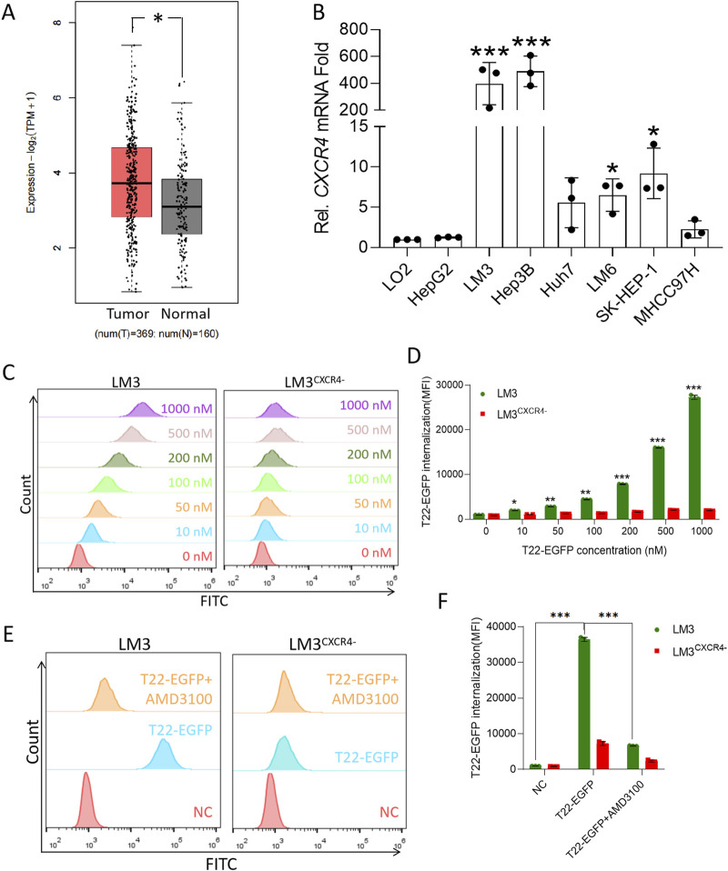
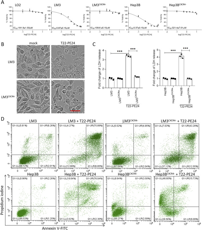
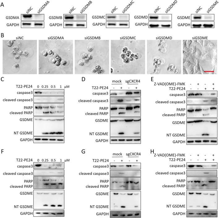
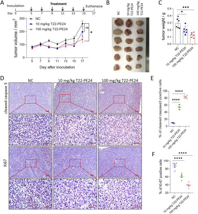
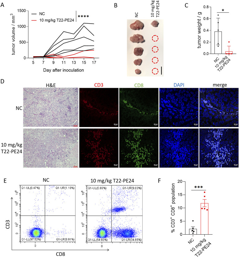
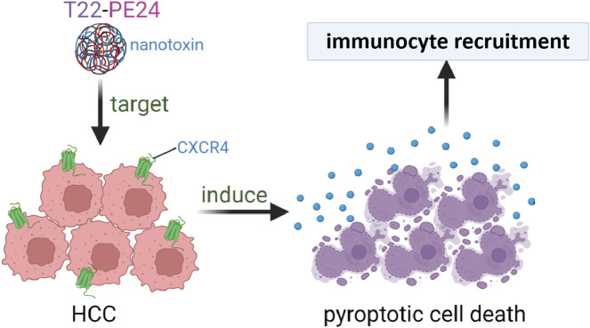
Introduction: Cytotoxic agents have shown limited benefits in hepatocellular carcinoma (HCC), mediated in part by the lack of targeting. As cell-penetrating peptides (CPPs) are capable of delivering various biologically active molecules into cells, including protein, peptides, small chemo-drugs, and nucleic acid with or without targeting, we developed T22-PE24, a CXCR4-targeted self-assembling cytotoxic nanotoxin, to effectively induce HCC pyroptosis. Methods: T22 incorporating enhanced green fluorescent protein (EGFP) or PE24 was purified from DE3 bacterial cells and characterized using transmission electron microscopy, the Zetasizer Nano®, and SEC-HPLC. The internalization effect of T22-EGFP was detected by flow cytometry system (FCS) in CXCR4+/LM3(CXCR4−) HCC cells. The CCK8, lactate dehydrogenase (LDH) release, Western blot, and nude mice HCC models were used to estimate the cell viability of T22-PE24. The complete-immunity HCC tumor-bearing mice model was used to assess the immune response of T22-PE24. Results: The round shape under transmission electron microscopy, 49.4 nm hydrodynamic diameter, and −33.33 mV zeta potential indicated that T22-PE24 self-assembled into nanoparticles. T22 incorporating EGFP selectively internalized in CXCR4+ HCC cells and showed no accumulation in CXCR4-knockout HCC cells. The T22-PE24 nanotoxin induced HCC pyroptosis via the caspase-3/GSDME signaling pathway and suppressed tumor growth in the absence of histological alterations in normal organs. Using the complete-immunity HCC tumor-bearing mice model, we found that T22-PE24 nanotoxin effectively induces the global reprogramming of cell components of the immune tumor microenvironment, leading to enhanced antitumor effects compared to those observed in immunodeficient mice. Conclusion: Our findings demonstrate the activation of the innate immune response in HCC by inducing pyroptosis with T22-PE24 nanotoxin treatment and support an implementation of this strategy for HCC treatment.
Overview
The claims and statistics of this paper haven’t been checked yet.
Run the check — free, no account needed. It reads the open-access text and checks the sources it cites.
Integrity signals
Registry
Cites
Statistics
Figures
Verification
Claims the paper makes
The claims and statistics of this paper haven’t been checked yet.
Run the check — free, no account needed. It reads the open-access text and checks the sources it cites.
Claims other papers make about this one
Each is checked against this paper’s own text: does it say what the citing paper says it does?
In the citing paper
“Beyond integrins, a CXCR4-targeted self-assembling nanotoxin (T22-PE24) selectively internalizes into CXCR4-positive hepatocellular carcinoma to trigger caspase-3/GSDME-dependent pyroptosis with immune remodeling [84, 85].”
From Overcoming tumor microenvironment barriers: transformable and bioinspired nanomedicine strategies for deep tumor penetration (2026) · cites this paper as [84]
Self-check
Percentages, sums and reported differences re-computed from the paper’s own numbers.
The self-check needs the paper’s full text.
Citations
Cites
Vogel, A., Meyer, T., Sapisochin, G., Salem, R., & Saborowski, A. (2022) · The Lancet · doi:10.1016/s0140-6736(22)01200-4 · Cited by 2,427
Research works printed but not linked: 34
- Anwanwan, 2020, Challenges in liver cancer and possible treatment approaches, Biochim. Biophys. Acta Rev. Cancer, 1873, 188314, 10.1016/j.bbcan.2019.188314 · DOI
- Cerella, 2014, From nature to bedside: pro-survival and cell death mechanisms as therapeutic targets in cancer treatment, Biotechnol. Adv., 32, 1111, 10.1016/j.biotechadv.2014.03.006 · DOI
- Chon, 2021, Hepatocellular carcinoma statistics in South Korea, Clin. Mol. Hepatol., 27, 512, 10.3350/cmh.2021.0171 · DOI
- El-Serag, 2008, Diagnosis and treatment of hepatocellular carcinoma, Gastroenterology, 134, 1752, 10.1053/j.gastro.2008.02.090 · DOI
- Falgas, Selective delivery of t22-pe24-h6 to cxcr4(+) diffuse large b-cell lymphoma cells leads to wide therapeutic index in a disseminated mouse model, Theranostics, 10, 5169, 10.7150/thno.43231 · DOI
- Falgas, A cxcr4-targeted nanocarrier achieves highly selective tumor uptake in diffuse large b-cell lymphoma mouse models, Haematologica, 105, 741, 10.3324/haematol.2018.211490 · DOI
- Gebhardt, 2014, Baculovirus resistance in codling moth is virus isolate-dependent and the consequence of a mutation in viral gene pe38, Proc. Natl. Acad. Sci. U. S. A., 111, 15711, 10.1073/pnas.1411089111 · DOI
- Gusarova, 2007, A cell-penetrating arf peptide inhibitor of foxm1 in mouse hepatocellular carcinoma treatment, J. Clin. Invest, 117, 99, 10.1172/JCI27527 · DOI
- Jin, 2018, Paclitaxel-loaded nanoparticles decorated with bivalent fragment hab18 f(ab')(2) and cell penetrating peptide for improved therapeutic effect on hepatocellular carcinoma, Artif. Cells Nanomed Biotechnol., 46, 1076, 10.1080/21691401.2017.1360325 · DOI
- Lee, 2019, Construction of an immunotoxin via site-specific conjugation of anti-her2 igg and engineered pseudomonas exotoxin a, J. Biol. Eng., 13, 56, 10.1186/s13036-019-0188-x · DOI
- Liu, 2016, Emerging landscape of cell penetrating peptide in reprogramming and gene editing, J. Control Release, 226, 124, 10.1016/j.jconrel.2016.02.002 · DOI
- Masuda, 1992, A novel anti-hiv synthetic peptide, t-22 ([tyr5,12,lys7]-polyphemusin ii), Biochem. Biophys. Res. Commun., 189, 845, 10.1016/0006-291x(92)92280-b · DOI
- Medina-Gutierrez, 2022, Potent anticancer activity of cxcr4-targeted nanostructured toxins in aggressive endometrial cancer models, Cancers (Basel), 15, 85, 10.3390/cancers15010085 · DOI
- Moradian, 2021, Pe38-based gene therapy of her2-positive breast cancer stem cells via vhh-redirected polyamidoamine dendrimers, Sci. Rep., 11, 15517, 10.1038/s41598-021-93972-5 · DOI
- Morgan, 2023, Gamma radiation coupled adp-ribosyl transferase activity of pseudomonas aeruginosa pe24 moiety, Appl. Microbiol. Biotechnol., 107, 1765, 10.1007/s00253-023-12401-x · DOI
- Murakami, 1999, Inhibitory mechanism of the cxcr4 antagonist t22 against human immunodeficiency virus type 1 infection, J. Virol., 73, 7489, 10.1128/JVI.73.9.7489-7496.1999 · DOI
- Nunez, 2023, T22-pe24-h6 nanotoxin selectively kills cxcr4-high expressing aml patient cells in vitro and potently blocks dissemination in vivo, Pharmaceutics, 15, 727, 10.3390/pharmaceutics15030727 · DOI
- Rioja-Blanco, Cxcr4-targeted nanotoxins induce gsdme-dependent pyroptosis in head and neck squamous cell carcinoma, J. Exp. Clin. Cancer Res., 41, 49, 10.1186/s13046-022-02267-8 · DOI
- Rioja-Blanco, Self-assembling protein nanocarrier for selective delivery of cytotoxic polypeptides to cxcr4(+) head and neck squamous cell carcinoma tumors, Acta Pharm. Sin. B, 12, 2578, 10.1016/j.apsb.2021.09.030 · DOI
- Sala, 2022, Gsdmd-dependent pyroptotic induction by a multivalent cxcr4-targeted nanotoxin blocks colorectal cancer metastases, Drug Deliv., 29, 1384, 10.1080/10717544.2022.2069302 · DOI
- Salem, 2022, Interventional treatment of hepatocellular carcinoma, J. Hepatol., 77, 1205, 10.1016/j.jhep.2022.03.037 · DOI
- Sanchez-Garcia, 2018, Self-assembling toxin-based nanoparticles as self-delivered antitumoral drugs, J. Control Release, 274, 81, 10.1016/j.jconrel.2018.01.031 · DOI
- Shao, 2021, Gasdermins: making pores for pyroptosis, Nat. Rev. Immunol., 21, 620, 10.1038/s41577-021-00602-2 · DOI
- Siegel, 2021, Cancer statistics, 2021, CA Cancer J. Clin., 71, 7, 10.3322/caac.21654 · DOI · Paper page
- Takeuchi, 2006, Direct and rapid cytosolic delivery using cell-penetrating peptides mediated by pyrenebutyrate, ACS Chem. Biol., 1, 299, 10.1021/cb600127m · DOI
- Wang, 2020, A bioorthogonal system reveals antitumour immune function of pyroptosis, Nature, 579, 421, 10.1038/s41586-020-2079-1 · DOI
- Wang, 2018, Lipid-modified cell-penetrating peptide-based self-assembly micelles for co-delivery of narciclasine and siulk1 in hepatocellular carcinoma therapy, Acta Biomater., 74, 414, 10.1016/j.actbio.2018.05.030 · DOI
- Yang, 2019, Tcf12 promotes the tumorigenesis and metastasis of hepatocellular carcinoma via upregulation of cxcr4 expression, Theranostics, 9, 5810, 10.7150/thno.34973 · DOI
- Yang, 2020, Cxcr4 mediates matrix stiffness-induced downregulation of ubtd1 driving hepatocellular carcinoma progression via yap signaling pathway, Theranostics, 10, 5790, 10.7150/thno.44789 · DOI
- Yu, Pyroptosis: mechanisms and diseases, Signal Transduct. Target Ther., 6, 128, 10.1038/s41392-021-00507-5 · DOI · Paper page
- Yu, Efficient intracellular delivery of proteins by a multifunctional chimaeric peptide in vitro and in vivo, Nat. Commun., 12, 5131, 10.1038/s41467-021-25448-z · DOI
- Zhang, 2020, Gasdermin e suppresses tumour growth by activating anti-tumour immunity, Nature, 579, 415, 10.1038/s41586-020-2071-9 · DOI
- Zhao, 2018, Cell-penetrating peptide-modified targeted drug-loaded phase-transformation lipid nanoparticles combined with low-intensity focused ultrasound for precision theranostics against hepatocellular carcinoma, Theranostics, 8, 1892, 10.7150/thno.22386 · DOI
- Zhao, 2023, A self-assembling cxcr4-targeted pyroptosis nanotoxin for melanoma therapy, Biomater. Sci., 11, 2200, 10.1039/d2bm02026b · DOI
Cited by
Sorted by citations
Sheng, J., Yuan, W., Zhang, M., Chen, Z., Qi, Y., Zhao, Y., & Zhang, S. (2026) · Journal of Nanobiotechnology · doi:10.1186/s12951-026-04028-7 · Cited by 35
Metadata
- Authors
- Huang, Y., Li, Y., He, R., Dong, S., Zhao, Z., & Jiao, X.
- Venue
- Frontiers in Bioengineering and Biotechnology
- Publisher
- Frontiers Media SA
- Published
- 2024-11-04
- Volume
- 12
- ISSN
- 2296-4185
- PMC
- PMC11570815
- Cited by
- 7
- References
- 1
About these checks
Registry, retraction, Cites and statistics results are computed, not judged, with one exception: a printed reference that no index lists is read once by a model before it counts as a citation error. Claims are extracted by a model and kept only when their quote appears verbatim in the text; each cited source is then checked, with a model’s help, for whether it says what the claim relies on. Those verdicts can be wrong — read the claim’s quote and the cited source before relying on one.
Statistics and arithmetic are recomputed, not judged.
Confidence bands are shown only for fields calibrated on our annotated test set. None is calibrated yet, so no band is shown.
What counts as a claim ›How we measure confidence ›How we check this ›
Are you an author, or do you see a problem with this page? Request a review
