

The DOI matches its Crossref record; 213 of the 216 references it cites were found in a registry or an index.
Hypoxia and Selective Autophagy in Cancer Development and Therapy
Open accessAbstract
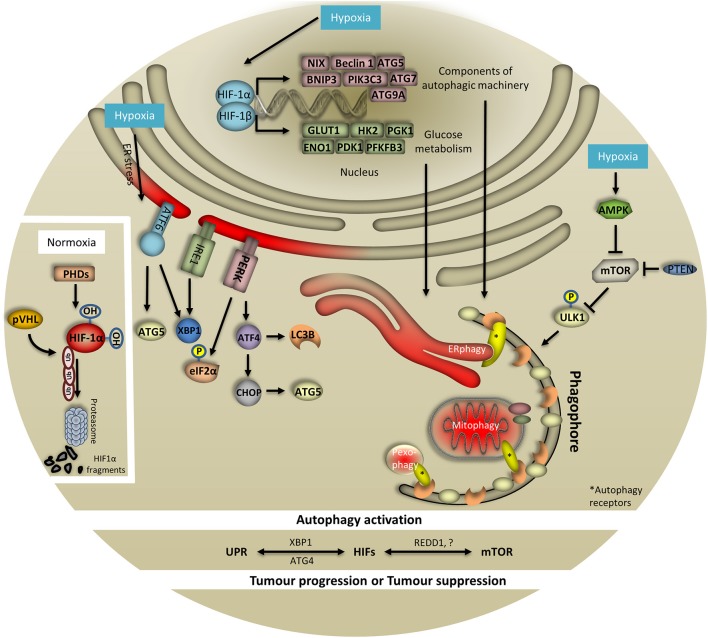
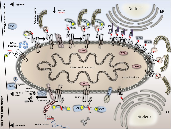
Low oxygen availability, a condition known as hypoxia, is a common feature of various pathologies including stroke, ischemic heart disease, and cancer. Hypoxia adaptation requires coordination of intricate pathways and mechanisms such as hypoxia-inducible factors (HIFs), the unfolded protein response (UPR), mTOR, and autophagy. Recently, great effort has been invested toward elucidating the interplay between hypoxia-induced autophagy and cancer cell metabolism. Although novel types of selective autophagy have been identified, including mitophagy, pexophagy, lipophagy, ERphagy and nucleophagy among others, their potential interface with hypoxia response mechanisms remains poorly understood. Autophagy activation facilitates the removal of damaged cellular compartments and recycles components, thus promoting cell survival. Importantly, tumor cells rely on autophagy to support self-proliferation and metastasis; characteristics related to poor disease prognosis. Therefore, a deeper understanding of the molecular crosstalk between hypoxia response mechanisms and autophagy could provide important insights with relevance to cancer and hypoxia-related pathologies. Here, we survey recent findings implicating selective autophagy in hypoxic responses, and discuss emerging links between these pathways and cancer pathophysiology.
Overview
The claims and statistics of this paper haven’t been checked yet.
Run the check — free, no account needed. It reads the open-access text and checks the sources it cites.
Integrity signals
Registry
Cites
Statistics
Figures
Verification
Claims the paper makes
The claims and statistics of this paper haven’t been checked yet.
Run the check — free, no account needed. It reads the open-access text and checks the sources it cites.
Claims other papers make about this one
Each is checked against this paper’s own text: does it say what the citing paper says it does?
In the citing paper
“Autophagy can be induced by certain treatments or microevironmental effects in relation to metabolic adaptation and resistance [45].”
From Hypoxia Signaling in Cancer: From Basics to Clinical Practice (2021) · cites this paper as [45]
Self-check
Percentages, sums and reported differences re-computed from the paper’s own numbers.
The self-check needs the paper’s full text.
Citations
Cites
3 retracted
Mitochondrial Autophagy Is an HIF-1-dependent Adaptive Metabolic Response to Hypoxia
Zhang, H., Bosch-Marce, M., Shimoda, L. A., Tan, Y. S., Baek, J. H., Wesley, J. B., Gonzalez, F. J., & Semenza, G. L. (2008) · Journal of Biological Chemistry · doi:10.1074/jbc.m800102200 · Cited by 1,601
RetractedRETRACTED: MiR‐137 functions as a tumor suppressor in pancreatic cancer by targeting MRGBP
Ding, F. H., Zhang, S., Gao, S., Shang, J., Li, Y., Cui, N., & Zhao, Q. (2018) · Journal of Cellular Biochemistry · doi:10.1002/jcb.26676 · Cited by 31
RetractedRen, C., Liao, J.-Y., Huang, J., Chen, W., Ma, X., & Luo, X. (2017) · Oncology Research Featuring Preclinical and Clinical Cancer Therapeutics · doi:10.3727/096504017x14954923820137 · Cited by 14
RetractedRepertoires of Autophagy in the Pathogenesis of Ocular Diseases
Li, Y., Jiang, Q. L., Cao, G.-F., Yao, J., & Yan, B. (2015) · Cellular Physiology and Biochemistry · doi:10.1159/000373980 · Cited by 16,351
AMPK and mTOR regulate autophagy through direct phosphorylation of Ulk1
Kim, J., Kundu, M., Viollet, B., & Guan, K.-L. (2011) · Nature Cell Biology · doi:10.1038/ncb2152 · Cited by 7,227
Targeting Hypoxia-Inducible Factors for the Treatment of Anemia in Chronic Kidney Disease Patients
Locatelli, F., Fishbane, S. N., Block, G. A., & Macdougall, I. C. (2017) · American Journal of Nephrology · doi:10.1159/000455166 · Cited by 4,836
Mechanism and medical implications of mammalian autophagy
Dikic, I., & Elazar, Z. (2018) · Nature Reviews Molecular Cell Biology · doi:10.1038/s41580-018-0003-4 · Cited by 2,771
Levy, J. M. M., Towers, C. G., & Thorburn, A. (2017) · Nature Reviews Cancer · doi:10.1038/nrc.2017.53 · Cited by 2,526
Hypoxia-inducible factors and the response to hypoxic stress
Majmundar, A. J., Wong, W. J., & Simon, M. C. (2010) · Molecular Cell · doi:10.1016/j.molcel.2010.09.022 · Cited by 2,380
HIF-1 mediates adaptation to hypoxia by actively downregulating mitochondrial oxygen consumption
Papandreou, I., Cairns, R. A., Fontana, L., Lim, A. L., & Denko, N. C. (2006) · Cell Metabolism · doi:10.1016/j.cmet.2006.01.012 · Cited by 2,267
Cited by
Sorted by citations
The Roles of Autophagy in Cancer
Yun, C. W., & Lee, S. H. (2018) · International Journal of Molecular Sciences · doi:10.3390/ijms19113466 · Cited by 965
Nedeljković, M., & Damjanović, A. (2019) · Cells · doi:10.3390/cells8090957 · Cited by 910
“These include: increased expression of ABC transporters (including ABCG2 and ABCC1) [105,106]; decreased proliferation [102]; complex modulation of cellular senescence and apoptosis [102]; induction of autophagy that aids in tumor survival [107]; enhanced genetic instability and subsequent clonal selection of aggressi…”
BackgroundGlioblastoma multiforme (GBM): An overview of current therapies and mechanisms of resistance
Wu, W. E., Klockow, J., Zhang, M., Lafortune, F., Chang, E., Jin, L., Wu, Y., & Daldrup-Link, H. E. (2021) · Pharmacological Research · doi:10.1016/j.phrs.2021.105780 · Cited by 877
Hypoxia signaling in human health and diseases: implications and prospects for therapeutics
Luo, Z., Tian, M., Yang, G., Tan, Q., Chen, Y., Li, G., Zhang, Q., Li, Y., Wan, P., & Wu, J. (2022) · Signal Transduction and Targeted Therapy · doi:10.1038/s41392-022-01080-1 · Cited by 499
“Hypoxiainduced autophagy performs a certain function in tumor progression.(86) Several hypoxia-responsive genes’ transcription is regulated by HIF-1 activation under hypoxia stress.”
BackgroundYao, R.-Q., Ren, C., Xia, Z.-F., & Yao, Y.-M. (2021) · Autophagy · doi:10.1080/15548627.2020.1725377 · Cited by 441
“etiology of various severe conditions, initiates ER stress and functional changes in mitochondria concurrently, accompanied by activation of a variety of organelle-specific autophagies [10].”
BackgroundKhandia, R., Dadar, M., Munjal, A. K., Dhama, K., Karthik, K., Tiwari, R., Yatoo, M. I., Iqbal, H. M. N., Singh, K. P., Joshi, S. K., et al. (2019) · Cells · doi:10.3390/cells8070674 · Cited by 337
The Double-Edge Sword of Autophagy in Cancer: From Tumor Suppression to Pro-tumor Activity
Chavez-Dominguez, R., Perez-Medina, M., López-González, J. S., Galicia-Velasco, M., & Aguilar‐Cázares, D. (2020) · Frontiers in Oncology · doi:10.3389/fonc.2020.578418 · Cited by 322
“Quin et al. reported in acute myeloid leukemia cells that PDK-1 associates with ULK-1 promoting its activation and leading to induction of autophagy (148).”
BackgroundInfluentialMechanism of interaction between autophagy and apoptosis in cancer
Das, S., Shukla, N., Singh, S. S., Kushwaha, S., & Shrivastava, R. (2021) · APOPTOSIS · doi:10.1007/s10495-021-01687-9 · Cited by 269
He, R., Wang, Z., Cui, M., Liu, S., Wu, W., Chen, M., Wu, Y., Qu, Y., Lin, H., Chen, S., et al. (2021) · Autophagy · doi:10.1080/15548627.2021.1872227 · Cited by 236
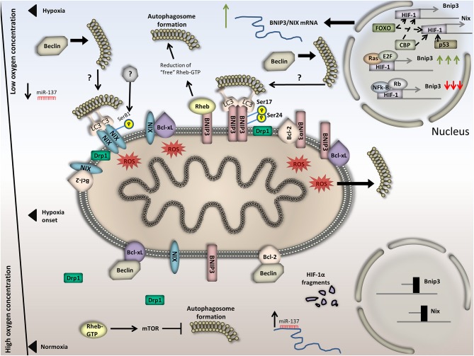
“BNIP3-BNIP3L-induced mitophagy upon hypoxia additionally requires the activity of essential autophagy components such as ATG7 and the interaction with LC3B [78,79].”
BackgroundSelective Autophagy Regulates Cell Cycle in Cancer Therapy
Zheng, K., He, Z., Kitazato, K., & Wang, Y. (2018) · Theranostics · doi:10.7150/thno.30308 · Cited by 229
Reggiori, F., & Molinari, M. (2022) · Physiological Reviews · doi:10.1152/physrev.00038.2021 · Cited by 232
Outstanding Questions in Mitophagy: What We Do and Do Not Know
Montava‐Garriga, L., & Ganley, I. G. (2019) · Journal of Molecular Biology · doi:10.1016/j.jmb.2019.06.032 · Cited by 218
“metabolic switch to upregulate glycolytic genes, attenuate mitochondria respiration and trigger mitophagy [88,90,91].”
BackgroundMolecular Mechanisms, Biomarkers and Emerging Therapies for Chemotherapy Resistant TNBC
Ferrari, P., Scatena, C., Ghilli, M., Bargagna, I., Lorenzini, G., & Nicolini, A. (2022) · International Journal of Molecular Sciences · doi:10.3390/ijms23031665 · Cited by 218
Role and regulation of autophagy in cancer
Rakesh, R., Dharshini, L. C. P., Sakthivel, K. M., & Rasmi, R. R. (2022) · Biochimica et Biophysica Acta (BBA) - Molecular Basis of Disease · doi:10.1016/j.bbadis.2022.166400 · Cited by 198
Nonsense-mediated RNA decay: an emerging modulator of malignancy
Tan, K., Stupack, D. G., & Wilkinson, M. (2022) · Nature reviews. Cancer · doi:10.1038/s41568-022-00481-2 · Cited by 154
Hypoxia-Driven Effects in Cancer: Characterization, Mechanisms, and Therapeutic Implications
Shi, R., Liao, C., & Zhang, Q. H. (2021) · Cells · doi:10.3390/cells10030678 · Cited by 151
Inflammation and Metabolism in Cancer Cell—Mitochondria Key Player
Neagu, M. T., Constantin, C., Popescu, I., Zipeto, D., Tzanakakis, G. N., Nikitovic, D., Fenga, C., Stratakis, C. A., Spandidos, D. A., & Tsatsakis, A. M. (2019) · Frontiers in Oncology · doi:10.3389/fonc.2019.00348 · Cited by 151
AGE-RAGE synergy influences programmed cell death signaling to promote cancer
Waghela, B. N., Vaidya, F. U., Ranjan, K., Chhipa, A. S., Tiwari, B. S., & Pathak, C. (2020) · Molecular and Cellular Biochemistry · doi:10.1007/s11010-020-03928-y · Cited by 135
“Hypoxia plays a key role in cancer metastasis and is also intricately related to autophagy machinery in cancer cells [95].”
BackgroundT cell exhaustion in malignant gliomas
Watowich, M., Gilbert, M. R., & Larion, M. (2023) · Trends in cancer · doi:10.1016/j.trecan.2022.12.008 · Cited by 140
Hypoxia-induced alternative splicing: the 11th Hallmark of Cancer
Farina, A. R., Cappabianca, L. A., Sebastiano, M., Zelli, V., Guadagni, S., & Mackay, A. R. (2020) · Journal of Experimental & Clinical Cancer Research · doi:10.1186/s13046-020-01616-9 · Cited by 134
“This caspase-independent cell death mechanism is characterized by cellular vacuolation, cellular swelling and necrotic cell lysis is mediated by RIPK1, RIPK3 and MLKL, induced by the UPR, and also involves unconventional XPB1s splicing and hypoxiainduced autophagy, which regulate autophagosome/lysosome fusion [5, 175].”
BackgroundHepatic Tumor Microenvironments and Effects on NK Cell Phenotype and Function
Fernández, J. P., Luddy, K. A., Harmon, C., & O’Farrelly, C. (2019) · International Journal of Molecular Sciences · doi:10.3390/ijms20174131 · Cited by 119
Niu, Y., Lin, Z., Wan, A., Sun, L., Yan, S., Liang, H., Zhan, S., Chen, D., Bu, X., Liu, P., et al. (2021) · Hepatology · doi:10.1002/hep.31846 · Cited by 115
“The homeostatic response to intratumoral hypoxic environment is mediated by hypoxia-inducible factor (HIF-1), consisting of HIF-1α and HIF-1β subunits (7), which triggers a variety of cellular responses such as angiogenesis (8), erythropoiesis (9), apoptosis (10), metastasis (11), drug-resistance (12), autophagy (13)…”
BackgroundTargeting Autophagy in Breast Cancer
Cocco, S., Leone, A., Piezzo, M., Caputo, R., Lauro, V. D., Rella, F. D., Fusco, G., Capozzi, M., Gioia, G. D., Budillon, A., et al. (2020) · International Journal of Molecular Sciences · doi:10.3390/ijms21217836 · Cited by 120
Hypoxia Signaling in Cancer: From Basics to Clinical Practice
Sebestyén, A., Kopper, L., Dankó, T., & Tímár, J. (2021) · Pathology and Oncology Research · doi:10.3389/pore.2021.1609802 · Cited by 110
“reduces oxygen consumption and/or induces autophagy [45] leading slower metabolic activity and forces cellular survival mechanisms with balanced bioenergetics [46].”
BackgroundPlasticity of cancer invasion and energy metabolism
Parlani, M., Jorgez, C. J., & Friedl, P. (2022) · Trends in Cell Biology · doi:10.1016/j.tcb.2022.09.009 · Cited by 106
Sirtuins' control of autophagy and mitophagy in cancer
Aventaggiato, M., Vernucci, E., Barreca, F., Russo, M. A., & Tafani, M. (2020) · Pharmacology & Therapeutics · doi:10.1016/j.pharmthera.2020.107748 · Cited by 103
Li, J., Feng, Y., Li, Y., He, P., Zhou, Q.-Y., Tian, Y., Yao, R.-Q., & Yao, Y.-M. (2024) · Cell Proliferation · doi:10.1111/cpr.13621 · Cited by 111
“36,37 While increasing knowledge continues to reveal that NCOA4-mediated ferritin degradation is regarded as conservative catabolism.”
BackgroundThe Interplay Between Tumor Suppressor p53 and Hypoxia Signaling Pathways in Cancer
Zhang, C., Liu, J., Wang, J., Zhang, T., Xu, D., Hu, W., & Feng, Z. (2021) · Frontiers in Cell and Developmental Biology · doi:10.3389/fcell.2021.648808 · Cited by 100
“In addition to apoptosis, hypoxia induces autophagy via both HIF-1-dependent and -independent pathways, such as the mTOR pathway, unfolded protein response (UPR) pathway and PKCδ-JNK1 pathway (Fang et al., 2015; Daskalaki et al., 2018).”
BackgroundRaudenská, M., Balvan, J., & Masařík, M. (2021) · Molecular Cancer · doi:10.1186/s12943-021-01423-6 · Cited by 99
Autophagy as a novel therapeutic target in vascular calcification
Phadwal, K., Feng, D., Zhu, D., & MacRae, V. E. (2019) · Pharmacology & Therapeutics · doi:10.1016/j.pharmthera.2019.107430 · Cited by 90
Oxygen releasing materials: Towards addressing the hypoxia-related issues in tissue engineering
Agarwal, T., Kazemi, S., Costantini, M., Perfeito, F. G., Correia, C. R., Gaspar, V. M., Montazeri, L., Maria, C. D., Mano, J. F., Vosough, M., et al. (2021) · Materials Science and Engineering C · doi:10.1016/j.msec.2021.111896 · Cited by 93
Hypoxia-inducible factor-1: Regulatory mechanisms and drug development in stroke
Pan, Z., Ma, G., Kong, L., & Du, G. (2021) · Pharmacological Research · doi:10.1016/j.phrs.2021.105742 · Cited by 93
Mitofusins: Disease Gatekeepers and Hubs in Mitochondrial Quality Control by E3 Ligases
Escobar‐Henriques, M., & Joaquim, M. (2019) · Frontiers in Physiology · doi:10.3389/fphys.2019.00517 · Cited by 85
Qian, X., Pan, Y., Huang, Q.-Y., Shi, Y., Huang, Q.-Y., Hu, Z., & Xiong, L. (2019) · OncoTargets and Therapy · doi:10.2147/ott.s191317 · Cited by 82
“ments and recycle components, thereby preventing cells from apoptosis and increasing survivability.(64)”
BackgroundThe Role of Ceramide Metabolism and Signaling in the Regulation of Mitophagy and Cancer Therapy
Sheridan, M., & Öğretmen, B. (2021) · Cancers · doi:10.3390/cancers13102475 · Cited by 82
Mitochondrial Perturbations Couple mTORC2 to Autophagy in C. elegans
Aspernig, H., Heimbucher, T., Qi, W., Gangurde, D., Curic, S., Yan, Y., von Gromoff, E. D., Baumeister, R., & Thien, A. (2019) · Cell Reports · doi:10.1016/j.celrep.2019.09.072 · Cited by 75
The role of autophagy in cancer: from molecular mechanism to therapeutic window
Jalali, P., Shahmoradi, A., Samii, A., Mazloomnejad, R., Hatamnejad, M. R., Saeed, A., Namdar, A., & Salehi, Z. (2025) · Frontiers in Immunology · doi:10.3389/fimmu.2025.1528230 · Cited by 89
“In the cancer microenvironment, autophagy provides cellular energy and inhibits cytotoxicity under stressful conditions (34, 35).”
Autophagy in cancer cell remodeling and quality control
Hernandez, G. A., & Perera, R. M. (2022) · Molecular Cell · doi:10.1016/j.molcel.2022.03.023 · Cited by 73
Autophagy: a double-edged sword in ischemia–reperfusion injury
Tang, L., Zhang, W., Yan, L., Wang, W., Deng, X., Wang, C., & Shi, W. (2025) · Cellular & Molecular Biology Letters · doi:10.1186/s11658-025-00713-x · Cited by 82
Castelli, S., Ciccarone, F., Tavian, D., & Ciriolo, M. R. (2021) · Journal of Experimental & Clinical Cancer Research · doi:10.1186/s13046-021-01887-w · Cited by 70
Autophagy and Breast Cancer: Connected in Growth, Progression, and Therapy
Wu, Q., & Sharma, D. (2023) · Cells · doi:10.3390/cells12081156 · Cited by 69
Lin, C., Blessing, A. M., Pulliam, T. L., Shi, Y., Wilkenfeld, S. R., Han, J. J., Murray, M. M., Pham, A. H., Duong, K., Brun, S. N., et al. (2021) · Oncogene · doi:10.1038/s41388-021-01658-z · Cited by 63
“Conversely, in more advanced cancers, 106 autophagy can enable cells to evade apoptosis in hypoxic and nutrient-deficient 107 environments as well as promote drug resistance(21-24).”
BackgroundRacanè, L., Ptiček, L., Fajdetić, G., Tralić‐Kulenović, V., Klobučar, M., Pavelić, S. K., Perić, M., Paljetak, H. Č., Verbanac, D., & Starčević, K. (2019) · Bioorganic Chemistry · doi:10.1016/j.bioorg.2019.103537 · Cited by 65
Ikeda, S., Abe, F., Matsuda, Y., Kitadate, A., Takahashi, N., & Tagawa, H. (2020) · Cancer Science · doi:10.1111/cas.14614 · Cited by 61
Cancer cells adapt FAM134B/BiP mediated ER-phagy to survive hypoxic stress
Chipurupalli, S., Ganesan, R., Martini, G., Mele, L. L., Reggio, A., Esposito, M., Kannan, E., Namasivayam, V., Grumati, P., Desiderio, V., et al. (2022) · Cell Death and Disease · doi:10.1038/s41419-022-04813-w · Cited by 62
“It is also speculated that its upregulation is HIF-1α dependent [57].”
BackgroundYang, T., Qu, X., Wang, X., Xu, D., Sheng, M., Lin, Y., Ke, M., Song, C., Xia, Q., Jiang, L., et al. (2023) · Hepatology · doi:10.1097/hep.0000000000000638 · Cited by 59
“As HIF-1 α stabilization and nuclear translocation are involved in autophagy induction, [36] we proposed that there might be a crosstalk between PD1 and HIF-1 α in response to fatty acid stimulation.”
Hypoxia and Extracellular Acidification as Drivers of Melanoma Progression and Drug Resistance
Dratkiewicz, E., Simiczyjew, A., Mazurkiewicz, J., Ziętek, M., Matkowski, R., & Nowak, D. (2021) · Cells · doi:10.3390/cells10040862 · Cited by 65
Shafabakhsh, R., Arianfar, F., Vosough, M., Mirzaei, H. R., Mahjoubin‐Tehran, M., Khanbabaei, H., Kowsari, H., Shojaie, L., Azar, M. E. F., Hamblin, M. R., et al. (2021) · Cancer Gene Therapy · doi:10.1038/s41417-020-00272-7 · Cited by 57
HIF-1a regulates hypoxia-induced autophagy via translocation of ANKRD37 in colon cancer
Deng, M., Zhang, W., Yuan, L., Tan, J., & Chen, Z. (2020) · Experimental Cell Research · doi:10.1016/j.yexcr.2020.112175 · Cited by 55
Rani, S., Roy, S., Singh, M., & Kaithwas, G. (2022) · Oxidative Medicine and Cellular Longevity · doi:10.1155/2022/2407223 · Cited by 57
“Interestingly, data revealed that the expression of BNIP3 is elevated in parallel with the transcriptional activities of HIF-1 α , whereas nuclear factor kappa-light-chain-enhancer of activated B cells (NF-κ B) are responsible for its downregulation [90, 91].”
Signs of ROS-Associated Autophagy in Testis and Sperm in a Rat Model of Varicocele
Sadeghi, N., Erfani-Majd, N., Tavalaee, M., Tabandeh, M. R., Drevet, J. R., & Nasr‐Esfahani, M. H. (2020) · Oxidative Medicine and Cellular Longevity · doi:10.1155/2020/5140383 · Cited by 53
“This catabolic process can be activated under various stress conditions such as oxidative stress [4], thermal stress [5, endoplasmic reticulum stress [6], hypoxia [7], and unbalanced diet [8].”
BackgroundRole of Mitofusins and Mitophagy in Life or Death Decisions
Joaquim, M., & Escobar‐Henriques, M. (2020) · Frontiers in Cell and Developmental Biology · doi:10.3389/fcell.2020.572182 · Cited by 50
“…and SIAH-1 (Liu et al., 2012; Fu et al., 2013; Cilenti et al., 2014; Yun et al., 2014; Tang et al., 2015; Mukherjee and Chakrabarti, 2016a,b; Szargel et al., 2016; Chen et al., 2017; Daskalaki et al., 2018; Di Rita et al., 2018; Ferrucci et al., 2018; Lampert et al., 2019; Shefa et al., 2019).”
Mitochondrial quality surveillance: mitophagy in cardiovascular health and disease
Diao, R. Y., & Gustafsson, Å. B. (2021) · American Journal of Physiology-Cell Physiology · doi:10.1152/ajpcell.00360.2021 · Cited by 50
Molecular Mechanism of Autophagy and Its Regulation by Cannabinoids in Cancer
Lee, X. C., Werner, E., & Falasca, M. (2021) · Cancers · doi:10.3390/cancers13061211 · Cited by 48
Targeting Cytoprotective Autophagy to Enhance Anticancer Therapies
Xiao, M., Benoit, A., Hasmim, M., Duhem, C., Vogin, G., Berchem, G., Noman, M. Z., & Janji, B. (2021) · Frontiers in Oncology · doi:10.3389/fonc.2021.626309 · Cited by 47
“Other factors interacting with the autophagy process could also be considered as potential targets to inhibit autophagy and overcome therapy resistance, such as AMPK and HIF-1a, which are reported as key inducers of autophagy through negative regulation of the mTOR pathways and inducing hypoxic conditions, respectivel…”
BackgroundPhysicochemical aspects of the tumour microenvironment as drivers of vasculogenic mimicry
Andreucci, E., Peppicelli, S., Ruzzolini, J., Bianchini, F., & Calorini, L. (2022) · Cancer and Metastasis Reviews · doi:10.1007/s10555-022-10067-x · Cited by 44
Autophagy and cancer: Basic mechanisms and inhibitor development
Hama, Y., Ogasawara, Y., & Noda, N. N. (2023) · Cancer Science · doi:10.1111/cas.15803 · Cited by 47
Subhan, M. A. (2022) · RSC Advances · doi:10.1039/d2ra02005j · Cited by 42
Shim, S. M., Choi, H. R., Kwon, S. C., Kim, H. Y., Sung, K. W., Jung, E. J., Mun, S. R., Bae, T. H., Kim, D. H., Son, Y. S., et al. (2022) · Autophagy · doi:10.1080/15548627.2022.2126617 · Cited by 39
Lyu, P., Wen, J., Zhang, W., Liu, N., Stolzer, I., Gießl, A., Jia, Y., Mauro, D., Zhang, F., Ciccia, F., et al. (2024) · Annals of the Rheumatic Diseases · doi:10.1136/ard-2023-224491 · Cited by 36
“33 34 .”
Multitarget Pharmacology of Sulfur–Nitrogen Heterocycles: Anticancer and Antioxidant Perspectives
Drakontaeidi, A., Papanotas, I., & Pontiki, E. A. (2024) · Antioxidants · doi:10.3390/antiox13080898 · Cited by 36
Tao, G., Wang, X., Wang, J., Ye, Y., Zhang, M., Yan, L., & Ding, S. (2024) · British Journal of Pharmacology · doi:10.1111/bph.17373 · Cited by 36
“Bnip3-Bnip3L-induced mitophagy upon hypoxia additionally requires the activity of essential autophagy components such as ATG7 and the interaction with LC3B (Daskalaki et al., 2018; Park et al., 2013).”
Greville, G., Llop, E., Huang, C., Creagh-Flynn, J., Pfister, S., O’Flaherty, R., Madden, S. F., Peracaula, R., Rudd, P. M., McCann, A. H., et al. (2020) · Frontiers in Oncology · doi:10.3389/fonc.2020.01218 · Cited by 35
“Tumor cells use autophagy to support self-proliferation and metastasis; leading to poor disease prognosis (52), which indicates that hypoxia aids to these tumor features.”
BackgroundAutophagy and senescence in cancer therapy
Patel, N. H., Bloukh, S., Alwohosh, E., Alhesa, A., Saleh, T., & Gewirtz, D. A. (2021) · Advances in cancer research · doi:10.1016/bs.acr.2021.01.002 · Cited by 36
Autophagy and Apoptosis: Current Challenges of Treatment and Drug Resistance in Multiple Myeloma
Al‐Odat, O. S., Guirguis, D. A., Schmalbach, N. K., Yao, G., Budak-Alpdoǧan, T., Jonnalagadda, S. C., & Pandey, M. K. (2022) · International Journal of Molecular Sciences · doi:10.3390/ijms24010644 · Cited by 34
Yang, H., Hu, T., Hu, P., Qi, C., & Lin, Q. (2021) · Molecular Medicine Reports · doi:10.3892/mmr.2021.11995 · Cited by 32
“Autophagy is a highly conserved cellular process, which is activated by various factors, such as hypoxia (38).”
The Peroxisome-Autophagy Redox Connection: A Double-Edged Sword?
Li, H., Lismont, C., Revenco, I., Hussein, M. A. F., Costa, C. F., & Fransen, M. (2021) · Frontiers in Cell and Developmental Biology · doi:10.3389/fcell.2021.814047 · Cited by 37
“However, under hypoxic conditions, these proteins are stabilized and translocated to the nucleus to initiate the transcription of genes involved in cellular adaptation and survival, including a set of genes essential for autophagy (e.g., ATG5, ATG7, ATG9A, BECN1, BNIP3, and BNI3L) (Daskalaki et al., 2018).”
BackgroundInfluentialPan, Y., Liu, Y., Wei, W., Yang, X., Wang, Z., & Xin, W. (2023) · Small · doi:10.1002/smll.202205739 · Cited by 32
The HIF target ATG9A is essential for epithelial barrier function and tight junction biogenesis
Dowdell, A. S., Cartwright, I. M., Goldberg, M. S., Kostelecky, R. E., Ross, T., Welch, N., Glover, L. E., & Colgan, S. P. (2020) · Molecular Biology of the Cell · doi:10.1091/mbc.e20-05-0291 · Cited by 30
“…autophagy can be induced through at least three separate pathways, namely, the unfolded protein response (e.g., protein misfolding in the ER), AMPK activation as a result of energetic stress, and stabilization of HIF-α isoforms and subsequent induction of autophagy genes (Daskalaki et al., 2018).”
BackgroundInfluentialZhu, F., Wang, X., Zeng, H., Yin, Z., & Ye, Z. (2019) · Oncology Letters · doi:10.3892/ol.2019.10881 · Cited by 29
“Among the five previously identified ARGs associated with MM, HIF1A is the most widely studied (59,60).”
BackgroundNucleic Acid Delivery with Red-Blood-Cell-Based Carriers
Pelle, G. D., & Kostevšek, N. (2021) · International Journal of Molecular Sciences · doi:10.3390/ijms22105264 · Cited by 31
Autophagic-Related Proteins in Brain Gliomas: Role, Mechanisms, and Targeting Agents
Pizzimenti, C., Fiorentino, V., Franchina, M., Martini, M., Giuffrè, G., Lentini, M., Silvestris, N., Pietro, M. D., Fadda, G., Tuccari, G., et al. (2023) · Cancers · doi:10.3390/cancers15092622 · Cited by 35
The Autophagy Machinery Contributes to E-cadherin Turnover in Breast Cancer
Damiano, V., Spessotto, P., Vanin, G., Perin, T., Maestro, R., & Santarosa, M. (2020) · Frontiers in Cell and Developmental Biology · doi:10.3389/fcell.2020.00545 · Cited by 28
“Mounting evidence suggests that autophagy endows tumor cells with the capability to survive anoikis, hypoxia and chemotherapy treatments (Levine and Kroemer, 2008; Levy et al., 2014; Daskalaki et al., 2018).”
BackgroundGuo, Y., Zhai, Y., Wu, L., Wang, Y., Wu, P., & Xiong, L. (2022) · International Journal of Molecular Sciences · doi:10.3390/ijms23062927 · Cited by 28
Autophagy and oxidative stress in solid tumors: Mechanisms and therapeutic opportunities
Carretero-Fernández, M., Cabrera-Serrano, A. J., Sánchez‐Maldonado, J. M., Ruiz-Durán, L., Jiménez-Romera, F., García‐Verdejo, F. J., González‐Olmedo, C., Cardús, A., Díaz-Beltrán, L., Gutiérrez‐Bautista, J. F., et al. (2025) · Critical Reviews in Oncology/Hematology · doi:10.1016/j.critrevonc.2025.104820 · Cited by 37
“On the other hand, HIF-1 α , which is stabilized under oxidative stress, has been shown to activate autophagy pathways, including the AMPK/mTOR signaling axis, highlighting the crucial role of ROS in modulating autophagic activity (Daskalaki et al., 2018; J. Li et al., 2022; Zhou et al., 2018).”
Roperto, S., Falco, F. D., Perillo, A., Cătoi, C., & Roperto, F. P. (2019) · Veterinary Microbiology · doi:10.1016/j.vetmic.2019.108396 · Cited by 27
Zhang, S., Gong, F., Liu, J., Liu, T., Yang, J., & Hu, J. (2022) · Biomedicine & Pharmacotherapy · doi:10.1016/j.biopha.2022.113004 · Cited by 27
Host cell egress of Brucella abortus requires BNIP3L‐mediated mitophagy
Verbeke, J., Fayt, Y., Martin, L. S., Yilmaz, O., Sędzicki, J., Reboul, A., Jadot, M., Renard, P., Dehio, C., Renard, H., et al. (2023) · The EMBO Journal · doi:10.15252/embj.2022112817 · Cited by 32
Zhu, X., Yang, C., Yu, Q., & Han, L. (2024) · Meat Science · doi:10.1016/j.meatsci.2024.109692 · Cited by 27
Beyond Self-Recycling: Cell-Specific Role of Autophagy in Atherosclerosis
Henderson, J. M., Weber, C., & Santovito, D. (2021) · Cells · doi:10.3390/cells10030625 · Cited by 27
HIF1α is required for NK cell metabolic adaptation during virus infection
Victorino, F., Bigley, T. M., Park, E., Yao, C.-H., Benoit, J. B., Yang, L., Piersma, S. J., Lauron, E. J., Davidson, R. M., Patti, G. J., et al. (2021) · eLife · doi:10.7554/elife.68484 · Cited by 24
“The 1 α KO NK cells had an increase in apoptosis as a result of decreased expression of glycolytic genes and abnormal glucose metabolism that have been previously associated with apoptosis ( Dengler et al., 2014 ; El Mjiyad et al., 2011 ; Greer et al., 2012 ; MacFarlane et al., 2012 ).”
InfluentialAutophagy and its role in regeneration and remodeling within invertebrate
Song, Q., Liu, H., Zhen, H., & Zhao, B. (2020) · Cell & Bioscience · doi:10.1186/s13578-020-00467-3 · Cited by 25
Chen, J., Wen, B., Wang, Y., Wu, S., Zhang, X., Gu, Y., Wang, Z., Wang, J., Zhang, W., & Yong, J. (2020) · Biomedicine & Pharmacotherapy · doi:10.1016/j.biopha.2020.110898 · Cited by 24
Jiao, B., Liu, S., Zhao, H., Zhuang, Y., Ma, S., Lin, C., Hu, J., & Liu, X. (2022) · Cell Death and Disease · doi:10.1038/s41419-022-05114-y · Cited by 25
“Additionally, HIF-1α and HIF-2α can transcribe independent but overlapping sets of target genes when hypoxia develops [3, 4] (Fig.”
BackgroundMechanistic Insights into Autophagy-Dependent Cell Death (ADCD): A Novel Avenue for Cancer Therapy
Rahman, M. A., Jalouli, M., Al-Zharani, M. M., Apu, E. H., & Harrath, A. H. (2025) · Cells · doi:10.3390/cells14141072 · Cited by 29
“In hypoxic situations, hypoxia-inducible factor 1-alpha (HIF-1 α ) stimulates autophagy to supply an alternate energy source [88].”
Friend or Foe: Paradoxical Roles of Autophagy in Gliomagenesis
Batara, D. C., Choi, M., Shin, H.-U., Kim, H., & Kim, S.-H. (2021) · Cells · doi:10.3390/cells10061411 · Cited by 24
“Phosphoglycerate kinase 1 (PGK1), glucose transporters 1/3 (GLUT1/3), lactate dehydrogenase (LDHA), enolase 1 (ENO1), hexokinases (HK1/2), pyruvate dehydrogenase kinase 1 (PDK1), and 6-Phosphofructokinase-2 (PFK-2) or fructose bisphosphatase-2 (FBPase-2) are all influenced by HIF-1 [86].”
BackgroundTargeting Autophagy in Thyroid Cancer: EMT, Apoptosis, and Cancer Stem Cells
Holm, T. M., Yeo, S. K., Turner, K. M., & Guan, J. (2022) · Frontiers in Cell and Developmental Biology · doi:10.3389/fcell.2022.821855 · Cited by 23
“…to men, the underlying molecular biology driving this mutation has not yet been elucidated and recent epidemiologic studies suggest that the observed disparity between men and women is due to overdiagnosis (Song et al., 2013; Meng et al., 2014; Daskalaki et al., 2018; Gugnoni et al., 2021).”
BackgroundThe Multifaceted Effects of Autophagy on the Tumor Microenvironment
Kang, R., Zeh, H. J., Lotze, M. T., & Tang, D. (2020) · Advances in experimental medicine and biology · doi:10.1007/978-3-030-35727-6_7 · Cited by 22
Focus on the Multimodal Role of Autophagy in Rheumatoid Arthritis
Chadha, S., Behl, T., BUNGAU, S. G., Kumar, A., Kaur, R., Venkatachalam, T., Gupta, A. B., Kandhwal, M., & Chandel, D. (2020) · Inflammation · doi:10.1007/s10753-020-01324-8 · Cited by 21
“From various studies, the role of autophagy is observed in rheumatoid arthritis as well as systemic lupus erythematosus [33, 34].”
BackgroundLiu, R., Tang, J., Ding, C., Liang, W., Zhang, L., Chen, T., Xiong, Y., Dai, X., Li, W., Xu, Y., et al. (2017) · Cancer Letters · doi:10.1016/j.canlet.2016.12.040 · Cited by 22
Pizzimenti, C., Fiorentino, V., Ruggeri, C., Franchina, M., Ercoli, A., Tuccari, G., & Ieni, A. (2024) · International Journal of Molecular Sciences · doi:10.3390/ijms252212118 · Cited by 25
Increased ATG5 Expression Predicts Poor Prognosis and Promotes EMT in Cervical Carcinoma
Zhou, S., Wang, X., Ding, J., Yang, H., & Xie, Y. (2021) · Frontiers in Cell and Developmental Biology · doi:10.3389/fcell.2021.757184 · Cited by 21
“Novel increasing evidence showed that the process of EMT could be promoted or inhibited by autophagy (Catalano et al., 2015; Dash et al., 2018; Daskalaki et al., 2018; Wang et al., 2019; Wu et al., 2019; Liu et al., 2020).”
BackgroundWang, N., Muhetaer, G., Zhang, X., Yang, B., Wang, C., Zhang, Y., Wang, X., Zhang, J., Wang, S., Zheng, Y., et al. (2020) · Frontiers in Pharmacology · doi:10.3389/fphar.2020.591400 · Cited by 20
“With regard to hypoxic stress, Hif-1α has been extensively explored in several studies regarding hypoxia-induced autophagy during cancer metastasis (Daskalaki et al., 2018).”
BackgroundJena, B. C., Rout, L., Dey, A., & Mandal, M. (2021) · Journal of Cellular Physiology · doi:10.1002/jcp.30419 · Cited by 20
“Moreover, TGF‐β and hypoxia reprogram the tumor cells towards self‐renewal of CSC and angiogenesis, resulting in EMT and tumor metastasis (Daskalaki et al., 2018; Hardy et al., 2017; Jiang et al., 2016).”
BackgroundFasting promotes acute hypoxic adaptation by suppressing mTOR-mediated pathways
Zhao, R., Zhao, X., Wang, X., Liu, Y., Yang, J., Jiang, S., Zhou, X., Jiao, B., Zhang, L., Liu, Y., et al. (2021) · Cell Death and Disease · doi:10.1038/s41419-021-04351-x · Cited by 21
“BNIP3 is a transmembrane protein in the outer mitochondrial membrane that contains an LIR motif region on the cytoplasmic side, which can be recognized by LC3 to form autophagosomes, engulfing mitochondria [27].”
BackgroundLee, J., Lee, S.-A., Son, S., Choi, J., Nguyen, T. D., Kim, J., Son, D., & Song, C. (2023) · Cell & Bioscience · doi:10.1186/s13578-023-01107-2 · Cited by 20
“In hypoxia, BNIP3 is the target gene of hypoxia-inducible factor-1-alpha (HIF1α) [28, 29]; however, it is not clear whether BNIP3 is regulated by HIF1α during Mtb infection in the absence of hypoxia.”
BackgroundResultInfluentialAutophagy Modulation by Viral Infections Influences Tumor Development
Leonardi, L., Sibéril, S., Alifano, M., Cremer, I., & Joubert, P.-E. (2021) · Frontiers in Oncology · doi:10.3389/fonc.2021.743780 · Cited by 16
Mechanisms of HIF-driven immunosuppression in tumour microenvironment
Bandopadhyay, S., & Patranabis, S. (2023) · Journal of the Egyptian National Cancer Institute · doi:10.1186/s43046-023-00186-z · Cited by 20
“This autophagic degradation of cellular components is attributed to hypoxic stress response [82].”
BackgroundInfluentialSATB2 knockdown decreases hypoxia‑induced autophagy and stemness in oral squamous cell carcinoma
Dong, W., Chen, Y., Qian, N., Sima, G., Zhang, J., Guo, Z., & Wang, C. (2020) · Oncology Letters · doi:10.3892/ol.2020.11589 · Cited by 16
“malignancy of cancer cells during hypoxic stress that ultimately leads to poor clinical outcomes (10,11).”
BackgroundFUNDC1-mediated mitophagy in bovine papillomavirus-infected urothelial cells
Roperto, S., Russo, V., Falco, F. D., Rosati, A., Cătoi, C., & Roperto, F. P. (2019) · Veterinary Microbiology · doi:10.1016/j.vetmic.2019.05.017 · Cited by 15
Metadata
- Authors
- Daskalaki, I., Gkikas, I., & Tavernarakis, N.
- Affiliations
- University of Crete (GR)
- Corresponding author
- Tavernarakis, Nektarios
- Venue
- Frontiers in Cell and Developmental Biology
- Publisher
- Frontiers Media SA
- Published
- 2018-09-10
- Volume
- 6
- Pages
- 104-104
- ISSN
- 2296-634X (ISSN-L)
- PMC
- PMC6139351
- OpenAlex
- W2890767393
- Cited by
- 188
- References
- 216
- Citations by year
- 2026: 17 · 2025: 20 · 2024: 21 · 2023: 19 · 2022: 31 · 2021: 39 · 2020: 22 · 2019: 15 · 2018: 3
- FWCI
- 2.82
- Type
- journal article
- Language
- en
- Open access
- gold
- doi.org
- Locations
- Frontiers in Cell and Developmental Biology (publishedVersion, cc-by)
- Europe PMC (PubMed Central) (publishedVersion, cc-by)
- PubMed Central (publishedVersion, cc-by)
- PubMed (publishedVersion)
- DOAJ (DOAJ: Directory of Open Access Journals) (submittedVersion, cc-by-sa)
- Funders
- European Commission
- H2020 European Research Council (695190, 737599)
- Topics
- Life Sciences › Biochemistry, Genetics and Molecular Biology › Cell Biology › Autophagy in Disease and Therapy
- Life Sciences › Biochemistry, Genetics and Molecular Biology › Cancer Research › Cancer, Hypoxia, and Metabolism
- Life Sciences › Biochemistry, Genetics and Molecular Biology › Molecular Biology › Ubiquitin and proteasome pathways
- Keywords
- hypoxia, selective autophagy, cancer, hypoxia-inducible factor, unfolded protein response, mTOR, cancer metabolism, mitophagy, pexophagy, lipophagy, nucleophagy, metastasis, tumor cell survival
About these checks
Registry, retraction, Cites and statistics results are computed, not judged, with one exception: a printed reference that no index lists is read once by a model before it counts as a citation error. Claims are extracted by a model and kept only when their quote appears verbatim in the text; each cited source is then checked, with a model’s help, for whether it says what the claim relies on. Those verdicts can be wrong — read the claim’s quote and the cited source before relying on one.
Statistics and arithmetic are recomputed, not judged.
Confidence bands are shown only for fields calibrated on our annotated test set. None is calibrated yet, so no band is shown.
What counts as a claim ›How we measure confidence ›How we check this ›
Are you an author, or do you see a problem with this page? Request a review
