Cited by 29

The DOI matches its Crossref record; 2 of the 2 references it cites were found in a registry or an index.

MiR-155 Induction by Microbes/Microbial Ligands Requires NF-κB-Dependent de novo Protein Synthesis

Cremer, Thomas J., Fatehchand, Kavin, Shah, Prexy, Gillette, Devyn, Patel, Hemal, Marsh, Rachel L., Besecker, Beth Y., Rajaram, Murugesan V. S., Cormet-Boyaka, Estelle, Kanneganti, Thirumala-Devi, Schlesinger, Larry S., Butchar, Jonathan P., Tridandapani, Susheela·2012·Frontiers in Cellular and Infection Microbiology

Overview

The claims and statistics of this paper haven’t been checked yet.

Run the check — free, no account needed. It reads the open-access text and checks the sources it cites.

A few free checks a day · papers already verified open instantly · results are public

Integrity signals

Registry

Crossref · 10.3389/fcimb.2012.00073

Cites

2 of 2 references verified0 citation errors · 0 not checkable

Statistics

Figures

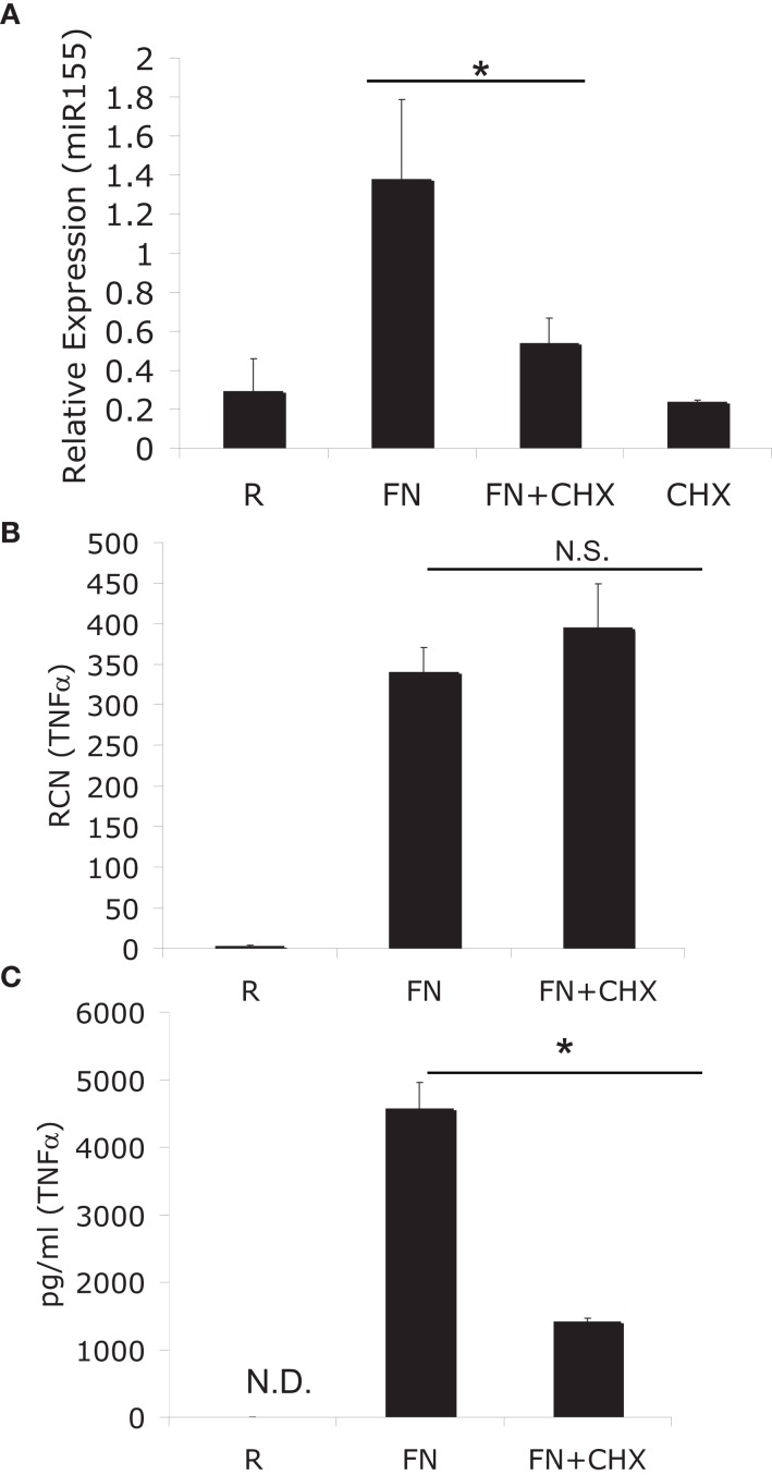
Figure 1 MiR-155 induction requires de novo host-protein synthesis. (A) PBM were infected with F. novicida (FN) at an MOI of 50 for 6 h in the presence or absence of 2 μM cycloheximide (CHX), and then RNA was isolated. “R” stands for resting/uninfected cells. Mature miR-155 expression was assayed by qRT-PCR and data expressed as relative expression from triplicate samples. (B) TNFα mRNA was assayed from the same samples as in (A). (C) Secreted TNFα was measured from the media of the cells in (A) by ELISA. *Designates a p value < 0.05 by Students t-test, while N.S. designates a p value > 0.05. These data are representative of three independent experiments.
Figure 1 MiR-155 induction requires de novo host-protein synthesis. (A) PBM were infected with F. novicida (FN) at an MOI of 50 for 6 h in the presence or absence of 2 μM cycloheximide (CHX), and then RNA was isolated. “R” stands for resting/uninfected cells. Mature miR-155 expression was assayed by qRT-PCR and data expressed as relative expression from triplicate samples. (B) TNFα mRNA was assayed from the same samples as in (A). (C) Secreted TNFα was measured from the media of the cells in (A) by ELISA. *Designates a p value < 0.05 by Students t-test, while N.S. designates a p value > 0.05. These data are representative of three independent experiments.CC BY-NC · PubMed Central
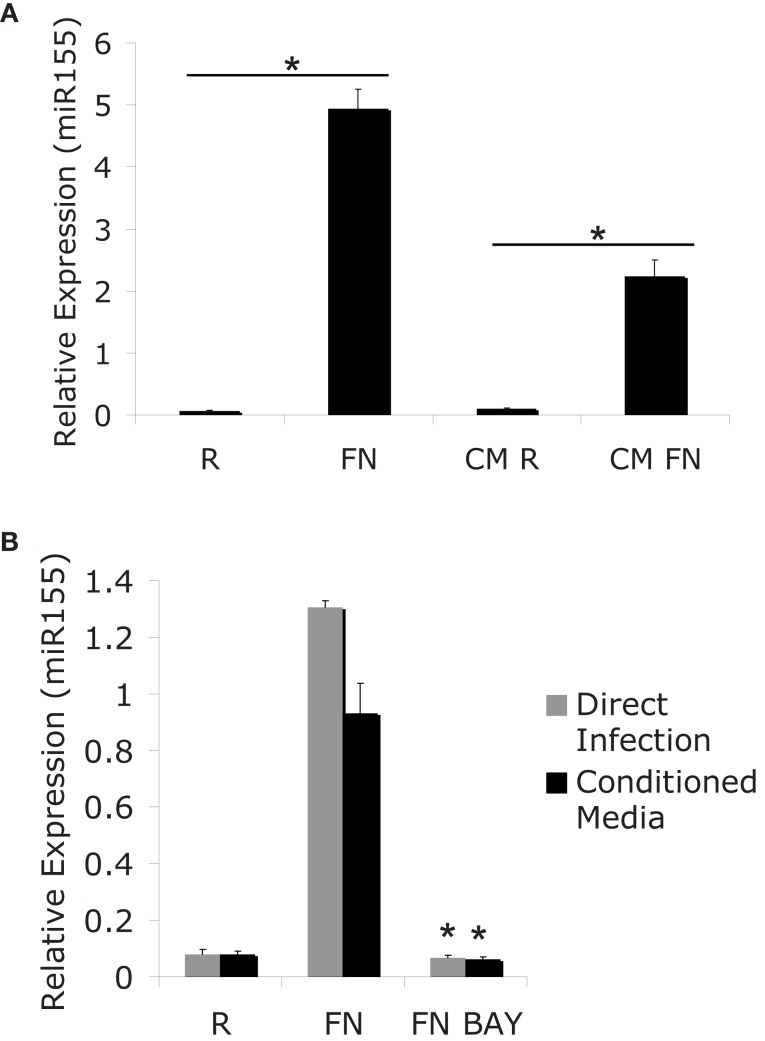
Figure 2 MiR-155 induction can be mediated indirectly though secreted factors. (A) PBM were infected with FN at an MOI of 50 for 24 h. Media from infected cells was sterile filtered to remove bacteria and RNA was isolated from the cells (R and Fn). New PBM were cultured in the conditioned media for 24 h (CM R and CM FN) followed by RNA isolation. Mature miR-155 expression was assayed by qRT-PCR. (B) PBM were infected for 24 h, then the media from the infected cells was sterile filtered to generate conditioned media. New PBM were infected with F.n. at an MOI of 50 for 6 h or treated with conditioned media for 6 h in the presence of BAY-7085-11 (5 μM) or DMSO control. MiR-155 expression was assayed by qRT-PCR. These data are representative of at least three independent experiments. *Designates a p value < 0.05.
Figure 2 MiR-155 induction can be mediated indirectly though secreted factors. (A) PBM were infected with FN at an MOI of 50 for 24 h. Media from infected cells was sterile filtered to remove bacteria and RNA was isolated from the cells (R and Fn). New PBM were cultured in the conditioned media for 24 h (CM R and CM FN) followed by RNA isolation. Mature miR-155 expression was assayed by qRT-PCR. (B) PBM were infected for 24 h, then the media from the infected cells was sterile filtered to generate conditioned media. New PBM were infected with F.n. at an MOI of 50 for 6 h or treated with conditioned media for 6 h in the presence of BAY-7085-11 (5 μM) or DMSO control. MiR-155 expression was assayed by qRT-PCR. These data are representative of at least three independent experiments. *Designates a p value < 0.05.CC BY-NC · PubMed Central
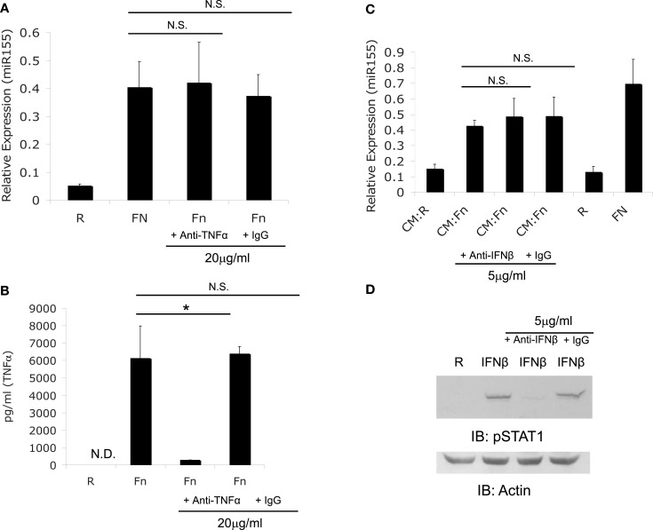
Figure 3 F. novicida induction miR-155 expression is not mediated by TNFα or IFNβ. (A) PBM were treated with TNFα neutralizing antibody or isotype control antibody, then infected with F.n. at an MOI of 50 for 6 h. RNA was collected and assayed for miR-155 expression. (B) The media from the samples in (A) was assayed for human TNFα by ELISA. (C) Conditioned media (CM) was obtained as in Figures 2B. Conditioned media from monocytes uninfected (CM:R) or infected (CM:Fn) with F.n. at an MOI of 50 was sterile filtered and incubated with neutralizing antibody or isotype control antibody at 37°C for 1 h. New PBM were then cultured in the conditioned media conditions, uninfected (R), or direct infection (FN) for 6 h. RNA was collected and miR-155 expression was assayed by qRT-PCR. (D) Effective neutralization of IFNβ was demonstrated by pre-treating media with recombinant IFNβ (500 U/ml) with or without neutralizing antibody or isotype control antibody. Then PBM were cultured in media alone, IFNβ, IFNβ plus neutralizing antibody, or IFNβ plus isotype control for 15 min. Cells were lysed and subject to western blot for phosphorylated STAT1, followed by Actin re-probe.
Figure 3 F. novicida induction miR-155 expression is not mediated by TNFα or IFNβ. (A) PBM were treated with TNFα neutralizing antibody or isotype control antibody, then infected with F.n. at an MOI of 50 for 6 h. RNA was collected and assayed for miR-155 expression. (B) The media from the samples in (A) was assayed for human TNFα by ELISA. (C) Conditioned media (CM) was obtained as in Figures 2B. Conditioned media from monocytes uninfected (CM:R) or infected (CM:Fn) with F.n. at an MOI of 50 was sterile filtered and incubated with neutralizing antibody or isotype control antibody at 37°C for 1 h. New PBM were then cultured in the conditioned media conditions, uninfected (R), or direct infection (FN) for 6 h. RNA was collected and miR-155 expression was assayed by qRT-PCR. (D) Effective neutralization of IFNβ was demonstrated by pre-treating media with recombinant IFNβ (500 U/ml) with or without neutralizing antibody or isotype control antibody. Then PBM were cultured in media alone, IFNβ, IFNβ plus neutralizing antibody, or IFNβ plus isotype control for 15 min. Cells were lysed and subject to western blot for phosphorylated STAT1, followed by Actin re-probe.CC BY-NC · PubMed Central
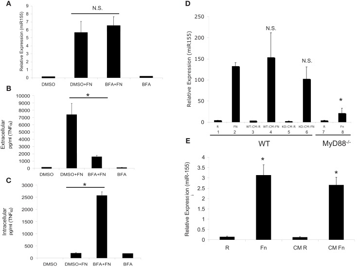
Figure 4 MiR-155 induction is not mediated through host cytokines, but rather by soluble bacterial factors. (A) PBM were infected with F.n. at an MOI of 50 for 16 h in the presence of Brefeldin A (BFA) or DMSO vehicle control. RNA was isolated and mature miR-155 expression was assayed by qRT-PCR. (B) Extracellular TNFα levels were measured by ELISA. (C) Intracellular TNFα levels were measured by ELISA from the cell lysate. (D) WT and MyD88−/− BMMs were infected for 24 h at an MOI of 50 and then sterile filtered to remove bacteria to generate conditioned media (CM). RNA was collected from the infected cells for qRT-PCR (Bars 1, 2, 7, 8). The conditioned media was then placed on new wild-type BMM for 24 h and RNA was subsequently collected for qRT-PCR (Bars 3, 4, 5, 6). These data are representative of three independent experiments. *Designates a p value < 0.05. (E) F.n. was suspended in RPMI-1640 with 10% heat-inactivated fetal bovine serum at a concentration of 3.5 × 10⁸ bacteria/ml (comparable to 5 × 10⁶ PBM infected at MOI 50 in 1 ml) and incubated at 37°C for 24 h. The media was sterile filtered to remove bacteria. PBM were directly infected with F.n. at an MOI of 50 or treated with the filtered media that previously contained F.n. for 16 h. RNA was isolated and miR-155 expression assayed by qRT-PCR. These data are representative of three independent experiments. *Designates a p value < 0.05.
Figure 4 MiR-155 induction is not mediated through host cytokines, but rather by soluble bacterial factors. (A) PBM were infected with F.n. at an MOI of 50 for 16 h in the presence of Brefeldin A (BFA) or DMSO vehicle control. RNA was isolated and mature miR-155 expression was assayed by qRT-PCR. (B) Extracellular TNFα levels were measured by ELISA. (C) Intracellular TNFα levels were measured by ELISA from the cell lysate. (D) WT and MyD88−/− BMMs were infected for 24 h at an MOI of 50 and then sterile filtered to remove bacteria to generate conditioned media (CM). RNA was collected from the infected cells for qRT-PCR (Bars 1, 2, 7, 8). The conditioned media was then placed on new wild-type BMM for 24 h and RNA was subsequently collected for qRT-PCR (Bars 3, 4, 5, 6). These data are representative of three independent experiments. *Designates a p value < 0.05. (E) F.n. was suspended in RPMI-1640 with 10% heat-inactivated fetal bovine serum at a concentration of 3.5 × 10⁸ bacteria/ml (comparable to 5 × 10⁶ PBM infected at MOI 50 in 1 ml) and incubated at 37°C for 24 h. The media was sterile filtered to remove bacteria. PBM were directly infected with F.n. at an MOI of 50 or treated with the filtered media that previously contained F.n. for 16 h. RNA was isolated and miR-155 expression assayed by qRT-PCR. These data are representative of three independent experiments. *Designates a p value < 0.05.CC BY-NC · PubMed Central
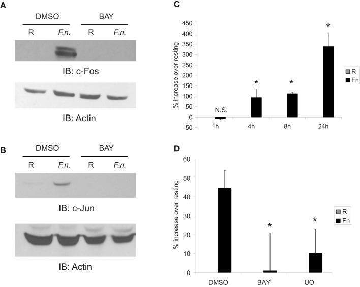
Figure 5 Fos and Jun expression is highly up-regulated by infection in an NF-κB-dependent mechanism to control AP-1 activity. (A) PBM were pre-treated with DMSO or BAY-11-7085 (5 μM; designated as BAY in the figure) for 30 min then infected with F.n. at an MOI of 50 for 4 h. Cells were lysed and subject to western blotting for c-Fos, followed by actin re-probe. (B) Protein-matched lysates were probed for c-Jun and re-probed with actin antibody. (C) RAW264.7 macrophages were transfected with an AP-1 luciferase reporter. 14 h post-transfection cells were uninfected or infected with F.n. at an MOI of 50 for 1, 4, 8, or 24 h. Cells were lysed and luciferase activity was measured by a luminometer in triplicate. Data are represented as % increase over uninfected control. (D) RAW264.7 macrophages were transfected with the AP-1 reporter as in (C). Fourteen hours post-transfection cells were pre-treated with DMSO vehicle control, BAY-11-7085, or U0126 then infected for 8 h. Data are represented as % increase over uninfected samples. These data are representative of three independent experiments. *Designates a p value < 0.05.
Figure 5 Fos and Jun expression is highly up-regulated by infection in an NF-κB-dependent mechanism to control AP-1 activity. (A) PBM were pre-treated with DMSO or BAY-11-7085 (5 μM; designated as BAY in the figure) for 30 min then infected with F.n. at an MOI of 50 for 4 h. Cells were lysed and subject to western blotting for c-Fos, followed by actin re-probe. (B) Protein-matched lysates were probed for c-Jun and re-probed with actin antibody. (C) RAW264.7 macrophages were transfected with an AP-1 luciferase reporter. 14 h post-transfection cells were uninfected or infected with F.n. at an MOI of 50 for 1, 4, 8, or 24 h. Cells were lysed and luciferase activity was measured by a luminometer in triplicate. Data are represented as % increase over uninfected control. (D) RAW264.7 macrophages were transfected with the AP-1 reporter as in (C). Fourteen hours post-transfection cells were pre-treated with DMSO vehicle control, BAY-11-7085, or U0126 then infected for 8 h. Data are represented as % increase over uninfected samples. These data are representative of three independent experiments. *Designates a p value < 0.05.CC BY-NC · PubMed Central
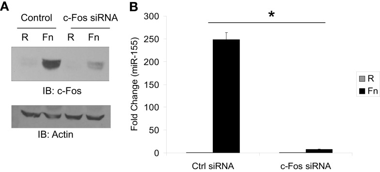
Figure 6 MiR-155 induction is dependent upon c-Fos. (A) RAW 264.7 macrophages were transfected with 4.0 μM siRNA control or siRNA against c-Fos. Thirty minutes post-transfection cells were left uninfected or infected with F.n. at an MOI of 50 for 4 h. Cells were then lysed and subject to western blotting for c-Fos. An antibody against Actin was used to re-probe the blots to ensure equal loading. (B) RAW 264.7 macrophages were transfected as done in part A and infected for 16 h. RNA was collected and miR-155 expression was assayed by qRT-PCR. These data are representative of three independent experiments. *Designates a p value < 0.05.
Figure 6 MiR-155 induction is dependent upon c-Fos. (A) RAW 264.7 macrophages were transfected with 4.0 μM siRNA control or siRNA against c-Fos. Thirty minutes post-transfection cells were left uninfected or infected with F.n. at an MOI of 50 for 4 h. Cells were then lysed and subject to western blotting for c-Fos. An antibody against Actin was used to re-probe the blots to ensure equal loading. (B) RAW 264.7 macrophages were transfected as done in part A and infected for 16 h. RNA was collected and miR-155 expression was assayed by qRT-PCR. These data are representative of three independent experiments. *Designates a p value < 0.05.CC BY-NC · PubMed Central
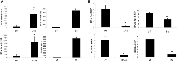
Figure 7 MiR-155 is induced by microbes/microbial ligands with a concomitant decrease in SHIP expression. Human PBM were treated with LPS (500 ng/ml), R848 (1 uM), or infected with B. cenocepacia (designated as Bc in the figure) at MOI 5, or M. smegmatis (designated as Ms in the figure) at MOI 5. mRNA was isolated from the cells 16 h later and assayed by qRT-PCR for (A) miR-155 and (B) SHIP expression. These data are representative of six independent experiments. *Designates a p value < 0.05.
Figure 7 MiR-155 is induced by microbes/microbial ligands with a concomitant decrease in SHIP expression. Human PBM were treated with LPS (500 ng/ml), R848 (1 uM), or infected with B. cenocepacia (designated as Bc in the figure) at MOI 5, or M. smegmatis (designated as Ms in the figure) at MOI 5. mRNA was isolated from the cells 16 h later and assayed by qRT-PCR for (A) miR-155 and (B) SHIP expression. These data are representative of six independent experiments. *Designates a p value < 0.05.CC BY-NC · PubMed Central
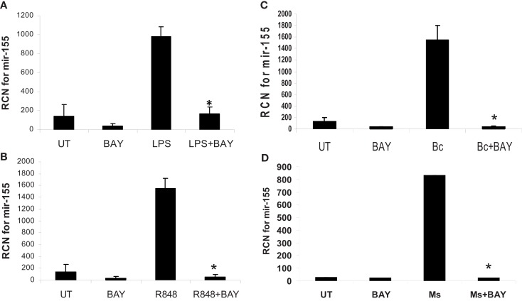
Figure 8 MiR-155 induction by microbes/microbial ligands is NF-κB-dependent. (A–D) Human PBM were pre-treated with or without the NF-κB inhibitor (BAY-11-7085, designated as BAY in the figure), and subsequently treated with (A) LPS (500 ng/ml), (B) R848 (1 uM), or infected with (C) B. cenocepacia (Bc) at MOI 5, or (D) M. smegmatis (Ms) at MOI 5. mRNA was isolated from the cells 16 h later and assayed for miR-155 by qRT-PCR. These data are representative of six independent experiments. *Designates a p value < 0.05.
Figure 8 MiR-155 induction by microbes/microbial ligands is NF-κB-dependent. (A–D) Human PBM were pre-treated with or without the NF-κB inhibitor (BAY-11-7085, designated as BAY in the figure), and subsequently treated with (A) LPS (500 ng/ml), (B) R848 (1 uM), or infected with (C) B. cenocepacia (Bc) at MOI 5, or (D) M. smegmatis (Ms) at MOI 5. mRNA was isolated from the cells 16 h later and assayed for miR-155 by qRT-PCR. These data are representative of six independent experiments. *Designates a p value < 0.05.CC BY-NC · PubMed Central
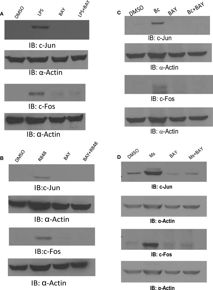
Figure 9 c-Jun and c-Fos are induced by microbes/microbial ligands in an NF-κB-dependent manner. (A–D) Human PBM were pre-treated with or without the NF-κB inhibitor (BAY-11-7085, designated as BAY in the figure), and subsequently treated with (A) LPS (500 ng/ml), (B) R848 (1 uM), or infected with (C) B. cenocepacia (Bc) at MOI 5, or (D) M. smegmatis (Ms) at MOI 5. Cell lysates were collected 16 h later. Protein-matched lysates were analyzed by Western blotting with c-Jun or c-Fos antibodies. All membranes were re-probed with Actin antibody to ensure equal loading of protein in all lanes. These data are representative of four independent experiments.
Figure 9 c-Jun and c-Fos are induced by microbes/microbial ligands in an NF-κB-dependent manner. (A–D) Human PBM were pre-treated with or without the NF-κB inhibitor (BAY-11-7085, designated as BAY in the figure), and subsequently treated with (A) LPS (500 ng/ml), (B) R848 (1 uM), or infected with (C) B. cenocepacia (Bc) at MOI 5, or (D) M. smegmatis (Ms) at MOI 5. Cell lysates were collected 16 h later. Protein-matched lysates were analyzed by Western blotting with c-Jun or c-Fos antibodies. All membranes were re-probed with Actin antibody to ensure equal loading of protein in all lanes. These data are representative of four independent experiments.CC BY-NC · PubMed Central
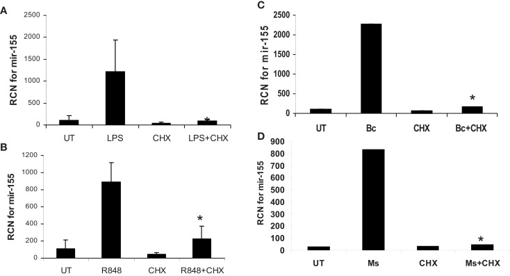
Figure 10 MiR-155 induction by microbes/microbial ligands requires de novo protein synthesis. (A–D) Human PBM were pre-treated with or without the protein synthesis inhibitor (CHX), and subsequently treated with (A) LPS (500 ng/ml), (B) R848 (1 uM), or infected with (C) B. cenocepacia (Bc) at MOI 5, or (D) M. smegmatis (Ms) at MOI 5. mRNA was isolated from the cells 16 h later and assayed for miR-155 by qRT-PCR. These data are representative of six independent experiments. *Designates a p value < 0.05.
Figure 10 MiR-155 induction by microbes/microbial ligands requires de novo protein synthesis. (A–D) Human PBM were pre-treated with or without the protein synthesis inhibitor (CHX), and subsequently treated with (A) LPS (500 ng/ml), (B) R848 (1 uM), or infected with (C) B. cenocepacia (Bc) at MOI 5, or (D) M. smegmatis (Ms) at MOI 5. mRNA was isolated from the cells 16 h later and assayed for miR-155 by qRT-PCR. These data are representative of six independent experiments. *Designates a p value < 0.05.CC BY-NC · PubMed Central

Verification

Claims the paper makes

The claims and statistics of this paper haven’t been checked yet.

Run the check — free, no account needed. It reads the open-access text and checks the sources it cites.

A few free checks a day · papers already verified open instantly · results are public

Claims other papers make about this one

Each is checked against this paper’s own text: does it say what the citing paper says it does?

Self-check

Percentages, sums and reported differences re-computed from the paper’s own numbers.

The self-check needs the paper’s full text.

Citations

Cites

Research works printed but not linked: 44

  1. Anest V. Cogswell P. C. Baldwin A. S. Jr. (2004). IkappaB kinase alpha and p65/RelA contribute to optimal epidermal growth factor-induced c-fos gene expression independent of IkappaBalpha degradation. J. Biol. Chem. 279, 31183– 31189 10.1074/jbc.M404380200 15155743 · DOI
  2. Bushati N. Cohen S. M. (2007). MicroRNA functions. Annu. Rev. Cell Dev. Biol. 23, 175– 205 10.1146/annurev.cellbio.23.090506.123406 17506695 · DOI · Paper page
  3. Bushati N. Cohen S. M. (2008). MicroRNAs in neurodegeneration. Curr. Opin. Neurobiol. 18, 292– 296 10.1016/j.conb.2008.07.001 18662781 · DOI
  4. Butchar J. P. Cremer T. J. Clay C. D. Gavrilin M. A. Wewers M. D. Marsh C. B. Schlesinger L. S. Tridandapani S. (2008). Microarray analysis of human monocytes infected with Francisella tularensis identifies new targets of host response subversion. PLoS ONE 3, e2924 10.1371/journal.pone.0002924 18698339 PMC2488368 · DOI
  5. Cole L. E. Shirey K. A. Barry E. Santiago A. Rallabhandi P. Elkins K. L. Puche A. C. Michalek S. M. Vogel S. N. (2007). Toll-like receptor 2-mediated signaling requirements for Francisella tularensis live vaccine strain infection of murine macrophages. Infect. Immun. 75, 4127– 4137 10.1128/IAI.01868-06 17517865 PMC1951974 · DOI
  6. Costinean S. Zanesi N. Pekarsky Y. Tili E. Volinia S. Heerema N. Croce C. M. (2006). Pre-B cell proliferation and lymphoblastic leukemia/high-grade lymphoma in E(mu)-miR155 transgenic mice. Proc. Natl. Acad. Sci. U.S.A. 103, 7024– 7029 10.1073/pnas.0602266103 16641092 PMC1459012 · DOI
  7. Cremer T. J. Ravneberg D. H. Clay C. D. Piper-Hunter M. G. Marsh C. B. Elton T. S. Gunn J. S. Amer A. Kanneganti T. D. Schlesinger L. S. Butchar J. P. Tridandapani S. (2009). MiR-155 induction by F. novicida but not the virulent F. tularensis results in SHIP down-regulation and enhanced pro-inflammatory cytokine response. PLoS ONE. 4, Fe8508 10.1371/journal.pone.0008508 PMC2794384 20041145 · DOI
  8. Dai Y. Diao Z. Sun H. Li R. Qiu Z. Hu Y. (2011). MicroRNA-155 is involved in the remodelling of human-trophoblast-derived HTR-8/SVneo cells induced by lipopolysaccharides. Hum. Reprod. 26, 1882– 1891 10.1093/humrep/der118 21515911 · DOI
  9. Eis P. S. Tam W. Sun L. Chadburn A. Li Z. M. Gomez F. Lund E. Dahlberg J. E. (2005). Accumulation of miR-155 and BIC RNA in human B cell lymphomas. Proc. Natl. Acad. Sci. U.S.A. 102, 3627– 3632 10.1073/pnas.0500613102 15738415 PMC552785 · DOI
  10. Ellis J. Oyston P. C. Green M. Titball R. W. (2002). Tularemia. Clin. Microbiol. Rev. 15, 631– 646 10.1128/CMR.15.4.631-646.2002 12364373 PMC126859 · DOI
  11. Gatto G. Rossi A. Rossi D. Kroening S. Bonatti S. Mallardo M. (2008). Epstein-Barr virus latent membrane protein 1 trans-activates miR-155 transcription through the NF-kappaB pathway. Nucleic Acids Res. 36, 6608– 6619 10.1093/nar/gkn666 18940871 PMC2582607 · DOI
  12. Gunn J. S. Ernst R. K. (2007). The structure and function of Francisella lipopolysaccharide. Ann. N. Y. Acad. Sci. 1105, 202– 218 10.1196/annals.1409.006 17395723 PMC2742961 · DOI
  13. Hambleton J. Weinstein S. L. Lem L. Defranco A. L. (1996). Activation of c-Jun N-terminal kinase in bacterial lipopolysaccharide-stimulated macrophages. Proc. Natl. Acad. Sci. U.S.A. 93, 2774– 2778 10.1073/pnas.93.7.2774 8610116 PMC39708 · DOI
  14. Hao S. Baltimore D. (2009). The stability of mRNA influences the temporal order of the induction of genes encoding inflammatory molecules. Nat. Immunol. 10, 281– 288 10.1038/ni.1699 19198593 PMC2775040 · DOI · Paper page
  15. Henke J. I. Goergen D. Zheng J. Song Y. Schuttler C. G. Fehr C. Junemann C. Niepmann M. (2008). MicroRNA-122 stimulates translation of hepatitis C virus RNA. EMBO J. 27, 3300– 3310 10.1038/emboj.2008.244 19020517 PMC2586803 · DOI
  16. Henry T. Brotcke A. Weiss D. S. Thompson L. J. Monack D. M. (2007). Type I interferon signaling is required for activation of the inflammasome during Francisella infection. J. Exp. Med. 204, 987– 994 10.1084/jem.20062665 17452523 PMC2118578 · DOI
  17. Hou J. Wang P. Lin L. Liu X. Ma F. An H. Wang Z. Cao X. (2009). MicroRNA-146a feedback inhibits RIG-I-dependent Type I IFN production in macrophages by targeting TRAF6, IRAK1, and IRAK2. J. Immunol. 183, 2150– 2158 10.4049/jimmunol.0900707 19596990 · DOI · Paper page
  18. Izar B. Mannala G. K. Mraheil M. A. Chakraborty T. Hain T. (2012). MicroRNA response to Listeria monocytogenes infection in epithelial cells. Int. J. Mol. Sci. 13, 1173– 1185 10.3390/ijms13011173 22312311 PMC3269745 · DOI
  19. Jiang X. Tsitsiou E. Herrick S. E. Lindsay M. A. (2010). MicroRNAs and the regulation of fibrosis. FEBS J. 277, 2015– 2021 10.1111/j.1742-4658.2009.07510.x 20412055 PMC2963651 · DOI
  20. Karin M. Liu Z. Zandi E. (1997). AP-1 function and regulation. Curr. Opin. Cell Biol. 9, 240– 246 10.1016/S0955-0674(97)80068-3 9069263 · DOI
  21. Kieffer T. L. Cowley S. Nano F. E. Elkins K. L. (2003). Francisella novicida LPS has greater immunobiological activity in mice than F. tularensis LPS, and contributes to F. novicida murine pathogenesis. Microbes Infect. 5, 397– 403 10.1016/S1286-4579(03)00052-2 12737995 · DOI
  22. Kohlhaas S. Garden O. A. Scudamore C. Turner M. Okkenhaug K. Vigorito E. (2009). Cutting edge: the Foxp3 target miR-155 contributes to the development of regulatory T cells. J. Immunol. 182, 2578– 2582 10.4049/jimmunol.0803162 19234151 · DOI
  23. Lee Y. S. Dutta A. (2009). MicroRNAs in cancer. Annu. Rev. Pathol. 4, 199– 227 10.1146/annurev.pathol.4.110807.092222 18817506 PMC2769253 · DOI
  24. Li H. Nookala S. Bina X. R. Bina J. E. Re F. (2006). Innate immune response to Francisella tularensis is mediated by TLR2 and caspase-1 activation. J. Leukoc. Biol. 80, 766– 773 10.1189/jlb.1105668 16895974 · DOI
  25. O’Connell R. M. Kahn D. Gibson W. S. Round J. L. Scholz R. L. Chaudhuri A. A. Kahn M. E. Rao D. S. Baltimore D. (2010). MicroRNA-155 promotes autoimmune inflammation by enhancing inflammatory T cell development. Immunity 33, 607– 619 10.1016/j.immuni.2010.09.009 20888269 PMC2966521 · DOI · Paper page
  26. O’Connell R. M. Taganov K. D. Boldin M. P. Cheng G. Baltimore D. (2007). MicroRNA-155 is induced during the macrophage inflammatory response. Proc. Natl. Acad. Sci. U.S.A. 104, 1604– 1609 10.1073/pnas.0610731104 17242365 PMC1780072 · DOI · Paper page
  27. Oyston P. C. Sjostedt A. Titball R. W. (2004). Tularaemia: bioterrorism defence renews interest in Francisella tularensis. Nat. Rev. Microbiol. 2, 967– 978 10.1038/nrmicro1045 15550942 · DOI
  28. Parsa K. V. Ganesan L. P. Rajaram M. V. Gavrilin M. A. Balagopal A. Mohapatra N. P. Wewers M. D. Schlesinger L. S. Gunn J. S. Tridandapani S. (2006). Macrophage pro-inflammatory response to Francisella novicida infection is regulated by SHIP. PLoS Pathog. 2, e71 10.1371/journal.ppat.0020071 16848641 PMC1513262 · DOI
  29. Pottier N. Maurin T. Chevalier B. Puissegur M. P. Lebrigand K. Robbe-Sermesant K. Bertero T. Lino Cardenas C. L. Courcot E. Rios G. Fourre S. Lo-Guidice J. M. Marcet B. Cardinaud B. Barbry P. Mari B. (2009). Identification of keratinocyte growth factor as a target of microRNA-155 in lung fibroblasts: implication in epithelial-mesenchymal interactions. PLoS ONE 4, e6718 10.1371/journal.pone.0006718 19701459 PMC2726943 · DOI
  30. Rajaram M. V. Butchar J. P. Parsa K. V. Cremer T. J. Amer A. Schlesinger L. S. Tridandapani S. (2009). Akt and SHIP modulate Francisella escape from the phagosome and induction of the Fas-mediated death pathway. PLoS ONE 4, e7919 10.1371/journal.pone.0007919 19936232 PMC2775408 · DOI
  31. Rajaram M. V. Ganesan L. P. Parsa K. V. Butchar J. P. Gunn J. S. Tridandapani S. (2006). Akt/Protein kinase B modulates macrophage inflammatory response to Francisella infection and confers a survival advantage in mice. J. Immunol. 177, 6317– 6324 17056562
  32. Santic M. Al Khodor S. Abu K. Y. (2010). Cell biology and molecular ecology of Francisella tularensis. Cell. Microbiol. 12, 129– 139 10.1111/j.1462-5822.2009.01400.x 19863554 · DOI
  33. Taganov K. D. Boldin M. P. Chang K. J. Baltimore D. (2006). NF-kappaB-dependent induction of microRNA miR-146, an inhibitor targeted to signaling proteins of innate immune responses. Proc. Natl. Acad. Sci. U.S.A. 103, 12481– 12486 10.1073/pnas.0605298103 16885212 PMC1567904 · DOI · Paper page
  34. Telepnev M. Golovliov I. Grundstrom T. Tarnvik A. Sjostedt A. (2003). Francisella tularensis inhibits Toll-like receptor-mediated activation of intracellular signalling and secretion of TNF-alpha and IL-1 from murine macrophages. Cell. Microbiol. 5, 41– 51 10.1046/j.1462-5822.2003.00251.x 12542469 · DOI
  35. Thai T. H. Calado D. P. Casola S. Ansel K. M. Xiao C. Xue Y. Murphy A. Frendewey D. Valenzuela D. Kutok J. L. Schmidt-Supprian M. Rajewsky N. Yancopoulos G. Rao A. Rajewsky K. (2007). Regulation of the germinal center response by microRNA-155. Science 316, 604– 608 10.1126/science.1141229 17463289 · DOI
  36. Thakran S. Li H. Lavine C. L. Miller M. A. Bina J. E. Bina X. R. Re F. (2008). Identification of Francisella tularensis lipoproteins that stimulate the toll-like receptor (TLR) 2/TLR1 heterodimer. J. Biol. Chem. 283, 3751– 3760 10.1074/jbc.M706854200 18079113 · DOI
  37. Tili E. Michaille J. J. Cimino A. Costinean S. Dumitru C. D. Adair B. Fabbri M. Alder H. Liu C. G. Calin G. A. Croce C. M. (2007). Modulation of miR-155 and miR-125b levels following lipopolysaccharide/TNF-alpha stimulation and their possible roles in regulating the response to endotoxin shock. J. Immunol. 179, 5082– 5089 17911593
  38. Toshchakov V. Jones B. W. Perera P. Y. Thomas K. Cody M. J. Zhang S. Williams B. R. Major J. Hamilton T. A. Fenton M. J. Vogel S. N. (2002). TLR4, but not TLR2, mediates IFN-beta-induced STAT1alpha/beta-dependent gene expression in macrophages. Nat. Immunol. 3, 392– 398 10.1038/ni774 11896392 · DOI
  39. van Rooij E. Marshall W. S. Olson E. N. (2008). Toward microRNA-based therapeutics for heart disease: the sense in antisense. Circ. Res. 103, 919– 928 10.1161/CIRCRESAHA.108.183426 18948630 PMC2725407 · DOI
  40. Vigorito E. Perks K. L. Abreu-Goodger C. Bunting S. Xiang Z. Kohlhaas S. Das P. P. Miska E. A. Rodriguez A. Bradley A. Smith K. G. Rada C. Enright A. J. Toellner K. M. MacLennan I. C. Turner M. (2007). MicroRNA-155 regulates the generation of immunoglobulin class-switched plasma cells. Immunity 27, 847– 859 10.1016/j.immuni.2007.10.009 18055230 PMC4135426 · DOI
  41. Xiao B. Liu Z. Li B. S. Tang B. Li W. Guo G. Shi Y. Wang F. Wu Y. Tong W. D. Guo H. Mao X. H. Zou Q. M. (2009). Induction of microRNA-155 during Helicobacter pylori infection and its negative regulatory role in the inflammatory response. J. Infect. Dis. 200, 916– 925 10.1086/605443 19650740 · DOI · Paper page
  42. Yin Q. McBride J. Fewell C. Lacey M. Wang X. Lin Z. Cameron J. Flemington E. K. (2008a). MicroRNA-155 is an Epstein-Barr virus-induced gene that modulates Epstein-Barr virus-regulated gene expression pathways. J. Virol. 82, 5295– 5306 10.1128/JVI.00493-08 18367535 PMC2395216 · DOI
  43. Yin Q. Wang X. McBride J. Fewell C. Flemington E. (2008b). B-cell receptor activation induces BIC/miR-155 expression through a conserved AP-1 element. J. Biol. Chem. 283, 2654– 2662 10.1074/jbc.M708218200 18048365 PMC2810639 · DOI
  44. Zhu F. G. Gomi K. Marshall J. S. (1998). Short-term and long-term cytokine release by mouse bone marrow mast cells and the differentiated KU-812 cell line are inhibited by brefeldin A. J. Immunol. 161, 2541– 2551 9725254

Cited by

Sorted by citations

See all 29

Metadata

Authors
Cremer, T. J., Fatehchand, K., Shah, P., Gillette, D., Patel, H., Marsh, R. L., Besecker, B. Y., Rajaram, M. V. S., Cormet-Boyaka, E., Kanneganti, T.-D., Schlesinger, L. S., Butchar, J. P., & Tridandapani, S.
Venue
Frontiers in Cellular and Infection Microbiology
Published
2012
Volume
2
ISSN
2235-2988
Cited by
29
References
2

About these checks

Registry, retraction, Cites and statistics results are computed, not judged, with one exception: a printed reference that no index lists is read once by a model before it counts as a citation error. Claims are extracted by a model and kept only when their quote appears verbatim in the text; each cited source is then checked, with a model’s help, for whether it says what the claim relies on. Those verdicts can be wrong — read the claim’s quote and the cited source before relying on one.

Statistics and arithmetic are recomputed, not judged.

Confidence bands are shown only for fields calibrated on our annotated test set. None is calibrated yet, so no band is shown.

What counts as a claim ›How we measure confidence ›How we check this ›

Are you an author, or do you see a problem with this page? Request a review