

The DOI matches its Crossref record; the references it cites are not checked yet.
Inflammatory Response of Human Peripheral Blood Mononuclear Cells and Osteoblasts Incubated With Metallic and Ceramic Submicron Particles
Open accessAbstract
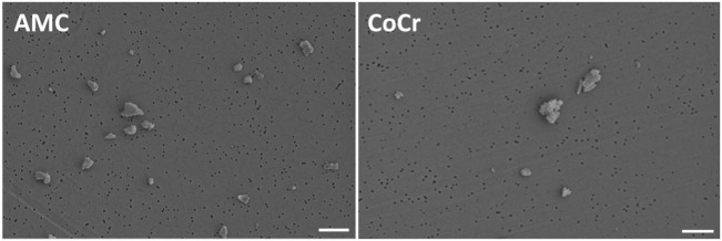
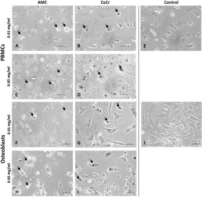
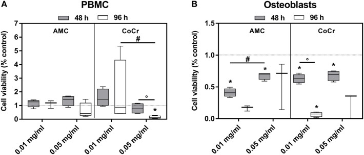
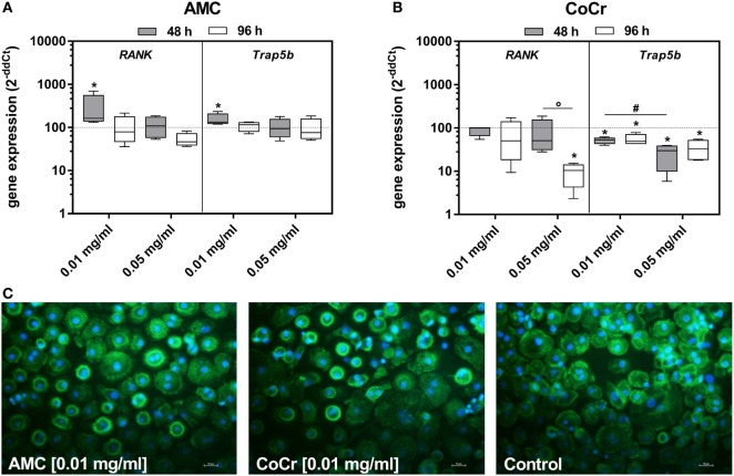
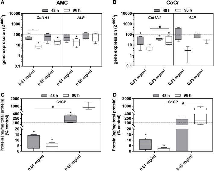
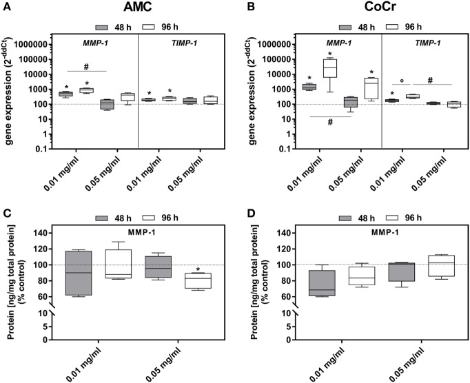
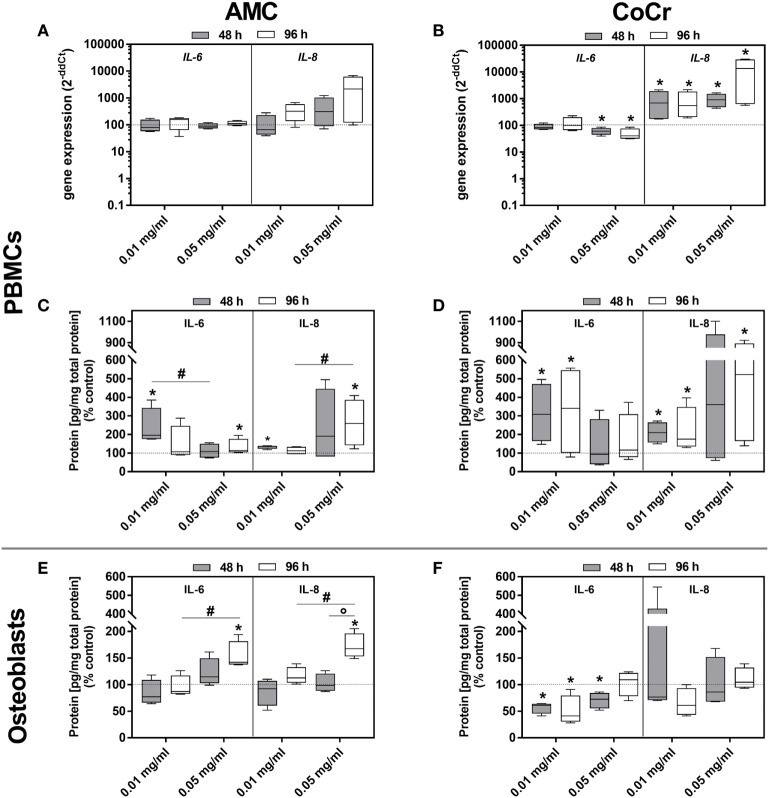
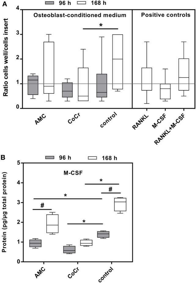
Inflammatory reactions associated with osteolysis and aseptic loosening are the result of wear particles generated at the articulating surfaces of implant components. The aim of the present study was to analyze the biological response of human osteoblasts and PBMCs after exposure to metallic and alumina ceramic particles regarding cellular differentiation, cytokine release and monocyte migration. Cells were exposed to particles (0.01 mg/ml, 0.05 mg/ml) from an alumina matrix composite (AMC) and a CoCr28Mo6 alloy with an average size of 0.5 µm over 48 h and 96 h. The expression rates of osteogenic (Col1A1, ALP) and pro-osteoclastic (RANK, Trap5b) differentiation markers as well as pro-osteolytic mediators (MMP-1, TIMP-1, IL-6, IL-8, MCP-1) were determined and soluble protein concentrations of active MMP-1, IL-6, IL-8 and pro-collagen type 1 in cell culture supernatants were evaluated. Additionally, the capacity of particle-treated osteoblasts to attract potentially pro-inflammatory cells to the site of particle exposure was investigated by migration assays using osteoblast-conditioned media. The cellular morphology and metabolism of human osteoblasts and adherent PBMCs were influenced by particle type and concentration. In human osteoblasts, Col1A1 expression rates and protein production were significantly reduced after exposing cells to the lower concentration of CoCr and AMC particles. Exposure to AMC particles (0.01 mg/ml) resulted in increased mRNA levels of RANK and Trap5b in adherent PBMCs. For MMP-1 gene expression, elevated levels were more prominent after incubation with CoCr compared to AMC particles in osteoblasts which was not reflected by the protein data. IL-6 and IL-8 mRNA and protein was induced in both cell types after treatment with AMC particles whereas exposure to CoCr particles resulted in significantly upregulated IL-6 and IL-8 protein contents in PBMCs only. Exposure of osteoblasts to CoCr particles reduced the chemoattractant potential of osteoblast-conditioned medium. Our results demonstrated distinct effects of AMC and CoCr particles in human osteoblasts and PBMCs. More complex cell models are required to further evaluate the impact of cellular interactions between different cell types during particle exposure.
Overview
The claims and statistics of this paper haven’t been checked yet.
Run the check — free, no account needed. It reads the open-access text and checks the sources it cites.
Integrity signals
Registry
Cites
Statistics
Figures
Verification
Claims the paper makes
The claims and statistics of this paper haven’t been checked yet.
Run the check — free, no account needed. It reads the open-access text and checks the sources it cites.
Claims other papers make about this one
Each is checked against this paper’s own text: does it say what the citing paper says it does?
The cited source says what the claim attributes to it.
In the citing paper
“Therefore, ceramic particles induce a lower immune response compared to Cr-Co particles [139].”
From Potential Impact of Prosthetic Biomaterials on the Periodontium: A Comprehensive Review (2023) · cites this paper as [139]
Self-check
Percentages, sums and reported differences re-computed from the paper’s own numbers.
The self-check needs the paper’s full text.
Citations
The citation lists haven’t been fetched yet
References 52 · Cited by 32
Research works printed but not linked: 48
- Wengler, 2014, Hip and knee replacement in Germany and the USA: analysis of individual inpatient data from German and US hospitals for the years 2005 to 2011, Dtsch Arztebl Int, 111, 407, 10.3238/arztebl.2014.0407 · DOI
- Hughes, 2017, Arthroplasty registries around the world: valuable sources of hip implant revision risk data, Curr Rev Musculoskelet Med, 10, 240, 10.1007/s12178-017-9408-5 · DOI
- Sadoghi, 2013, Revision surgery after total joint arthroplasty: a complication-based analysis using worldwide arthroplasty registers, J Arthroplasty, 28, 1329, 10.1016/j.arth.2013.01.012 · DOI
- Man, 2017, Immunological responses to total hip arthroplasty, J Funct Biomater, 8, E33, 10.3390/jfb8030033 · DOI
- Bitar, 2015, Biological response to prosthetic debris, World J Orthop, 6, 172, 10.5312/wjo.v6.i2.172 · DOI
- Gallo, 2014, Contributions of human tissue analysis to understanding the mechanisms of loosening and osteolysis in total hip replacement, Acta Biomater, 10, 2354, 10.1016/j.actbio.2014.02.003 · DOI
- Goodman, 1998, Cellular profile and cytokine production at prosthetic interfaces. Study of tissues retrieved from revised hip and knee replacements, J Bone Joint Surg Br, 80, 531, 10.1302/0301-620X.80B3.0800531 · DOI
- Green, 2000, Effect of size and dose on bone resorption activity of macrophages by in vitro clinically relevant ultra high molecular weight polyethylene particles, J Biomed Mater Res, 53, 490, 10.1002/1097-4636(200009)53:5<490::AID-JBM7>3.0.CO;2-7 · DOI
- Gallo, 2013, Particle disease: biologic mechanisms of periprosthetic osteolysis in total hip arthroplasty, Innate Immun, 19, 213, 10.1177/1753425912451779 · DOI
- Nich, 2013, Macrophages—key cells in the response to wear debris from joint replacements, J Biomed Mater Res A, 101, 3033, 10.1002/jbm.a.34599 · DOI
- Jämsen, 2014, Characterization of macrophage polarizing cytokines in the aseptic loosening of total hip replacements, J Orthop Res, 32, 1241, 10.1002/jor.22658 · DOI
- Hallab, 2009, Biologic effects of implant debris, Bull NYU Hosp Jt Dis, 67, 182
- Davidson, 1994, Abrasive wear of ceramic, metal, and UHMWPE bearing surfaces from third-body bone, PMMA bone cement, and titanium debris, Biomed Mater Eng, 4, 213
- Sonntag, 2012, What’s next? Alternative materials for articulation in total joint replacement, Acta Biomater, 8, 2434, 10.1016/j.actbio.2012.03.029 · DOI
- Xing, 2002, The effect of polyethylene particle phagocytosis on the viability of mature human macrophages, J Biomed Mater Res, 61, 619, 10.1002/jbm.10078 · DOI
- Germain, 2003, Comparison of the cytotoxicity of clinically relevant cobalt-chromium and alumina ceramic wear particles in vitro, Biomaterials, 24, 469, 10.1016/S0142-9612(02)00360-5 · DOI
- Liu, 2015, The biological response to nanometre-sized polymer particles, Acta Biomater, 23, 38, 10.1016/j.actbio.2015.05.016 · DOI
- Neale, 2000, The effect of particle phagocytosis and metallic wear particles on osteoclast formation and bone resorption in vitro, J Arthroplasty, 15, 654, 10.1054/arth.2000.6622 · DOI
- Athanasou, 2016, The pathobiology and pathology of aseptic implant failure, Bone Joint Res, 5, 162, 10.1302/2046-3758.55.BJR-2016-0086 · DOI
- Lohmann, 2000, Phagocytosis of wear debris by osteoblasts affects differentiation and local factor production in a manner dependent on particle composition, Biomaterials, 21, 551, 10.1016/S0142-9612(99)00211-2 · DOI
- Lohmann, 2002, Ceramic and PMMA particles differentially affect osteoblast phenotype, Biomaterials, 23, 1855, 10.1016/S0142-9612(01)00312-X · DOI
- Fritz, 2006, Chemokine gene activation in human bone marrow-derived osteoblasts following exposure to particulate wear debris, J Biomed Mater Res A, 77, 192, 10.1002/jbm.a.30609 · DOI
- Fritz, 2002, Titanium particles induce the immediate early stress responsive chemokines IL-8 and MCP-1 in osteoblasts, J Orthop Res, 20, 490, 10.1016/S0736-0266(01)00154-1 · DOI
- Lochner, 2011, The potential role of human osteoblasts for periprosthetic osteolysis following exposure to wear particles, Int J Mol Med, 28, 1055, 10.3892/ijmm.2011.778 · DOI
- Braux, 2016, Strontium-substituted bioceramics particles: a new way to modulate MCP-1 and Gro-α production by human primary osteoblastic cells, Materials (Basel), 9, E985, 10.3390/ma9120985 · DOI
- Waugh, 2008, The interleukin-8 pathway in cancer, Clin Cancer Res, 14, 6735, 10.1158/1078-0432.CCR-07-4843 · DOI · Paper page
- Deshmane, 2009, Monocyte chemoattractant protein-1 (MCP-1): an overview, J Interferon Cytokine Res, 29, 313, 10.1089/jir.2008.0027 · DOI · Paper page
- Gallo, 2012, Advantages and disadvantages of ceramic on ceramic total hip arthroplasty: a review, Biomed Pap Med Fac Univ Palacky Olomouc Czech Repub, 156, 204, 10.5507/bp.2012.063 · DOI
- Jeffers, 2012, Ceramic-on-ceramic bearings in hip arthroplasty: state of the art and the future, J Bone Joint Surg Br, 94, 735, 10.1302/0301-620X.94B6.28801 · DOI
- Laquerriere, 2003, Importance of hydroxyapatite particles characteristics on cytokines production by human monocytes in vitro, Biomaterials, 24, 2739, 10.1016/S0142-9612(03)00089-9 · DOI
- Laquerriere, 2004, MMP-2, MMP-9 and their inhibitors TIMP-2 and TIMP-1 production by human monocytes in vitro in the presence of different forms of hydroxyapatite particles, Biomaterials, 25, 2515, 10.1016/j.biomaterials.2003.09.034 · DOI
- Grandjean-Laquerriere, 2006, Influence of the zinc concentration of sol-gel derived zinc substituted hydroxyapatite on cytokine production by human monocytes in vitro, Biomaterials, 27, 3195, 10.1016/j.biomaterials.2006.01.024 · DOI
- Lange, 2011, Size dependent induction of proinflammatory cytokines and cytotoxicity of particulate beta-tricalciumphosphate in vitro, Biomaterials, 32, 4067, 10.1016/j.biomaterials.2011.02.039 · DOI
- Lange, 2009, Proinflammatory and osteoclastogenic effects of beta-tricalciumphosphate and hydroxyapatite particles on human mononuclear cells in vitro, Biomaterials, 30, 5312, 10.1016/j.biomaterials.2009.06.023 · DOI
- Laquerriere, 2001, Effect of hydroxyapatite sintering temperature on intracellular ionic concentrations of monocytes: a TEM-cryo-X-ray microanalysis study, J Biomed Mater Res, 58, 238, 10.1002/1097-4636(2001)58:3<238::AID-JBM1012>3.0.CO;2-I · DOI
- Jonitz-Heincke, 2017, The impact of metal ion exposure on the cellular behavior of human osteoblasts and PBMCs: in vitro analyses of osteolytic processes, Materials, 10, E734, 10.3390/ma10070734 · DOI
- Schulze, 2013, Cell viability, collagen synthesis and cytokine expression in human osteoblasts following incubation with generated wear particles using different bone cements, Int J Mol Med, 32, 227, 10.3892/ijmm.2013.1383 · DOI
- Jonitz-Heincke, 2016, Contribution of human osteoblasts and macrophages to bone matrix degradation and proinflammatory cytokine release after exposure to abrasive endoprosthetic wear particles, Mol Med Rep, 14, 1491, 10.3892/mmr.2016.5415 · DOI
- Goodman, 2013, The basic science of periprosthetic osteolysis, Instr Course Lect, 62, 201
- Yagil-Kelmer, 2004, Comparison of the response of primary human blood monocytes and the U937 human monocytic cell line to two different sizes of alumina ceramic particles, J Orthop Res, 22, 832, 10.1016/j.orthres.2003.10.022 · DOI
- Scharf, 2014, Molecular analysis of chromium and cobalt-related toxicity, Sci Rep, 4, 5729, 10.1038/srep05729 · DOI
- Kim, 2005, PGE2 induces the gene expression of bone matrix metalloproteinase-1 in mouse osteoblasts by cAMP-PKA signaling pathway, Int J Biochem Cell Biol, 37, 375, 10.1016/j.biocel.2004.07.013 · DOI
- Samelko, 2016, Cobalt alloy implant debris induces inflammation and bone loss primarily through danger signaling, not TLR4 activation: implications for DAMP-ening implant related inflammation, PLoS One, 11, e0160141, 10.1371/journal.pone.0160141 · DOI
- Burton, 2013, Orthopedic wear debris mediated inflammatory osteolysis is mediated in part by NALP3 inflammasome activation, J Orthop Res, 31, 73, 10.1002/jor.22190 · DOI
- Queally, 2009, Cobalt ions induce chemokine secretion in primary human osteoblasts, J Orthop Res, 27, 855, 10.1002/jor.20837 · DOI
- Johnson, 2014, The primary function of gp130 signaling in osteoblasts is to maintain bone formation and strength, rather than promote osteoclast formation, J Bone Miner Res, 29, 1492, 10.1002/jbmr.2159 · DOI
- Schmidt, 2003, IL-6 and PGE2 release by human osteoblasts on implant materials, Biomaterials, 24, 4191, 10.1016/S0142-9612(03)00317-X · DOI
- Goodman, 2010, Cellular chemotaxis induced by wear particles from joint replacements, Biomaterials, 31, 5045, 10.1016/j.biomaterials.2010.03.046 · DOI
Metadata
- Authors
- Klinder, A., Seyfarth, A., Hansmann, D., Bader, R., & Jonitz-Heincke, A.
- Affiliations
- Universitätsmedizin Rostock (DE)
- Venue
- Frontiers in Immunology
- Publisher
- Frontiers Media SA
- Published
- 2018-04-25
- Volume
- 9
- Pages
- 831-831
- ISSN
- 1664-3224 (ISSN-L)
- PMC
- PMC5996910
- OpenAlex
- W2801578735
- Cited by
- 32
- References
- 52
- Citations by year
- 2026: 3 · 2025: 3 · 2024: 3 · 2023: 5 · 2022: 3 · 2021: 3 · 2020: 5 · 2019: 5 · 2018: 2
- FWCI
- 3.90
- Type
- journal article
- Language
- en
- Open access
- gold
- doi.org
- Locations
- Frontiers in Immunology (publishedVersion, cc-by)
- Europe PMC (PubMed Central) (publishedVersion, cc-by)
- PubMed Central (publishedVersion, cc-by)
- PubMed (publishedVersion)
- DOAJ (DOAJ: Directory of Open Access Journals) (submittedVersion)
- Funders
- Deutsche Forschungsgemeinschaft (BA 3347/12-1)
- Topics
- Health Sciences › Medicine › Surgery › Orthopaedic implants and arthroplasty
- Health Sciences › Medicine › Surgery › Orthopedic Infections and Treatments
- Physical Sciences › Engineering › Biomedical Engineering › Bone Tissue Engineering Materials
- Keywords
- wear debris, osteolysis, aseptic loosening, alumina matrix composites, submicron particles, osteoblasts, peripheral blood mononuclear cells, inflammation, osteogenic differentiation, RANK, MMP-1, interleukin-6, interleukin-8, monocyte migration, chemoattractant activity
- MeSH terms
- Adult, Aged, Aged, 80 and over, Aluminum Oxide, Biocompatible Materials, Cell Survival, Cells, Cultured, Ceramics, Cobalt, Female, Humans, Leukocytes, Mononuclear, Male, Materials Testing, Middle Aged, Osteoblasts, Titanium, Interleukin-6, Interleukin-8, Culture Media, Conditioned, Matrix Metalloproteinase 1
About these checks
Registry, retraction, Cites and statistics results are computed, not judged, with one exception: a printed reference that no index lists is read once by a model before it counts as a citation error. Claims are extracted by a model and kept only when their quote appears verbatim in the text; each cited source is then checked, with a model’s help, for whether it says what the claim relies on. Those verdicts can be wrong — read the claim’s quote and the cited source before relying on one.
Statistics and arithmetic are recomputed, not judged.
Confidence bands are shown only for fields calibrated on our annotated test set. None is calibrated yet, so no band is shown.
What counts as a claim ›How we measure confidence ›How we check this ›
Are you an author, or do you see a problem with this page? Request a review
How to cite
Cite CoChat for the checks on this page, and the paper for its own findings.
- This page
- CoChat. (2026). Citation check: Inflammatory Response of Human Peripheral Blood Mononuclear Cells and Osteoblasts Incubated With Metallic and Ceramic Submicron Particles. https://app.cochat.ai/papers/10.3389/fimmu.2018.00831
- The paper
- Klinder, A., Seyfarth, A., Hansmann, D., Bader, R., & Jonitz-Heincke, A. (2018). Inflammatory Response of Human Peripheral Blood Mononuclear Cells and Osteoblasts Incubated With Metallic and Ceramic Submicron Particles. Frontiers in Immunology. https://doi.org/10.3389/fimmu.2018.00831
