

The DOI matches its Crossref record; 136 of the 136 references it cites were found in a registry or an index.
Bridging the Gap: Modulatory Roles of the Grb2-Family Adaptor, Gads, in Cellular and Allergic Immune Responses
Abstract
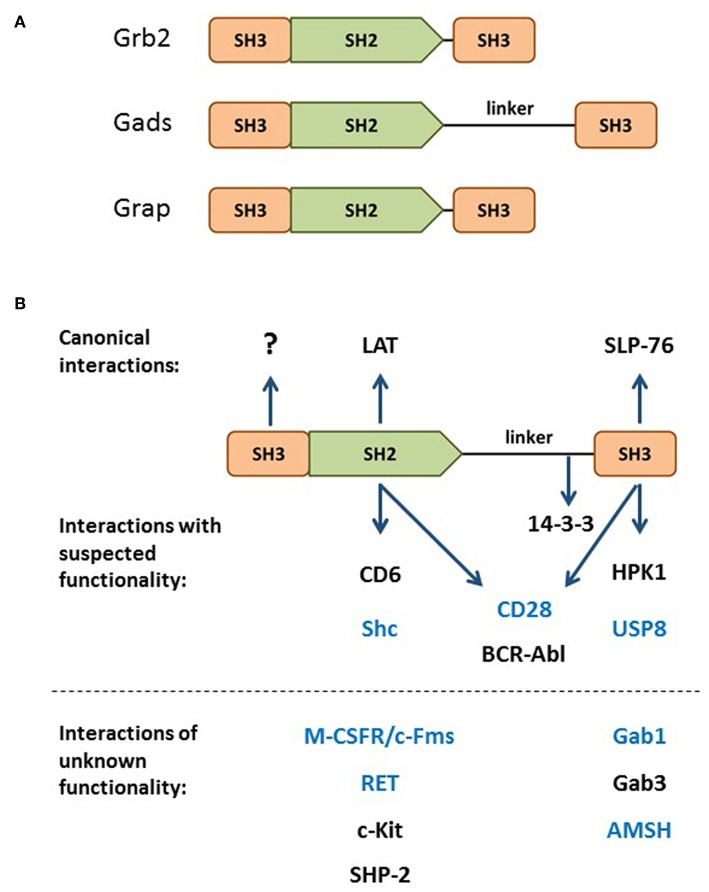
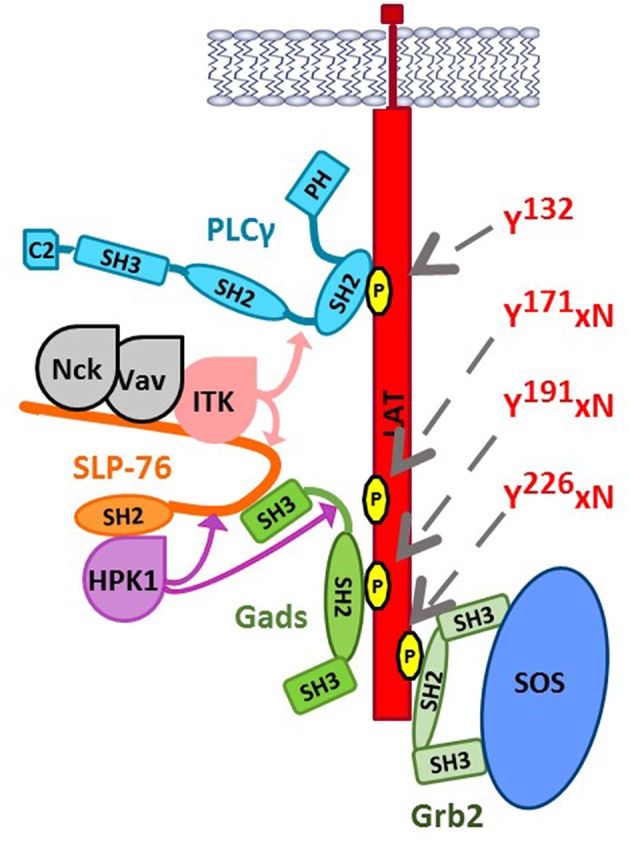
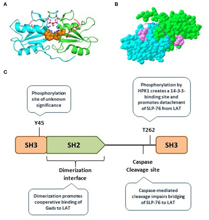
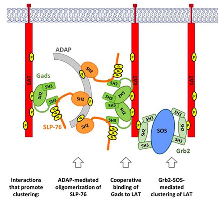
Antigen receptor signaling pathways are organized by adaptor proteins. Three adaptors, LAT, Gads, and SLP-76, form a heterotrimeric complex that mediates signaling by the T cell antigen receptor (TCR) and by the mast cell high affinity receptor for IgE (FcεRI). In both pathways, antigen recognition triggers tyrosine phosphorylation of LAT and SLP-76. The recruitment of SLP-76 to phospho-LAT is bridged by Gads, a Grb2 family adaptor composed of two SH3 domains flanking a central SH2 domain and an unstructured linker region. The LAT-Gads-SLP-76 complex is further incorporated into larger microclusters that mediate antigen receptor signaling. Gads is positively regulated by dimerization, which promotes its cooperative binding to LAT. Negative regulation occurs via phosphorylation or caspase-mediated cleavage of the linker region of Gads. FcεRI-mediated mast cell activation is profoundly impaired in LAT- Gads- or SLP-76-deficient mice. Unexpectedly, the thymic developmental phenotype of Gads-deficient mice is much milder than the phenotype of LAT- or SLP-76-deficient mice. This distinction suggests that Gads is not absolutely required for TCR signaling, but may modulate its sensitivity, or regulate a particular branch of the TCR signaling pathway; indeed, the phenotypic similarity of Gads- and Itk-deficient mice suggests a functional connection between Gads and Itk. Additional Gads binding partners include costimulatory proteins such as CD28 and CD6, adaptors such as Shc, ubiquitin regulatory proteins such as USP8 and AMSH, and kinases such as HPK1 and BCR-ABL, but the functional implications of these interactions are not yet fully understood. No interacting proteins or function have been ascribed to the evolutionarily conserved N-terminal SH3 of Gads. Here we explore the biochemical and functional properties of Gads, and its role in regulating allergy, T cell development and T-cell mediated immunity.
Overview
The claims and statistics of this paper haven’t been checked yet.
Run the check — free, no account needed. It reads the open-access text and checks the sources it cites.
Integrity signals
Registry
Cites
Statistics
Figures
Verification
Claims the paper makes
The claims and statistics of this paper haven’t been checked yet.
Run the check — free, no account needed. It reads the open-access text and checks the sources it cites.
Claims other papers make about this one
Each is checked against this paper’s own text: does it say what the citing paper says it does?
In the citing paper
“As a result, activated ZAP−70 will promote linker of activated T cells (LAT) and SLP76 signalling in T cell, crucial for T cell activation and development [23,24,25,36].”
From The Role of RhoH in TCR Signalling and Its Involvement in Diseases (2021) · cites this paper as [36]
Self-check
Percentages, sums and reported differences re-computed from the paper’s own numbers.
The self-check needs the paper’s full text.
Citations
Cites
Positive and negative selection of the T cell repertoire: what thymocytes see (and don't see)
Klein, L., Kyewski, B., Allen, P. M., & Hogquist, K. A. (2014) · Nature reviews. Immunology · doi:10.1038/nri3667 · Cited by 1,519
Phase separation of signaling molecules promotes T cell receptor signal transduction
Su, X., Ditlev, J. A., Hui, E., Xing, W., Banjade, S., Okrut, J., King, D. S., Taunton, J., Rosen, M. K., & Vale, R. D. (2016) · Science · doi:10.1126/science.aad9964 · Cited by 1,246
CD28 Costimulation: From Mechanism to Therapy
Esensten, J. H., Helou, Y. A., Chopra, G., Weiss, A., & Bluestone, J. A. (2016) · Immunity · doi:10.1016/j.immuni.2016.04.020 · Cited by 937
Specificity in Signal Transduction
Pawson, T. (2004) · Cell · doi:10.1016/s0092-8674(03)01077-8 · Cited by 816
T cell receptor ligation induces the formation of dynamically regulated signaling assemblies
Bunnell, S. C., Hong, D. I., Kardon, J. R., Yamazaki, T., McGlade, C. J., Barr, V. A., & Samelson, L. E. (2002) · The Journal of Cell Biology · doi:10.1083/jcb.200203043 · Cited by 632
Essential Role of LAT in T Cell Development
Zhang, W., Sommers, C. L., Burshtyn, D. N., Stebbins, C. C., Dejarnette, J. B., Trible, R. P., Grinberg, A., Tsay, H. C., Jacobs, H. M., Kessler, C. M., et al. (1999) · Immunity · doi:10.1016/s1074-7613(00)80032-1 · Cited by 547
LAT Is Required for TCR-Mediated Activation of PLCγ1 and the Ras Pathway
Finco, T. S., Kadlecek, T. A., Zhang, W., Samelson, L. E., & Weiss, A. (1998) · Immunity · doi:10.1016/s1074-7613(00)80659-7 · Cited by 497
Wardenburg, J. B., Fu, C., Jackman, J. K., Flotow, H., Wilkinson, S. E., Williams, D. H., Johnson, R., Kong, G., Chan, A. C., & Findell, P. R. (1996) · Journal of Biological Chemistry · doi:10.1074/jbc.271.33.19641 · Cited by 464
T cell receptor signalling networks: branched, diversified and bounded
Brownlie, R. J., & Zamoyska, R. (2013) · Nature Reviews Immunology · doi:10.1038/nri3403 · Cited by 453
Uncoupling of Nonreceptor Tyrosine Kinases from PLC-γ1 in an SLP-76-Deficient T Cell
Yablonski, D., Kuhné, M. R., Kadlecek, T. A., & Weiss, A. (1998) · Science · doi:10.1126/science.281.5375.413 · Cited by 413
Self-citation
Research works printed but not linked: 1
- Su, 1999, Interaction of SLP adaptors with the SH2 domain of Tec family kinases, Eur J Immunol., 29, 3702, 10.1002/(SICI)1521-4141(199911)29:11<3702::AID-IMMU3702>3.0.CO;2-R · DOI
Cited by
Zhang, J. Z., Nguyen, W. H., Greenwood, N., Rose, J. C., Ong, S.-E., Maly, D. J., & Baker, D. (2024) · Nature Biotechnology · doi:10.1038/s41587-023-02107-w · Cited by 37
The Role of RhoH in TCR Signalling and Its Involvement in Diseases
Ahmad Mokhtar, A. M., Hashim, I. F., Mohd Zaini Makhtar, M., Salikin, N. H., & Amin-Nordin, S. (2021) · Cells · doi:10.3390/cells10040950 · Cited by 25
Structural and mechanistic insights into the constitutive Themis–Grb2 complex in T cell signalling
Clancy, D. M., Sanz-Sanjuan, A., Gilis, E., Tougaard, P., Velghe, I., Van Droogenbroeck, Y., Felix, J., Bloch, Y., Cuadrado, Á. F., Merceron, R., et al. (2026) · Nature Communications · doi:10.1038/s41467-026-73359-8
Metadata
- Authors
- Yablonski, D.
- Venue
- Frontiers in Immunology
- Publisher
- Frontiers Media SA
- Published
- 2019-07-25
- Volume
- 10
- ISSN
- 1664-3224
- PMC
- PMC6669380
- Cited by
- 53
- References
- 136
- Funders
- Israel Science Foundation
- United States - Israel Binational Science Foundation
About these checks
Registry, retraction, Cites and statistics results are computed, not judged, with one exception: a printed reference that no index lists is read once by a model before it counts as a citation error. Claims are extracted by a model and kept only when their quote appears verbatim in the text; each cited source is then checked, with a model’s help, for whether it says what the claim relies on. Those verdicts can be wrong — read the claim’s quote and the cited source before relying on one.
Statistics and arithmetic are recomputed, not judged.
Confidence bands are shown only for fields calibrated on our annotated test set. None is calibrated yet, so no band is shown.
What counts as a claim ›How we measure confidence ›How we check this ›
Are you an author, or do you see a problem with this page? Request a review
Data sources
Bibliographic data: Semantic Scholar, OpenAlex, Crossref.
Contains information from the Semantic Scholar Academic Graph Datasets, made available under the ODC Attribution License (ODC-BY 1.0).
Semantic Scholar’s data is described in: Kinney, R., et al. (2023). The Semantic Scholar Open Data Platform. arXiv:2301.10140.
