

The DOI matches its Crossref record; 188 of the 188 references it cites were found in a registry or an index.
Microglia and Neuroinflammation: Crucial Pathological Mechanisms in Traumatic Brain Injury-Induced Neurodegeneration
Open accessAbstract
Traumatic brain injury (TBI) is one of the most common diseases in the central nervous system (CNS) with high mortality and morbidity. Patients with TBI usually suffer many sequelae in the life time post injury, including neurodegenerative disorders such as Alzheimer’s disease (AD) and Parkinson’s disease (PD). However, the pathological mechanisms connecting these two processes have not yet been fully elucidated. It is important to further investigate the pathophysiological mechanisms underlying TBI and TBI-induced neurodegeneration, which will promote the development of precise treatment target for these notorious neurodegenerative consequences after TBI. A growing body of evidence shows that neuroinflammation is a pivotal pathological process underlying chronic neurodegeneration following TBI. Microglia, as the immune cells in the CNS, play crucial roles in neuroinflammation and many other CNS diseases. Of interest, microglial activation and functional alteration has been proposed as key mediators in the evolution of chronic neurodegenerative pathology following TBI. Here, we review the updated studies involving phenotypical and functional alterations of microglia in neurodegeneration after injury, survey key molecules regulating the activities and functional responses of microglia in TBI pathology, and explore their potential implications to chronic neurodegeneration after injury. The work will give us a comprehensive understanding of mechanisms driving TBI-related neurodegeneration and offer novel ideas of developing corresponding prevention and treatment strategies for this disease.
Overview
The claims and statistics of this paper haven’t been checked yet.
Run the check — free, no account needed. It reads the open-access text and checks the sources it cites.
Integrity signals
Registry
Cites
Statistics
Figures
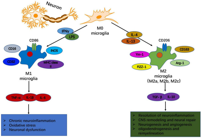
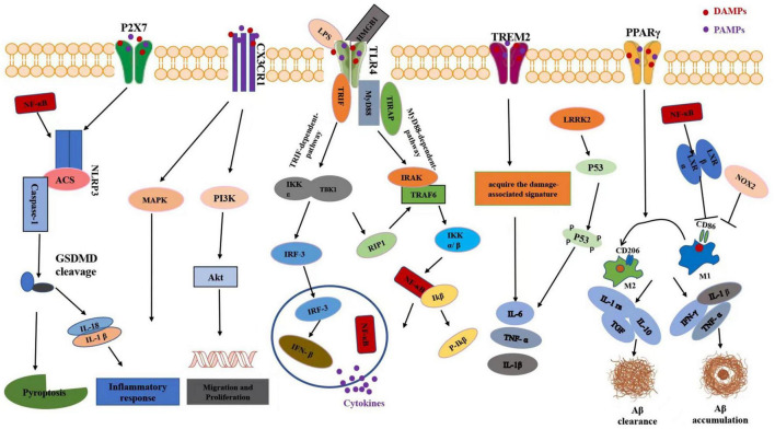
Verification
Claims the paper makes
The claims and statistics of this paper haven’t been checked yet.
Run the check — free, no account needed. It reads the open-access text and checks the sources it cites.
Claims other papers make about this one
Each is checked against this paper’s own text: does it say what the citing paper says it does?
The cited source says what the claim attributes to it.
In the citing paper
“Head trauma is known to cause microglial activation and neuroinflammation [498,499,500,501]”
From Revisiting the Pathogenesis of X-Linked Adrenoleukodystrophy (2025) · cites this paper as [498]
Self-check
Percentages, sums and reported differences re-computed from the paper’s own numbers.
The self-check needs the paper’s full text.
Citations
Cites
Pattern Recognition Receptors and Inflammation
Takeuchi, O., & Akira, S. (2010) · Cell · doi:10.1016/j.cell.2010.01.022 · Cited by 8,895
A Unique Microglia Type Associated with Restricting Development of Alzheimer’s Disease
Keren-Shaul, H., Spinrad, A., Weiner, A., Matcovitch-Natan, O., Dvir-Szternfeld, R., Ulland, T. K., David, E., Baruch, K., Lara-Astaiso, D., Toth, B., et al. (2017) · Cell · doi:10.1016/j.cell.2017.05.018 · Cited by 5,493
Zhang, Y., Chen, K., Sloan, S. A., Bennett, M. L., Scholze, A. R., O'Keeffe, S., Phatnani, H. P., Guarnieri, P., Caneda, C., Ruderisch, N., et al. (2014) · The Journal of Neuroscience · doi:10.1523/jneurosci.1860-14.2014 · Cited by 4,875
Microglia-mediated neurotoxicity: uncovering the molecular mechanisms
Block, M. L., Zecca, L., & Hong, J.-S. (2007) · Nature Reviews Neuroscience · doi:10.1038/nrn2038 · Cited by 4,110
ATP mediates rapid microglial response to local brain injury in vivo
Davalos, D., Grutzendler, J., Yang, G., Kim, J. V., Zuo, Y., Jung, S., Littman, D. R., Dustin, M. L., & Gan, W.-B. (2005) · Nature Neuroscience · doi:10.1038/nn1472 · Cited by 3,913
Kettenmann, H., Hanisch, U.-K., Noda, M., & Verkhratsky, A. (2011) · Physiological Reviews · doi:10.1152/physrev.00011.2010 · Cited by 3,547
Estimating the global incidence of traumatic brain injury
Dewan, M. C., Rattani, A., Gupta, S., Baticulon, R. E., Hung, Y., Punchak, M. A., Agrawal, A., Adeleye, A. O., Shrime, M. G., Rubiano, A. M., et al. (2018) · Journal of neurosurgery · doi:10.3171/2017.10.jns17352 · Cited by 3,459
TREM2 Variants in Alzheimer's Disease
Guerreiro, R., Wojtas, A., Bras, J., Carrasquillo, M., Rogaeva, E., Majounie, E., Cruchaga, C., Sassi, C., Kauwe, J. S. K., Younkin, S., et al. (2013) · New England Journal of Medicine · doi:10.1056/nejmoa1211851 · Cited by 3,020
Microglia Function in the Central Nervous System During Health and Neurodegeneration
Colonna, M., & Butovsky, O. (2017) · Annual Review of Immunology · doi:10.1146/annurev-immunol-051116-052358 · Cited by 2,894
Traumatic brain injury: integrated approaches to improve prevention, clinical care, and research
Maas, A. I. R., Menon, D. K., Adelson, P. D., Andelic, N., Bell, M. J., Belli, A., Bragge, P., Brazinova, A., Büki, A., Chesnut, R. M., et al. (2017) · The Lancet Neurology · doi:10.1016/s1474-4422(17)30371-x · Cited by 2,609
Cited by
Sorted by citations
Items 201–249 of 249
Experimental Models of Stroke: Seeing the Unseen
Yassaad, H., Kahoul, M., Ali, M., Balouta, L., & Mohamed, W. (2024) · Nutritional neurosciences · doi:10.1007/978-981-97-6818-9_3
Afridi, R., Bhusal, A., Lee, S. E., Hwang, E. M., Ryu, H., Kim, J.-H., & Suk, K. (2025) · Molecular and Cellular Neuroscience · doi:10.1016/j.mcn.2025.103994
Zhang, L., Ma, H., Zhang, X., Zhou, Y., Shen, Y., Tang, H., Shao, J., & Zhang, D. (2025) · Medicine · doi:10.1097/md.0000000000042271
Mitochondrial Preparation from Microglia for Glycan Analysis
Madabhushi, M., Palomino, T. V., Patil, M. N., Muddiman, D. C., Tyrrell, D. J., & Samal, J. (2025) · Journal of Visualized Experiments · doi:10.3791/68179
Gao, X., Li, M., Sun, Z., Wu, D., Xu, X., Zhang, H., Yang, Z., Xie, S., Guo, W., Chen, P., et al. (2025) · Molecular Nutrition & Food Research · doi:10.1002/mnfr.70006
Niazi, N. U. K., & Parveen, R. (2025) · Neuroscience Letters · doi:10.1016/j.neulet.2025.138314 · Cited by 1
Repurposing Immunomodulatory Drugs Targeting Microglia for Amyotrophic Lateral SclerosisPreprint
Briedé, J. J., & Maes, K. J. H. (2025) · SSRN Electronic Journal · doi:10.2139/ssrn.5334299
Manipulating Beneficial Gut Bacteria in Obesity-Related Emotional and Cognitive Dysfunction
Jefry Winner, G. (2025) · Oxford University Press eBooks · doi:10.1093/oxfordhb/9780190931544.013.16
Zhuo, H., Li, G., Li, X., Yu, Y., Su, J., Chen, J., Ming, L., & Huang, H. (2025) · Free Radical Biology and Medicine · doi:10.1016/j.freeradbiomed.2025.08.031
Interactions between heavy metals and neuroinflammatory processes
Choudhury, D. R., & Kumar, P. (2025) · Elsevier eBooks · doi:10.1016/b978-0-443-36575-1.00007-8
Impact Induces Phagocytic Defect in Reactive MicrogliaPreprint
Yu, R., Rogers, E. A., Manchanda, P., Beveridge, C. H., Randolph, C. E., Beauclair, T., Jethava, K. P., Shi, R., & Chopra, G. (2025) · bioRxiv (Cold Spring Harbor Laboratory) · doi:10.1101/2025.09.14.676171
Systemic Complications Leading to MODS in a Trauma Patient: A Case-Based Pathophysiologic Analysis
Benibol, Y., & Erol, A. H. (2025) · Kocaeli Üniversitesi Sağlık Bilimleri Dergisi · doi:10.30934/kusbed.1696112
Al-Rubaye, T., Isa, Z., Erenkol, D., Tarahomi, E., & Söğünmez, N. (2025) · Genes & Genomics · doi:10.1007/s13258-025-01687-y
Ye, M., & Shim, I. (2025) · Frontiers in Psychiatry · doi:10.3389/fpsyt.2025.1694735
Ren, X., Su, J., Shi, W., Wang, J., & Yang, X. (2025) · Acta Neurologica Belgica · doi:10.1007/s13760-025-02948-z
Copper-Induced Microglial Activation Involves PKC/NOX/PARP-1/TRPM2 Signaling Pathway
Chew, T. W., Zahir, N. Z. M., Syahir, A., & Mortadza, S. A. S. (2025) · Advances in experimental medicine and biology · doi:10.1007/978-3-032-03398-7_32
Jiang, G., Zhao, L., Liu, C., Tong, J.-M., Zhou, J., Xu, Q., Zhang, C., Xu, X., Tang, X.-P., Yang, H., et al. (2025) · Tungsten · doi:10.1007/s42864-025-00363-8
Riabinina, D., Khamidova, A., Abramova, O. V., Zorkina, Y., Reznik, A., Mukhin, V., Georgij, C., Georgy, M., Karine, L., Pavlova, O., et al. (2025) · SAGE Open Medicine · doi:10.1177/20503121251375865
Dogan, E. O., Bouley, J., Zhong, J., Harkins, A. L., Keeler, A. M., Bosco, D. A., Brown, R. H., & Henninger, N. (2024) · Figshare · doi:10.6084/m9.figshare.26656819
Sun, B., Zhang, A., Feng, S., Jamali, A. W., Jia, Z., Xu, Y., Wang, Z., Zhao, J., Sun, G., & Liu, L. D. (2026) · International Immunopharmacology · doi:10.1016/j.intimp.2026.116181
Devlin, P. J., Khan, R., Do, T. H., West, B. E., Korf, J. M., Guzman, G. U., Tan, C., Ahn, J., Flores, R., Maniskas, M. E., et al. (2026) · bioRxiv (Cold Spring Harbor Laboratory) · doi:10.64898/2026.01.12.699107
Introduction to Neurodegenerative Diseases
Bele, T. N., Devarshi, S. P., Kamble, R. V., Walvekar, A. P., & Chouhan, P. S. (2026) · IGI Global eBooks · doi:10.4018/979-8-3373-1365-8.ch001
Ibeanu, G. C., Eze, D. U., Eze, A., Atoyebi, S. O., Akingbade, T. O., Folarin, A. A., Okoli, J. C., Rowaiye, O. I., & Rowaiye, A. B. (2026) · The Journal of Nutritional Physiology · doi:10.1016/j.jnphys.2026.100019
Kuo, C.-W., Nguyen, T. X. D., Chen, K., Liu, H.-L., Wu, C.-K., Peng, C., Chang, M., & Hsieh, T.-H. (2026) · Journal of Integrative Neuroscience · doi:10.31083/jin48187
Ghafouri, S. N., Dastyafteh, N., Iraji, A., Mahdavi, M. R., & Saeedi, M. R. G. (2026) · Mini-Reviews in Medicinal Chemistry · doi:10.2174/0113895575439030260122105248
Visioli, F., & Tomé‐Carneiro, J. (2026) · Nutrition Research · doi:10.1016/j.nutres.2026.04.005
余丽梅, Wang, Y., Wu, W., An, P., & Yu, Z. (2026) · Neurological Research · doi:10.1080/01616412.2026.2663484
Kara, G., Ali, Y., Espinosa, J. L., Park, P., Holcomb, M., Flinn, H., Taylor, N., Galbraith, T., Leonard, F., & Villapol, S. (2026) · bioRxiv (Cold Spring Harbor Laboratory) · doi:10.64898/2026.04.25.720847
Repeated head impacts establish a senescent cranial bone marrow niche that impairs brain metabolism
Devlin, P., Khan, R., Do, T., West, B. E., Korf, J. M., Guzman, G. U., Ahn, J., Saltzman, A. B., Jain, A., Tan, C., et al. (2026) · Journal of Neuroinflammation · doi:10.1186/s12974-026-03846-8
Xing, Y., Do, V., Xu, Z., Do, C., Yang, C., Zhu, Z., & Gao, J. (2026) · ACS Nano Medicine · doi:10.64898/2026.05.27.727842
Wang, Y., Ren, C., Dong, W., Li, Q., Deng, F., Zhao, F., Cheng, Y., Sun, P., Li, H., & Wang, Y. (2026) · Chinese Medicine · doi:10.1186/s13020-026-01436-7
Targeting mGluR1-Mediated Microglial ER Stress Mitigates Copper-Induced TBI Neuroinflammation
Wang, D., Jiawei, Z., Zhou, Z., Cui, D., Liu, F. M., & Zhang, B. L. (2026) · Molecular Neurobiology · doi:10.1007/s12035-026-05958-x
Liu, P., Wu, X., Liu, X., Wang, Y., Jiang, S., Wu, K., Li, G., Ding, J., Wu, C., Wei, D. M., et al. (2026) · Advanced Science · doi:10.1002/advs.76220
Onigbinde, S. B., Solomon, J., Sandilya, V., Daramola, O. E., Fowowe, M., Adeniyi, M., Kobeissy, F. H., Mondello, S., Puccio, A. M., & Mechref, Y. S. (2026) · Analytica Chimica Acta · doi:10.1016/j.aca.2026.345882
Gerin, D. R., Medeiros, T. B., Palmero, C. Y., & de Barros, C. M. (2026) · International Journal of Biological Macromolecules · doi:10.1016/j.ijbiomac.2026.153259
Song, W., Li, E., Dai, R., Chu, B., Yuan, H., Ji, H., Dai, J., Li, L., Feng, X., Gu, J., et al. (2026) · International Journal of Biological Macromolecules · doi:10.1016/j.ijbiomac.2026.153357
Thant, M. T., Kongkatitham, V., Khine, H. E. E., Parichatikanond, W., Jayashan, S. S., Poldorn, P., Wongnongwa, Y., Towiwat, P., Böttcher, C., & Sritularak, B. (2026) · ACS Omega · doi:10.1021/acsomega.6c01737
Ashkhotov, A. V., Shnayder, N. A., Trefilova, V. V., & Nasyrova, R. F. (2026) · Vestnik of North-Eastern Federal University Medical Sciences · doi:10.25587/2587-5590-2026-2-15-29
Hydrogel design at anatomical extremes: Lessons learned from ocular and intraperitoneal applications
Haegebaert, R., Guerassimoff, L., Paoletti, L., Li, W., Sauvage, F., & Remaut, K. (2026) · Journal of Controlled Release · doi:10.1016/j.jconrel.2026.115248
Gut microbiota dysbiosis-induced neuroinflammation and IL-22/STAT3 activation in emotional disorders
Feng, X., Hu, Y., Zhu, L., Lu, X., Sun, L., Liu, J., Jin, J., Ji, X., & Zhao, L. (2026) · Brain Behavior and Immunity · doi:10.1016/j.bbi.2026.106979
Kara, G., Holcomb, M., Hijazi, A. A., Ali, Y., Espinosa, J. L., Cruz-Pineda, L., Park, P., Flinn, H., Taylor, N., Galbraith, T., et al. (2026) · Biomedical Microdevices · doi:10.1007/s10544-026-00843-9
Ali, W., Ahmad, I., Rahman, N. U., Khan, J., Alam, W., Rabbi, F., Shams, B., Rahim, T., Rehman, H., & Shams, H. (2026) · Molecular Neurobiology · doi:10.1007/s12035-026-06179-y
Understanding the immunological components of brain disorders
Ali, S., Acharya, S., & Ghosh, A. (2026) · Elsevier eBooks · doi:10.1016/b978-0-443-44631-3.00005-6
Apigenin as a Potential Modulator of Neuroinflammation in Multiple Sclerosis: A Review
de Santana, M. R., Silva, N. N., de Oliveira, J. V. R., Victor, M. M., Butt, A. M., & Costa, S. L. (2026) · Biology · doi:10.3390/biology15171545
Ramos, A. J., & Villarreal, A. (2026) · Frontiers in Network Physiology · doi:10.3389/fnetp.2026.1895622
Calabrò, R. S., Calderone, A., Ardizzone, A., Ciancarelli, I., Morone, G., Cammaroto, S., Rifici, C., & Quartarone, A. (2026) · Frontiers in Neurology · doi:10.3389/fneur.2026.1854973
Lien, Y. R., Mueller, C., Sharma, A. A., Mitchell, B. S., Nenert, R. E., Moyana, A., Pilkington, J., Martin, R. C., Morgan, C. J., Hoef, L. V., et al. (2026) · Epilepsia Open · doi:10.1002/epi4.70366
5-HT1A Antagonist WAY-100635 Prevents Chronic Neurodegeneration and Immune Activation After TBIPreprint
Dutta, S., Pradhan, L. K., Chen, J., Wang, N., Gao, X., & Das, A. (2026) · bioRxiv (Cold Spring Harbor Laboratory) · doi:10.64898/2026.10.11.758371
Photobiomodulation in Parkinson’s disease: From pathogenesis to intervention
Shao, J., Deng, Q., & Yang, L. (2026) · Experimental Neurology · doi:10.1016/j.expneurol.2026.116088
Metadata
- Authors
- Shao, F., Wang, X., Wu, H., Wu, Q., & Zhang, J.
- Affiliations
- First Affiliated Hospital Zhejiang University (CN)
- Second Affiliated Hospital of Zhejiang University (CN)
- Corresponding author
- Wu, Qun
- Zhang, Jianmin
- Venue
- Frontiers in Aging Neuroscience
- Publisher
- Frontiers Media SA
- Published
- 2022-03-25
- Volume
- 14
- Pages
- 825086-825086
- ISSN
- 1663-4365 (ISSN-L)
- PMC
- PMC8990307
- OpenAlex
- W4220730682
- Cited by
- 250
- References
- 188
- Citations by year
- 2026: 47 · 2025: 85 · 2024: 75 · 2023: 31 · 2022: 11
- FWCI
- 5.21
- Type
- journal article
- Language
- en
- Open access
- gold
- doi.org
- Licence
- Locations
- Frontiers in Aging Neuroscience (publishedVersion, cc-by)
- PubMed Central (publishedVersion, cc-by)
- PubMed (publishedVersion)
- DOAJ (DOAJ: Directory of Open Access Journals) (submittedVersion)
- Topics
- Life Sciences › Neuroscience › Neurology › Neuroinflammation and Neurodegeneration Mechanisms
- Health Sciences › Medicine › Clinical Neurology › Traumatic Brain Injury and Neurovascular Disturbances
- Health Sciences › Medicine › Clinical Neurology › Traumatic Brain Injury Research
- Keywords
- traumatic brain injury, microglia, neuroinflammation, neurodegeneration, microglial activation, Alzheimer's disease, Parkinson's disease, chronic neurodegeneration, microglial phenotype, treatment options
About these checks
Registry, retraction, Cites and statistics results are computed, not judged, with one exception: a printed reference that no index lists is read once by a model before it counts as a citation error. Claims are extracted by a model and kept only when their quote appears verbatim in the text; each cited source is then checked, with a model’s help, for whether it says what the claim relies on. Those verdicts can be wrong — read the claim’s quote and the cited source before relying on one.
Statistics and arithmetic are recomputed, not judged.
Confidence bands are shown only for fields calibrated on our annotated test set. None is calibrated yet, so no band is shown.
What counts as a claim ›How we measure confidence ›How we check this ›
Are you an author, or do you see a problem with this page? Request a review
