

The DOI matches its Crossref record; 221 of the 222 references it cites were found in a registry or an index.
Shaping Synapses by the Neural Extracellular Matrix
Open accessAbstract
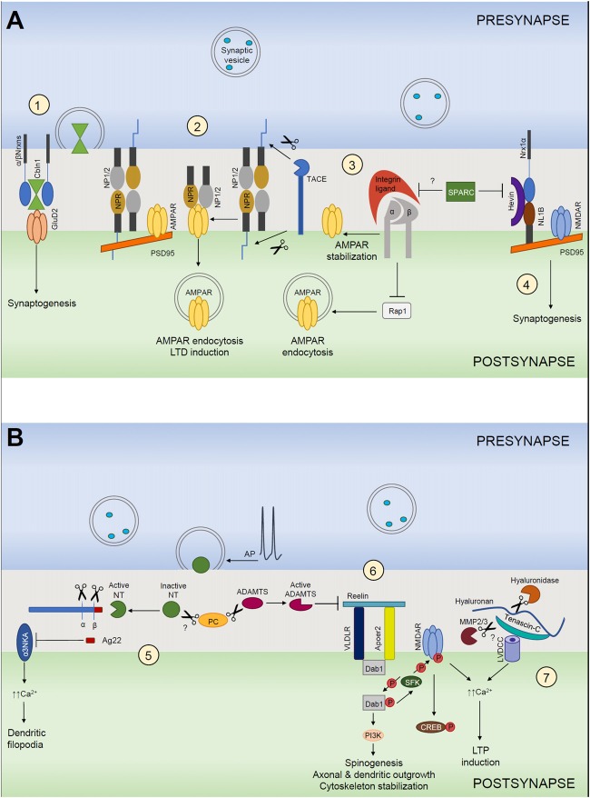
Accumulating data support the importance of interactions between pre- and postsynaptic neuronal elements with astroglial processes and extracellular matrix (ECM) for formation and plasticity of chemical synapses, and thus validate the concept of a tetrapartite synapse. Here we outline the major mechanisms driving: (i) synaptogenesis by secreted extracellular scaffolding molecules, like thrombospondins (TSPs), neuronal pentraxins (NPs) and cerebellins, which respectively promote presynaptic, postsynaptic differentiation or both; (ii) maturation of synapses via reelin and integrin ligands-mediated signaling; and (iii) regulation of synaptic plasticity by ECM-dependent control of induction and consolidation of new synaptic configurations. Particularly, we focused on potential importance of activity-dependent concerted activation of multiple extracellular proteases, such as ADAMTS4/5/15, MMP9 and neurotrypsin, for permissive and instructive events in synaptic remodeling through localized degradation of perisynaptic ECM and generation of proteolytic fragments as inducers of synaptic plasticity.
Overview
The claims and statistics of this paper haven’t been checked yet.
Run the check — free, no account needed. It reads the open-access text and checks the sources it cites.
Integrity signals
Registry
Cites
Statistics
Figures
Verification
Claims the paper makes
The claims and statistics of this paper haven’t been checked yet.
Run the check — free, no account needed. It reads the open-access text and checks the sources it cites.
Claims other papers make about this one
Each is checked against this paper’s own text: does it say what the citing paper says it does?
The source backs part of the claim, not all of it — or backs it for a different population, condition or comparison.
In the citing paper
“TNXB encodes tenascin proteins, which are extracellular matrix glycoproteins demonstrated to modulate synaptic plasticity in the brain [83].”
From Sex-specific DNA methylation differences in Alzheimer’s disease pathology (2021) · cites this paper as [83]
Experimental — accuracy not yet measured
Self-check
Percentages, sums and reported differences re-computed from the paper’s own numbers.
The self-check needs the paper’s full text.
Citations
Cites
Polazzi, E., Mengoni, I., Caprini, M., Peña‐Altamira, E., Kurtys, E., & Monti, B. (2012) · Neurosignals · doi:10.1159/000337115 · Cited by 9,023
Shrestha, S., & Offer, S. M. (2016) · Medical Epigenetics · doi:10.1159/000444713 · Cited by 8,951
How Matrix Metalloproteinases Regulate Cell Behavior
Sternlicht, M. D., & Werb, Z. (2001) · Annual Review of Cell and Developmental Biology · doi:10.1146/annurev.cellbio.17.1.463 · Cited by 3,959
Tripartite synapses: glia, the unacknowledged partner
Araque, A., Parpura, V., Sanzgiri, R. P., & Haydon, P. G. (1999) · Trends in Neurosciences · doi:10.1016/s0166-2236(98)01349-6 · Cited by 2,529
Neuroligins and neurexins link synaptic function to cognitive disease
Südhof, T. C. (2008) · Nature · doi:10.1038/nature07456 · Cited by 1,849 · cited 2×
“Mutations in cerebellins and their presynaptic neurexin receptors have been associated with multiple neurodevelopmental disorders, especially with autism spectrum disorders (ASDs), Tourette syndrome and schizophrenia (Südhof, 2008; Clarke et al., 2012; Bourgeron, 2015; Pendyala et al., 2017).”
BackgroundSynaptic scaling mediated by glial TNF-α
Stellwagen, D., & Malenka, R. C. (2006) · Nature · doi:10.1038/nature04671 · Cited by 1,715
NMDA receptor subunits: diversity, development and disease
Cull-Candy, S., Brickley, S., & Farrant, M. (2001) · Current Opinion in Neurobiology · doi:10.1016/s0959-4388(00)00215-4 · Cited by 1,694
Thrombospondins are astrocyte-secreted proteins that promote CNS synaptogenesis
Christopherson, K. S., Ullian, E. M., Stokes, C. C. A., Mullowney, C. E., Hell, J. W., Agah, A., Lawler, J., Mosher, D. F., Bornstein, P., & Barres, B. A. (2005) · Cell · doi:10.1016/j.cell.2004.12.020 · Cited by 1,688
Wake, H., Moorhouse, A. J., Jinno, S., Kohsaka, S., & Nabekura, J. (2009) · The Journal of Neuroscience · doi:10.1523/jneurosci.4363-08.2009 · Cited by 1,619
Protoplasmic Astrocytes in CA1 Stratum Radiatum Occupy Separate Anatomical Domains
Bushong, E. A., Martone, M. E., Jones, Y. Z., & Ellisman, M. H. (2002) · The Journal of Neuroscience · doi:10.1523/jneurosci.22-01-00183.2002 · Cited by 1,578
Research works printed but not linked: 1
- Jones, 2000, The tenascin family of ECM glycoproteins: structure, function, and regulation during embryonic development and tissue remodeling, Dev. Dyn., 218, 235, 10.1002/(SICI)1097-0177(200006)218:2<235::AID-DVDY2>3.0.CO;2-G · DOI
Cited by
Sorted by citations
Items 101–170 of 170
The extracellular matrix as a dynamic regulator of brain function and plasticity
Ge, C., & Tajerian, M. (2026) · Neurobiology of Disease · doi:10.1016/j.nbd.2026.107374 · Cited by 8
Extracellular matrix remodelling in neurological diseases
Kwok, J. C. F., & Dityatev, A. E. (2026) · Nature Reviews Neurology · doi:10.1038/s41582-026-01209-8 · Cited by 9
Self-citationMüller, P., Dietrich, D., Schoch, S., Pitsch, J., Becker, A. J., & Cases‐Cunillera, S. (2023) · Neurobiology of Disease · doi:10.1016/j.nbd.2023.106364 · Cited by 8
Perineuronal Net Microscopy: From Brain Pathology to Artificial Intelligence
Paveliev, M., Egorchev, A. A., Musin, F., Lipachev, N., Melnikova, A., Gimadutdinov, R. M., Kashipov, A., Molotkov, D., Chickrin, D. E., & Aganov, A. V. (2024) · International Journal of Molecular Sciences · doi:10.3390/ijms25084227 · Cited by 8
Richard, A., & Lu, X. (2019) · Neural Regeneration Research · doi:10.4103/1673-5374.247459 · Cited by 6
Lipachev, N., Melnikova, A., Fedosimova, S. V., Arnst, N., Kochneva, A., Shaikhutdinov, N., Dvoeglazova, A., Андреевна, Т. А., Олегович, М. М., Aganov, A. V., et al. (2022) · Neuroscience Research · doi:10.1016/j.neures.2022.06.005 · Cited by 6
John, U., Patro, N., & Patro, I. K. (2023) · Journal of Chemical Neuroanatomy · doi:10.1016/j.jchemneu.2023.102286 · Cited by 6
Gregorio, R. D., Chen, X., Petit, E., Dobrenis, K., & Sze, J. Y. (2019) · Cerebral Cortex · doi:10.1093/cercor/bhz191 · Cited by 6
Perineuronal nets: Cruise from a honeycomb to the safety nets
John, U., Patro, N., & Patro, I. K. (2022) · Brain Research Bulletin · doi:10.1016/j.brainresbull.2022.10.004 · Cited by 6
Loupy, K. M., Dawud, L. M., Zambrano, C. A., Lee, T., Heinze, J. D., Elsayed, A. I., Hassell, J. E., D'Angelo, H. M., Frank, M. G., Maier, S. F., et al. (2025) · NeuroImmunoModulation · doi:10.1159/000544842 · Cited by 5
Scaffold Materials and Toxicity
Nair, S. A., & Gayathri, V. (2023) · Springer eBooks · doi:10.1007/978-981-19-7834-0_20 · Cited by 5
Miyata, S., Tsuda, M., & Mitsui, S. (2023) · Molecular Neurobiology · doi:10.1007/s12035-023-03890-y · Cited by 5
Leveraging Biomaterial Platforms to Study Aging-Related Neural and Muscular Degeneration
Hidalgo‐Alvarez, V., & Madl, C. M. (2024) · Biomolecules · doi:10.3390/biom14010069 · Cited by 5
Distribution of the Extracellular Matrix in the Pararubral Area of the Rat
Szarvas, D., Gaál, B., Matesz, C., & Rácz, É. (2018) · Neuroscience · doi:10.1016/j.neuroscience.2018.10.027 · Cited by 5
Gaál, B., Magyar, Á., Rácz, É., Matesz, C., Wolf, E., & Kiss, P. (2021) · Neural Regeneration Research · doi:10.4103/1673-5374.320988 · Cited by 6
Chondroitin Sulphate Proteoglycan Axonal Coats in the Human Mediodorsal Thalamic Nucleus
Pantazopoulos, H., Hossain, N. M., Chelini, G., Durning, P. T., Barbas, H., Zikopoulos, B., & Berretta, S. (2022) · Frontiers in Integrative Neuroscience · doi:10.3389/fnint.2022.934764 · Cited by 5
Electron Microscopy of Neurons on Biomimetic Substrates
Bovio, C. L., Mollo, V., Mariano, A., & Santoro, F. (2024) · Methods in molecular biology · doi:10.1007/978-1-0716-3969-6_2 · Cited by 4
The long-loop recycling (LLR) of synaptic components as a question of economics
Georgiev, S. V., & Rizzoli, S. O. (2023) · Molecular and Cellular Neuroscience · doi:10.1016/j.mcn.2023.103862 · Cited by 4
Schreurs, B. G., O’Dell, D. E., & Wang, D. (2024) · Biology · doi:10.3390/biology13030200 · Cited by 4
DNA Methylation in Alzheimer’s Disease
Weymouth, L. S., Smith, A. R., & Lunnon, K. (2024) · Current topics in behavioral neurosciences · doi:10.1007/7854_2024_530 · Cited by 4
Yang, S., Gigout, S., Molinaro, A., Naito-Matsui, Y., Hilton, S., Foscarin, S., Nieuwenhuis, B., Verhaagen, J., Pizzorusso, T., Saksida, L. M., et al. (2020) · bioRxiv (Cold Spring Harbor Laboratory) · doi:10.1101/2020.01.03.894188 · Cited by 4
Ritok, A., Kiss, P., Zaher, A., Wolf, E., Ducza, L., Bácskai, T., Matesz, C., & Gaál, B. (2022) · Scientific Reports · doi:10.1038/s41598-022-25692-3 · Cited by 3
Catudio-Garrett, E. L., Bielawski, A. M., Ruchti, E., Sherer, L. M., Waghmare, I., Hess-Homeier, D. L., McCabe, B. D., Stowers, R. S., & Certel, S. J. (2023) · Genetics · doi:10.1093/genetics/iyac190 · Cited by 3
MR Imaging of Neurofluids in the Developing Brain
Agarwal, N., Klein, W. M., & Tuura, R. (2025) · Neuroimaging Clinics of North America · doi:10.1016/j.nic.2024.12.005 · Cited by 2
Genoud, S., Chaichim, C., Porto, R. R., Tomanić, T., Stefen, H., Parić, E., Sarkar, S., Yoo, D., Gao, W., Hardeman, E. C., et al. (2025) · Molecular Neurobiology · doi:10.1007/s12035-025-05259-9 · Cited by 3
Collagens as New Players in Nervous System Diseases
Heikkinen, A., Fox, M. A., & Pihlajaniemi, T. A. (2021) · Biology of extracellular matrix · doi:10.1007/978-3-030-67592-9_8 · Cited by 3
Czuba, E., Głowiński, S., Wojcik, S. P., Lietzau, G., Zabielska-Kaczorowska, M. A., & Kowiański, P. (2023) · Molecular and Cellular Neuroscience · doi:10.1016/j.mcn.2023.103860 · Cited by 3
Neural extracellular matrix regulates visual sensory motor integration
Reinhard, J., Mueller-Buehl, C., Wiemann, S., Roll, L., Luft, V., Shabani, H., Rathbun, D. L., Gan, L., Kuo, C., Franzen, J., et al. (2024) · iScience · doi:10.1016/j.isci.2024.108846 · Cited by 3
Chen, F., Wang, L., Liu, H., Wang, Y., Tang, B., & Bao, N. (2026) · Materials Today Bio · doi:10.1016/j.mtbio.2026.102822 · Cited by 4
Huang, Y.-F., Liu, C., Liu, Z., Zhuang, X., Chen, J.-H., Zhang, Z.-N., Shen, C., & Hu, Y. (2026) · Brain · doi:10.1093/brain/awag058 · Cited by 3
Neural extracellular matrix regulates visual sensory motor integrationPreprint
Reinhard, J., Mueller-Buehl, C., Wiemann, S., Roll, L., Luft, V., Shabani, H., Rathbun, D. L., Gan, L., Kuo, C., Franzen, J., et al. (2023) · bioRxiv (Cold Spring Harbor Laboratory) · doi:10.1101/2023.04.17.537074 · Cited by 2
von Kügelgen, N., Ludwik, K. A., Mendonsa, S., Römer, C., Becher, E., Breimann, L., Strauch, M., Mari, T., Mongellaz, S., Zuckerman, B., et al. (2025) · NAR Molecular Medicine · doi:10.1093/narmme/ugaf005 · Cited by 1
Chisholm, A., Ferland, J. N., Ellis, R. J., & Hurd, Y. L. (2025) · Biological Psychiatry · doi:10.1016/j.biopsych.2025.08.023 · Cited by 1
Yaylacı, M., Doğan, Ö., Kırşan, F. Z., & Öztop, D. B. (2025) · Journal of Molecular Neuroscience · doi:10.1007/s12031-025-02415-7 · Cited by 1
Campbell, A. S., Minařík, M., Buckley, D., Anand, T., Gela, D., Pšenička, M., & Baker, C. V. H. (2025) · Journal of Anatomy · doi:10.1111/joa.70061 · Cited by 1
Yue, L., Craig, M. T., & Morris, B. J. (2025) · Molecular Neurobiology · doi:10.1007/s12035-025-05369-4 · Cited by 2
Remodeling of Perineuronal Nets in the Striato-Cortical Axis in L-DOPA-Induced Dyskinesia Rat Model
Bilbay, N. T., Tel, B. C., Akkuş, G., Cakir-Aktas, C., Solakoğlu, S. T., Çakmaklı, G. Y., & Elibol, B. (2025) · International Journal of Molecular Sciences · doi:10.3390/ijms262311726 · Cited by 1
Roles and therapeutic prospects of the laminin family in disorders of the nervous system
Zhang, X., Li, X., Gao, L., Qiao, Y., Lv, B., Zhang, Q. D., Zhang, M., Yu, J., Li, H., Bi, H., et al. (2026) · Journal of Translational Medicine · doi:10.1186/s12967-026-08410-9 · Cited by 2
Impact of Reck expression and promoter activity in neuronal in vitro differentiation
Trombetta-Lima, M., Assis-Ribas, T., Cintra, R. C., Campeiro, J. D., Guerreiro, J. R., Winnischofer, S. M. B., Nascimento, I. C., Ulrich, H., Hayashi, M. A. F., & Sogayar, M. C. (2021) · Molecular Biology Reports · doi:10.1007/s11033-021-06175-6 · Cited by 1
Rozhnova, M. A., Kazantsev, V., & Pankratova, E. V. (2021) · Scientific School Dynamics of Complex Networks and their Applications (DCNA) · doi:10.1109/dcna53427.2021.9586855 · Cited by 1
Szabó, F., Sigutova, V., Schneider, A. M., Hoerder‐Suabedissen, A., & Molnár, Z. (2024) · bioRxiv (Cold Spring Harbor Laboratory) · doi:10.1101/2024.05.06.592829 · Cited by 1
[Research progress on the role of thrombospondin in synapse formation].
Gong, C., Xiang, G., Liu, K., & Zhang, H. (2019) · Europe PMC (PubMed Central) · doi:10.7507/1002-1892.201809006
PROFILE OF EXTRACELLULAR MATRIX COMPONENTS IN NEURONS IN VITRO AT INDUCED NEUROINFLAMMATION
Dominova, I. N., & Turkin, A. (2020) · Современные проблемы науки и образования · doi:10.17513/spno.29780
Sex-biased gene expression in rhesus macaque and human brainsPreprint
DeCasien, A. R., Sherwood, C. C., & Higham, J. P. (2020) · bioRxiv (Cold Spring Harbor Laboratory) · doi:10.1101/2020.07.17.208785
Assessment of CSF biomarkers and microRNA-mediated disease mechanisms in SMAPreprint
Welby, E., Rehborg, R., Harmelink, M. M., & Ebert, A. D. (2021) · bioRxiv (Cold Spring Harbor Laboratory) · doi:10.1101/2021.05.19.444816
Análise do papel do gene supressor de tumor RECK durante a diferenciação neurogênica
Ribas, T. D. A. (2019) · Digital Library of Theses and Dissertations (Universidade de São Paulo) · doi:10.11606/d.87.2019.tde-13012021-103739
Zhao, X., Zhang, F., Kandel, S. R., Brau, F., & He, J. J. (2021) · bioRxiv (Cold Spring Harbor Laboratory) · doi:10.1101/2021.11.22.469603
Zhao, X., Zhang, F., Kandel, S. R., Brau, F., & He, J. J. (2021) · SSRN Electronic Journal · doi:10.2139/ssrn.3985171
Nairn, A. C., & Leslie, S. N. (2020) · Faculty Opinions – Post-Publication Peer Review of the Biomedical Literature · doi:10.3410/f.738571114.793579225
Dionne, O., & Corbin, F. (2021) · Research Square · doi:10.21203/rs.3.rs-146075/v1
MMP-9 and TIMP-1 Levels in Children and Adolescents with Obsessive Compulsive Disorder
Dalyan, S., Çoban, Ö. G., Erdem, B. K., Yolcu, Ö., Önder, A., Adanır, A. S., Nasıroğlu, S., & Akbaş, S. H. (2023) · Neurochemical Journal · doi:10.1134/s1819712423010038
Ahmadpour, S. (2023) · The Egyptian Journal of Neurology Psychiatry and Neurosurgery · doi:10.1186/s41983-023-00719-2
Additional file 2 of Sex-specific DNA methylation differences in Alzheimer’s disease pathology
Zhang, L., Young, J. I., Gomez, L., Silva, T. C., Schmidt, M. A., Cai, J., Chen, X., Martin, E. R., & Wang, L. (2021) · Figshare · doi:10.6084/m9.figshare.14490464
Szabó, F., Sigutova, V., Schneider, A. M., Hoerder‐Suabedissen, A., & Molnár, Z. (2024) · Journal of Anatomy · doi:10.1111/joa.14181 · Cited by 1
Campbell, A. S., Minařík, M., Buckley, D., Anand, T., Gela, D., Pšenička, M., & Baker, C. V. H. (2025) · bioRxiv (Cold Spring Harbor Laboratory) · doi:10.1101/2025.02.04.636467
Mohamaddokht, M., Falck, J. E., Öktem, S. K., Rizzu, P., Friedrich, K., Heutink, P., Rosenmund, C., Garner, C. C., Schmitz, D., & Ackermann, F. (2025) · bioRxiv (Cold Spring Harbor Laboratory) · doi:10.1101/2025.07.03.662734
Extracellular Origins of Cognition: Lessons from Primate Neocortical ECM
Stern, S. (2025) · PROTEOMICS · doi:10.1002/pmic.13979
Fan, Z., Yao, S., Gong, X., Xu, H., & Chen, G. (2025) · Journal of Pharmaceutical and Biomedical Analysis · doi:10.1016/j.jpba.2025.117148
Additional file 1 of Sex-specific DNA methylation differences in Alzheimer’s disease pathology
Zhang, L., Young, J. I., Gomez, L., Silva, T. C., Schmidt, M. A., Cai, J., Chen, X., Martin, E. R., & Wang, L. (2021) · Figshare · doi:10.6084/m9.figshare.14490461
Devienne, G. F. (2019) · theses.fr (ABES) · doi:10.70675/453418efzc908z49e0zba6fz71fefaa50691
Etude du rôle du Syndécan à la jonction neuromusculaire de Caenorhabditis elegans
Vachon, C. (2020) · theses.fr (ABES) · doi:10.70675/4f34af1dzeed7z426dzbab2zb1e21a201491
Idziak, A. (2022) · theses.fr (ABES) · doi:10.70675/91c65c6ez289bz41b2za50azc10a875e4675
Molecular mechanisms underlying the surface organization of the NMDA receptors during development
Bénac, N. (2024) · theses.fr (ABES) · doi:10.70675/e3508a2ezb62ez4f99zb27bz156201271974
Fujita, E., Hadano, J., Hashimoto, J., & Matsumoto, K. (2026) · Behavioural Brain Research · doi:10.1016/j.bbr.2026.116225
Vasoya, D., Keavey, L., Levit, C., Watzeels, T., Heron, S., Cholewa-Waclaw, J., Dando, O., Mancuso, R., & Bowles, K. R. (2026) · bioRxiv (Cold Spring Harbor Laboratory) · doi:10.64898/2026.06.30.735652 · Cited by 1
Transcriptomic Profiling of the Amygdala of Children With Autism Spectrum Disorder
Babu, J., Lal, A., Challagundla, L., Allen, O., Griffin, M. L., Gisabella, B., & Pantazopoulos, H. (2026) · Autism Research · doi:10.1002/aur.70333
Li, Y., Park, R., Krishnamachary, B., Lee, H., Lei, Z., Li, H., & Wu, J. (2026) · International Journal of Molecular Sciences · doi:10.64898/2026.07.31.742052
Melrose, J. (2026) · Research Square · doi:10.21203/rs.3.rs-10723418/v1
Differences between astrocytes and fibroblasts in the effects of TGFβ on fibronectin matrix assembly
Sun, Y., & Schwarzbauer, J. E. (2026) · Matrix Biology · doi:10.1016/j.matbio.2026.102051
Metadata
- Authors
- Ferrer-Ferrer, M., & Dityatev, A.
- Affiliations
- German Center for Neurodegenerative Diseases (DE)
- Corresponding author
- Dityatev, Alexander
- ORCID
- Dityatev, Alexander
- Venue
- Frontiers in Neuroanatomy
- Publisher
- Frontiers Media SA
- Published
- 2018-05-15
- Volume
- 12
- Pages
- 40-40
- ISSN
- 1662-5129 (ISSN-L)
- PMC
- PMC5962695
- OpenAlex
- W2804637583
- Cited by
- 169
- References
- 222
- Citations by year
- 2026: 11 · 2025: 16 · 2024: 18 · 2023: 19 · 2022: 26 · 2021: 34 · 2020: 19 · 2019: 21 · 2018: 5
- FWCI
- 2.59
- Type
- journal article
- Language
- en
- Open access
- gold
- doi.org
- Locations
- Frontiers in Neuroanatomy (publishedVersion, cc-by)
- Europe PMC (PubMed Central) (publishedVersion, cc-by)
- PubMed Central (publishedVersion, cc-by)
- PubMed (publishedVersion)
- DOAJ (DOAJ: Directory of Open Access Journals) (submittedVersion, cc-by-sa)
- DZNE Pub (submittedVersion, cc-by)
- Funders
- Seventh Framework Programme (Initial Training Network ExtraBrain)
- Topics
- Life Sciences › Neuroscience › Cellular and Molecular Neuroscience › Neuroscience and Neuropharmacology Research
- Life Sciences › Neuroscience › Neurology › Neuroinflammation and Neurodegeneration Mechanisms
- Life Sciences › Biochemistry, Genetics and Molecular Biology › Genetics › Genetics and Neurodevelopmental Disorders
- Keywords
- synaptogenesis, synaptic plasticity, thrombospondin, cerebellin, reelin, integrin signaling, extracellular protease, ADAMTS, MMP-9, synaptic remodeling
About these checks
Registry, retraction, Cites and statistics results are computed, not judged, with one exception: a printed reference that no index lists is read once by a model before it counts as a citation error. Claims are extracted by a model and kept only when their quote appears verbatim in the text; each cited source is then checked, with a model’s help, for whether it says what the claim relies on. Those verdicts can be wrong — read the claim’s quote and the cited source before relying on one.
Statistics and arithmetic are recomputed, not judged.
Confidence bands are shown only for fields calibrated on our annotated test set. None is calibrated yet, so no band is shown.
What counts as a claim ›How we measure confidence ›How we check this ›
Are you an author, or do you see a problem with this page? Request a review
