

The DOI matches its Crossref record; 25 of the 27 references it cites were found in a registry or an index.
Absence of chronic traumatic encephalopathy in retired football players with multiple concussions and neurological symptomatology
Open accessAbstract
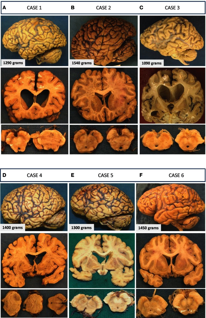
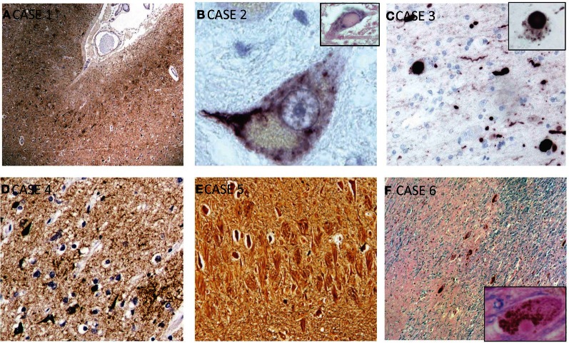
BACKGROUND: Chronic traumatic encephalopathy (CTE) is the term coined for the neurodegenerative disease often suspected in athletes with histories of repeated concussion and progressive dementia. Histologically, CTE is defined as a tauopathy with a distribution of tau-positive neurofibrillary tangles (NFTs) that is distinct from other tauopathies, and usually shows an absence of beta-amyloid deposits, in contrast to Alzheimer's disease (AD). Although the connection between repeated concussions and CTE-type neurodegeneration has been recently proposed, this causal relationship has not yet been firmly established. Also, the prevalence of CTE among athletes with multiple concussions is unknown. METHODS: We performed a consecutive case series brain autopsy study on six retired professional football players from the Canadian Football League (CFL) with histories of multiple concussions and significant neurological decline. RESULTS: All participants had progressive neurocognitive decline prior to death; however, only 3 cases had post-mortem neuropathological findings consistent with CTE. The other 3 participants had pathological diagnoses of AD, amyotrophic lateral sclerosis (ALS), and Parkinson's disease (PD). Moreover, the CTE cases showed co-morbid pathology of cancer, vascular disease, and AD. DISCUSSION: Our case studies highlight that not all athletes with history of repeated concussions and neurological symptomology present neuropathological changes of CTE. These preliminary findings support the need for further research into the link between concussion and CTE as well as the need to expand the research to other possible causes of taupathy in athletes. They point to a critical need for prospective studies with good sampling methods to allow us to understand the relationship between multiple concussions and the development of CTE.
Overview
The claims and statistics of this paper haven’t been checked yet.
Run the check — free, no account needed. It reads the open-access text and checks the sources it cites.
Integrity signals
Registry
Cites
Statistics
Figures
Verification
Claims the paper makes
The claims and statistics of this paper haven’t been checked yet.
Run the check — free, no account needed. It reads the open-access text and checks the sources it cites.
Claims other papers make about this one
Each is checked against this paper’s own text: does it say what the citing paper says it does?
The source backs part of the claim, not all of it — or backs it for a different population, condition or comparison.
In the citing paper
“It is now well established that a large percentage of former athletes identified as having CTE neuropathology also have neuropathology associated with age related neurodegenerative diseases such as AD, PD, Lewy body disease, hippocampal sclerosis, and ALS [14, 41–44] to the extent that they meet full neuropathological criteria for an alternative, well-characterized neurodegenerative disease [16].”
From The Need to Separate Chronic Traumatic Encephalopathy Neuropathology from Clinical Features (2017) · cites this paper as [41]
Experimental — accuracy not yet measuredIn the citing paper
“It is also important to note that CTE-consistent pathology has been reported in cases with no history of head trauma, further indicating that it may be a common pathology within the general population and not specific to the aftermath of mTBI [55, 69, 89, 119].”
From DNA repair deficiency and senescence in concussed professional athletes involved in contact sports (2019) · cites this paper as [55]
In the citing paper
“In different autopsy series, the percentage of former professional American football players who have died and donated their brains to research varies from 50 % [88], 80 % [146], to 97 %) [128].”
From The neuropathology of sport (2014) · cites this paper as [88]
In the citing paper
“Although CTE was first identified in association with boxing, CTE has been diagnosed neuropathologically in American football players, professional and amateur boxers, ice hockey players, professional wrestlers, soccer players, rugby players, and a professional baseball player (Figs. 3, 4, 5, 6) [38, 65, 66, 88, 91, 127, 128, 146–148].”
From The neuropathology of sport (2014) · cites this paper as [88]
Self-check
Percentages, sums and reported differences re-computed from the paper’s own numbers.
The self-check needs the paper’s full text.
Citations
Cites
Hyman, B. T., Phelps, C. H., Beach, T. G., Bigio, E. H., Cairns, N. J., Carrillo, M. C., Dickson, D. W., Duyckaerts, C., Frosch, M. P., Masliah, E., et al. (2012) · Alzheimer's & Dementia · doi:10.1016/j.jalz.2011.10.007 · Cited by 3,092
Chronic traumatic encephalopathy in athletes: Progressive tauopathy after repetitive head injury
McKee, A. C., Cantu, R. C., Nowinski, C. J., Hedley-Whyte, E. T., Gavett, B. E., Budson, A. E., Santini, V. E., Lee, H.-S., Kubilus, C. A., & Stern, R. A. (2009) · Journal of Neuropathology & Experimental Neurology · doi:10.1097/nen.0b013e3181a9d503 · Cited by 2,295
The spectrum of disease in chronic traumatic encephalopathy
McKee, A. C., Stein, T. D., Nowinski, C. J., Stern, R. A., Daneshvar, D. H., Alvarez, V. E., Lee, H.-S., Hall, G., Wojtowicz, S. M., Baugh, C. M., et al. (2013) · Brain · doi:10.1093/brain/aws307 · Cited by 2,098
Parkinson's Disease and Parkinsonism: Neuropathology
Dickson, D. W. (2012) · Cold Spring Harbor Perspectives in Medicine · doi:10.1101/cshperspect.a009258 · Cited by 847
Corsellis, J. A. N., Bruton, C. J., & Freeman-Browne, D. (1973) · Psychological Medicine · doi:10.1017/s0033291700049588 · Cited by 836
Gavett, B. E., Stern, R. A., & McKee, A. C. (2011) · Clinics in Sports Medicine · doi:10.1016/j.csm.2010.09.007 · Cited by 696
CHRONIC TRAUMATIC ENCEPHALOPATHY IN A NATIONAL FOOTBALL LEAGUE PLAYER: PART II
Omalu, B. I., DeKosky, S. T., Hamilton, R. L., Minster, R. L., Kamboh, M. I., Shakir, A. M., & Wecht, C. H. (2006) · Neurosurgery · doi:10.1227/01.neu.0000245601.69451.27 · Cited by 694
Chronic Traumatic Encephalopathy in a National Football League Player
Kelly, D. F. (2006) · Neurosurgery · doi:10.1097/00006123-200605000-00037 · Cited by 694
TDP-43 proteinopathy and motor neuron disease in chronic traumatic encephalopathy
McKee, A. C., Gavett, B. E., Stern, R. A., Nowinski, C. J., Cantu, R. C., Kowall, N. W., Perl, D. P., Hedley-Whyte, E. T., Price, B., Sullivan, C., et al. (2010) · Journal of Neuropathology & Experimental Neurology · doi:10.1097/nen.0b013e3181ee7d85 · Cited by 659
Long‐term Consequences of Repetitive Brain Trauma: Chronic Traumatic Encephalopathy
Stern, R. A., Riley, D., Daneshvar, D. H., Nowinski, C. J., Cantu, R. C., & McKee, A. C. (2011) · PM&R · doi:10.1016/j.pmrj.2011.08.008 · Cited by 598
Cited by
Sorted by citations
Epidemiology of mild traumatic brain injury and neurodegenerative disease
Gardner, R. C., & Yaffe, K. (2015) · Molecular and Cellular Neuroscience · doi:10.1016/j.mcn.2015.03.001 · Cited by 673
A systematic review of potential long-term effects of sport-related concussion.
Manley, G., Gardner, A. J., Schneider, K. J., Guskiewicz, K. M., Bailes, J., Cantu, R. C., Castellani, R. J., Turner, M., Jordan, B. D., Randolph, C., et al. (2017) · British Journal of Sports Medicine · doi:10.1136/bjsports-2017-097791 · Cited by 663
The Neuropathology of Chronic Traumatic Encephalopathy
McKee, A. C., Stein, T. D., Kiernan, P. T., & Alvarez, V. E. (2015) · Brain Pathology · doi:10.1111/bpa.12248 · Cited by 585
McKee, A. C., Daneshvar, D. H., Alvarez, V. E., & Stein, T. D. (2014) · Acta Neuropathologica · doi:10.1007/s00401-013-1230-6 · Cited by 422
Putukian, M. (2015) · British Journal of Sports Medicine · doi:10.1136/bjsports-2015-095586 · Cited by 342
Metadata
- Authors
- Hazrati, L.-N., Tartaglia, M. C., Diamandis, P., Davis, K. D., Green, R. E., Wennberg, R., Wong, J. C., Ezerins, L., & Tator, C. H.
- Affiliations
- University of Toronto (CA)
- University Health Network (CA)
- Corresponding author
- Hazrati, Lili-Naz
- ORCID
- Venue
- Frontiers in Human Neuroscience
- Publisher
- Frontiers Media SA
- Published
- 2013
- Volume
- 7
- Pages
- 222-222
- ISSN
- 1662-5161 (ISSN-L)
- PMC
- PMC3662898
- OpenAlex
- W2127938589
- Cited by
- 146
- References
- 27
- Citations by year
- 2026: 1 · 2025: 2 · 2024: 8 · 2023: 7 · 2022: 10 · 2021: 6 · 2020: 7 · 2019: 10 · 2018: 15 · 2017: 16 · 2016: 17 · 2015: 26 · 2014: 13 · 2013: 7
- FWCI
- 15.33
- Type
- journal article
- Language
- en
- Open access
- gold
- doi.org
- Licence
- cc-by
- Locations
- Frontiers in Human Neuroscience (publishedVersion, cc-by)
- Europe PMC (PubMed Central) (publishedVersion, cc-by)
- PubMed Central (publishedVersion, cc-by)
- PubMed (publishedVersion)
- DOAJ (DOAJ: Directory of Open Access Journals) (submittedVersion, cc-by-sa)
- eScholarship@McGill (McGill) (submittedVersion)
- eScholarship@McGill (McGill) (submittedVersion, cc-by)
- Funders
- Physicians' Services Incorporated Foundation
- Topics
- Health Sciences › Medicine › Clinical Neurology › Traumatic Brain Injury Research
- Life Sciences › Neuroscience › Cellular and Molecular Neuroscience › Alzheimer's disease research and treatments
- Health Sciences › Medicine › Clinical Neurology › Traumatic Brain Injury and Neurovascular Disturbances
- Keywords
- chronic traumatic encephalopathy, repeated concussions, Canadian Football League, tauopathy, neurofibrillary tangles, Alzheimer's disease, amyotrophic lateral sclerosis, Parkinson's disease, postmortem neuropathology, neurocognitive decline
About these checks
Registry, retraction, Cites and statistics results are computed, not judged, with one exception: a printed reference that no index lists is read once by a model before it counts as a citation error. Claims are extracted by a model and kept only when their quote appears verbatim in the text; each cited source is then checked, with a model’s help, for whether it says what the claim relies on. Those verdicts can be wrong — read the claim’s quote and the cited source before relying on one.
Statistics and arithmetic are recomputed, not judged.
Confidence bands are shown only for fields calibrated on our annotated test set. None is calibrated yet, so no band is shown.
What counts as a claim ›How we measure confidence ›How we check this ›
Are you an author, or do you see a problem with this page? Request a review
