

The DOI matches its Crossref record; 75 of the 76 references it cites were found in a registry or an index.
Stress modulates intestinal secretory immunoglobulin A
Open accessAbstract
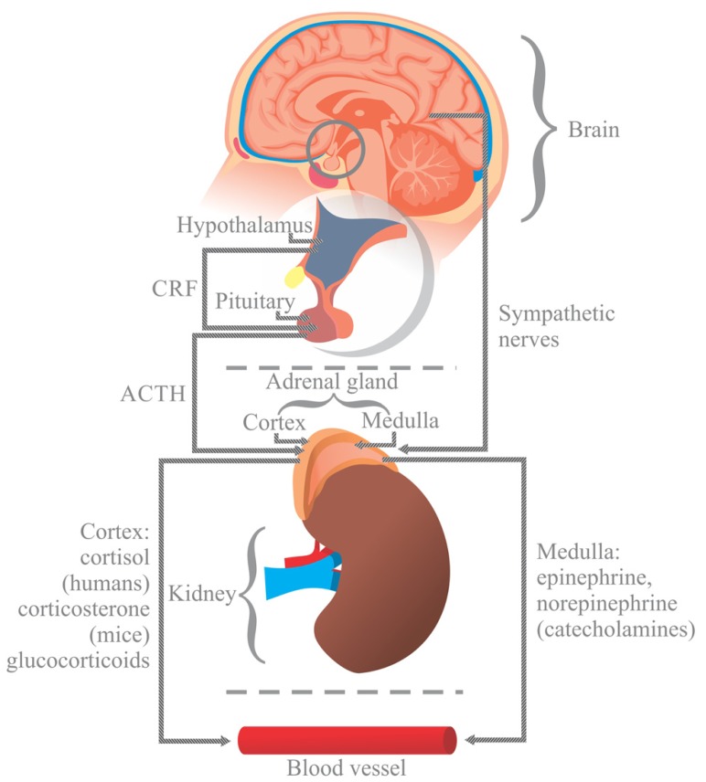
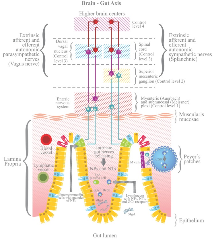
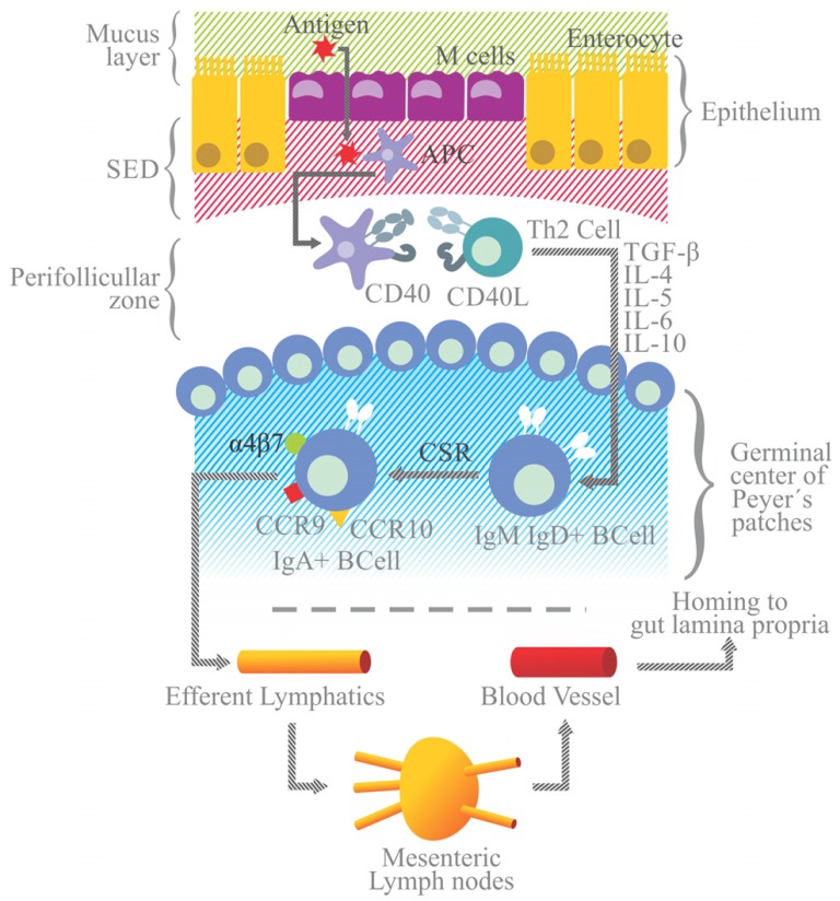
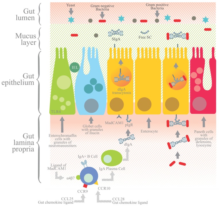
Stress is a response of the central nervous system to environmental stimuli perceived as a threat to homeostasis. The stress response triggers the generation of neurotransmitters and hormones from the hypothalamus pituitary adrenal axis, sympathetic axis and brain gut axis, and in this way modulates the intestinal immune system. The effects of psychological stress on intestinal immunity have been investigated mostly with the restraint/immobilization rodent model, resulting in an up or down modulation of SIgA levels depending on the intensity and time of exposure to stress. SIgA is a protein complex formed by dimeric (dIgA) or polymeric IgA (pIgA) and the secretory component (SC), a peptide derived from the polymeric immunoglobulin receptor (pIgR). The latter receptor is a transmembrane protein expressed on the basolateral side of gut epithelial cells, where it uptakes dIgA or pIgA released by plasma cells in the lamina propria. As a result, the IgA-pIgR complex is formed and transported by vesicles to the apical side of epithelial cells. pIgR is then cleaved to release SIgA into the luminal secretions of gut. Down modulation of SIgA associated with stress can have negative repercussions on intestinal function and integrity. This can take the form of increased adhesion of pathogenic agents to the intestinal epithelium and/or an altered balance of inflammation leading to greater intestinal permeability. Most studies on the molecular and biochemical mechanisms involved in the stress response have focused on systemic immunity. The present review analyzes the impact of stress (mostly by restraint/immobilization, but also with mention of other models) on the generation of SIgA, pIgR and other humoral and cellular components involved in the intestinal immune response. Insights into these mechanisms could lead to better therapies for protecting against pathogenic agents and avoiding epithelial tissue damage by modulating intestinal inflammation.
Overview
The claims and statistics of this paper haven’t been checked yet.
Run the check — free, no account needed. It reads the open-access text and checks the sources it cites.
Integrity signals
Registry
Cites
Statistics
Figures
Verification
Claims the paper makes
The claims and statistics of this paper haven’t been checked yet.
Run the check — free, no account needed. It reads the open-access text and checks the sources it cites.
Claims other papers make about this one
Each is checked against this paper’s own text: does it say what the citing paper says it does?
The source backs part of the claim, not all of it — or backs it for a different population, condition or comparison.
In the citing paper
“Like cortisol, IgA fluctuations can indicate positive and negative welfare states (105, 106) and be measured non-invasively.”
From Comparison of animal welfare assessment tools and methodologies: need for an effective approach for captive elephants in Asia (2024) · cites this paper as [106]
Experimental — accuracy not yet measured
Self-check
Percentages, sums and reported differences re-computed from the paper’s own numbers.
The self-check needs the paper’s full text.
Citations
Cites
1 retracted
Zhang, Y., Zhang, Y., Miao, J., Hanley, G., Stuart, C. A., Sun, X., Chen, T., & Yin, D. (2008) · Journal of Neuroimmunology · doi:10.1016/j.jneuroim.2008.08.011 · Cited by 58
RetractedPathways of Antigen Processing
Blum, J. S., Wearsch, P. A., & Cresswell, P. (2013) · Annual Review of Immunology · doi:10.1146/annurev-immunol-032712-095910 · Cited by 1,635
Dhabhar, F. S., & McEwen, B. S. (1997) · Brain Behavior and Immunity · doi:10.1006/brbi.1997.0508 · Cited by 1,014
Dhabhar, F. S. (2009) · Neuroimmunomodulation · doi:10.1159/000216188 · Cited by 903
The regulation of IgA class switching
Cerutti, A. (2008) · Nature reviews. Immunology · doi:10.1038/nri2322 · Cited by 777
Aberrant expansion of segmented filamentous bacteria in IgA-deficient gut
Suzuki, K., Meek, B., Doi, Y., Muramatsu, M., Chiba, T., Honjo, T., & Fagarasan, S. (2004) · Proceedings of the National Academy of Sciences · doi:10.1073/pnas.0307317101 · Cited by 721
Mucosal Immunity: Induction, Dissemination, and Effector Functions
Brandtzaeg, P. (2009) · Scandinavian Journal of Immunology · doi:10.1111/j.1365-3083.2009.02319.x · Cited by 483
PPARγ as a new therapeutic target in inflammatory bowel diseases
Dubuquoy, L., Rousseaux, C., Thuru, X., Peyrin-Biroulet, L., Romano, O., Chavatte, P., Chamaillard, M., & Desreumaux, P. (2006) · Gut · doi:10.1136/gut.2006.093484 · Cited by 450
Johansen, F.-E., & Kaetzel, C. S. (2011) · Mucosal Immunology · doi:10.1038/mi.2011.37 · Cited by 368
Schmidt, S. U., Rainer, J., Ploner, C., Presul, E., Riml, S., & Kofler, R. (2004) · Cell Death and Differentiation · doi:10.1038/sj.cdd.4401456 · Cited by 364
Cited by
Sorted by citations
Stress and the Microbiota–Gut–Brain Axis in Visceral Pain: Relevance to Irritable Bowel Syndrome
Moloney, R. D., Johnson, A. C., O’Mahony, S. M., Dinan, T. G., Meerveld, B. G., & Cryan, J. F. (2015) · CNS Neuroscience & Therapeutics · doi:10.1111/cns.12490 · Cited by 412
Environmental risk factors and cardiovascular diseases: a comprehensive expert review
Münzel, T. F., Hahad, O., Sørensen, M., Lelieveld, J., Duerr, G. D., Nieuwenhuijsen, M. J., & Daiber, A. (2021) · Cardiovascular Research · doi:10.1093/cvr/cvab316 · Cited by 404
Glutamatergic Signaling Along The Microbiota-Gut-Brain Axis
Baj, A., Moro, E., Bistoletti, M., Orlandi, V. T., Crema, F., & Giaroni, C. (2019) · International Journal of Molecular Sciences · doi:10.3390/ijms20061482 · Cited by 339
The microbiota–gut–brain axis in gastrointestinal disorders: stressed bugs, stressed brain or both?
Palma, G. D., Collins, S. M., Berčík, P., & Verdu, E. F. (2014) · The Journal of Physiology · doi:10.1113/jphysiol.2014.273995 · Cited by 309
Stull, V. J., Finer, E., Bergmans, R. S., Febvre, H. P., Longhurst, C. A., Manter, D. K., Patz, J. A., & Weir, T. L. (2018) · Scientific Reports · doi:10.1038/s41598-018-29032-2 · Cited by 269
Metadata
- Authors
- Campos-Rodríguez, R., Godínez-Victoria, M., Abarca-Rojano, E., Pacheco-Yépez, J., Reyna-Garfias, H., Barbosa-Cabrera, R. E., & Drago-Serrano, M. E.
- Affiliations
- Instituto Politécnico Nacional (MX)
- Venue
- Frontiers in Integrative Neuroscience
- Publisher
- Frontiers Media SA
- Published
- 2013
- Volume
- 7
- Pages
- 86-86
- ISSN
- 1662-5145 (ISSN-L)
- PMC
- PMC3845795
- OpenAlex
- W2168128529
- Cited by
- 119
- References
- 76
- Citations by year
- 2026: 9 · 2025: 9 · 2024: 15 · 2023: 11 · 2022: 8 · 2021: 15 · 2020: 11 · 2019: 7 · 2018: 6 · 2017: 5 · 2016: 6 · 2015: 11 · 2014: 6
- FWCI
- 0.63
- Type
- journal article
- Language
- en
- Open access
- gold
- doi.org
- Licence
- cc-by
- Locations
- Frontiers in Integrative Neuroscience (publishedVersion, cc-by)
- Europe PMC (PubMed Central) (publishedVersion, cc-by)
- PubMed Central (publishedVersion, cc-by)
- PubMed (publishedVersion)
- DOAJ (DOAJ: Directory of Open Access Journals) (submittedVersion, cc-by-sa)
- Topics
- Life Sciences › Neuroscience › Behavioral Neuroscience › Stress Responses and Cortisol
- Health Sciences › Medicine › Gastroenterology › Gastrointestinal motility and disorders
- Life Sciences › Neuroscience › Cellular and Molecular Neuroscience › Neuropeptides and Animal Physiology
- Keywords
- psychological stress, secretory IgA, intestinal immunity, polymeric immunoglobulin receptor, polymeric IgA, dimeric IgA, secretory component, hypothalamic-pituitary-adrenal axis, gut-brain axis, restraint stress, intestinal permeability, intestinal inflammation, intestinal epithelium, pathogen adhesion
About these checks
Registry, retraction, Cites and statistics results are computed, not judged, with one exception: a printed reference that no index lists is read once by a model before it counts as a citation error. Claims are extracted by a model and kept only when their quote appears verbatim in the text; each cited source is then checked, with a model’s help, for whether it says what the claim relies on. Those verdicts can be wrong — read the claim’s quote and the cited source before relying on one.
Statistics and arithmetic are recomputed, not judged.
Confidence bands are shown only for fields calibrated on our annotated test set. None is calibrated yet, so no band is shown.
What counts as a claim ›How we measure confidence ›How we check this ›
Are you an author, or do you see a problem with this page? Request a review
