

The DOI matches its Crossref record; 2 of the 2 references it cites were found in a registry or an index.
Beyond Colonoscopy: Exploring New Cell Surface Biomarkers for Detection of Early, Heterogenous Colorectal Lesions
Abstract
Colorectal cancer (CRC) is the third leading cause of cancer-related deaths among both men and women in the United States. Early detection and surgical removal of high-risk lesions in the colon can prevent disease from developing and spreading. Despite implementation of programs aimed at early detection, screening colonoscopies fail to detect a fraction of potentially aggressive colorectal lesions because of their location or nonobvious morphology. Optical colonoscopies, while highly effective, rely on direct visualization to detect changes on the surface mucosa that are consistent with dysplasia. Recent advances in endoscopy techniques and molecular imaging permit microscale visualization of the colonic mucosa. These technologies can be combined with various molecular probes that recognize and target heterogenous lesion surfaces to achieve early, real-time, and potentially non-invasive, detection of pre-cancerous lesions. The primary goal of this review is to contextualize existing and emergent CRC surface biomarkers and assess each’s potential as a candidate marker for early marker-based detection of CRC lesions. CRC markers that we include were stratified by the level of support gleaned from peer-reviewed publications, abstracts, and databases of both CRC and other cancers. The selected biomarkers, accessible on the cell surface and preferably on the luminal surface of the colon tissue, are organized into three categories: (1) established biomarkers (those with considerable data and high confidence), (2) emerging biomarkers (those with increasing research interest but with less supporting data), and (3) novel candidates (those with very recent data, and/or supportive evidence from other tissue systems). We also present an overview of recent advances in imaging techniques useful for visual detection of surface biomarkers, and discuss the ease with which these methods can be combined with microscopic visualization.
Overview
The claims and statistics of this paper haven’t been checked yet.
Run the check — free, no account needed. It reads the open-access text and checks the sources it cites.
Integrity signals
Registry
Cites
Statistics
Figures
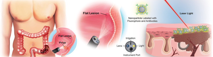
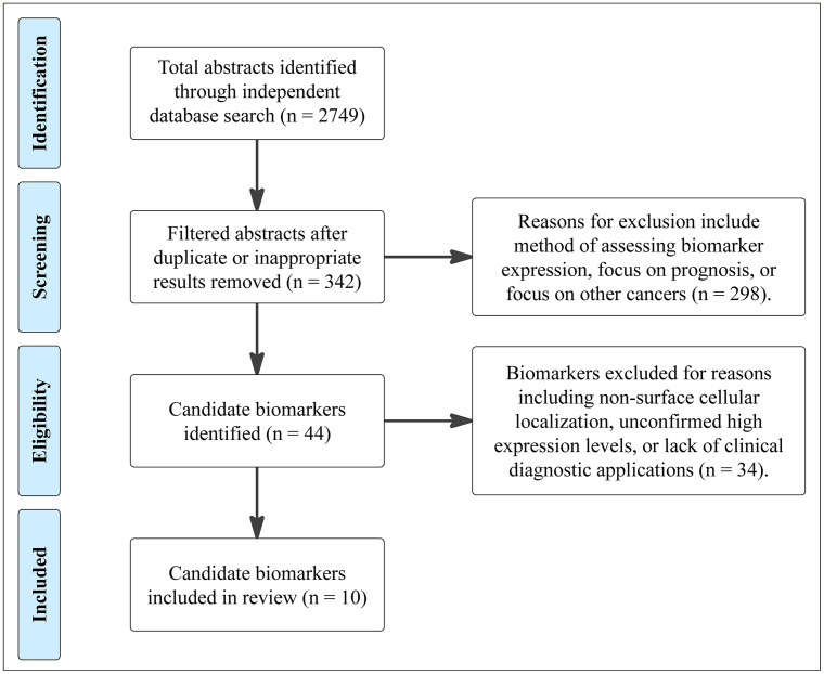
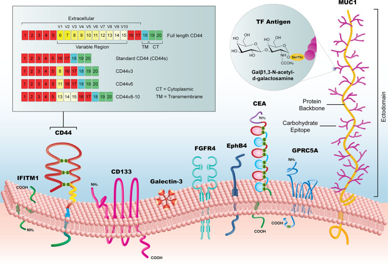
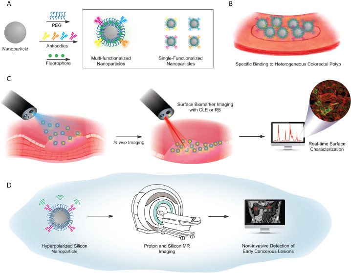
Verification
Claims the paper makes
The claims and statistics of this paper haven’t been checked yet.
Run the check — free, no account needed. It reads the open-access text and checks the sources it cites.
Claims other papers make about this one
Each is checked against this paper’s own text: does it say what the citing paper says it does?
The check wasn’t confident either way, so it gives no verdict — never disputed.
In the citing paper
“However, ASGPR expression in CRC is heterogeneous within CRC lesions [94]. Therefore, while Gal enhances uptake in some CRC models, its broader applicability may be limited.”
From Chitosan-Based Drug Delivery Systems for Targeted Chemotherapy in Colorectal Cancer: A Scoping Review (2025) · cites this paper as [94]
Experimental — accuracy not yet measured
Self-check
Percentages, sums and reported differences re-computed from the paper’s own numbers.
The self-check needs the paper’s full text.
Citations
Cites
Comprehensive review of targeted therapy for colorectal cancer
Xie, Y.-H., Chen, Y.-X., & Fang, J.-Y. (2020) · Signal Transduction and Targeted Therapy · doi:10.1038/s41392-020-0116-z · Cited by 1,687
Turkington, R. C., Longley, D. B., Allen, W. L., Stevenson, L., McLaughlin, K., Dunne, P. D., Blayney, J. K., Salto-Tellez, M., Van Schaeybroeck, S., & Johnston, P. G. (2014) · Cell Death & Disease · doi:10.1038/cddis.2014.10 · Cited by 98
Research works printed but not linked: 148
- Siegel, 2020, Cancer Statistics, 2020, CA Cancer J Clin, 70, 7, 10.3322/caac.21590 · DOI · Paper page
- Bénard, 2018, Systematic Review of Colorectal Cancer Screening Guidelines for Average-Risk Adults: Summarizing the Current Global Recommendations, World J Gastroenterol, 24, 10.3748/wjg.v24.i1.124 · DOI
- Fass, 2020, Demographics Predict Stage III/IV Colorectal Cancer in Individuals Under Age 50, J Clin Gastroenterol, 54, 10.1097/MCG.0000000000001374 · DOI
- Zhao, 2019, Magnitude, Risk Factors, and Factors Associated With Adenoma Miss Rate of Tandem Colonoscopy: A Systematic Review and Meta-Analysis, Gastroenterology, 156, 10.1053/j.gastro.2019.01.260 · DOI · Paper page
- D’Andrea, 2020, Quantifying the Impact of Adherence to Screening Strategies on Colorectal Cancer Incidence and Mortality, Cancer Med, 9, 10.1002/cam4.2735 · DOI
- Roth, 2019, Clinical and Budget Impact of Increasing Colorectal Cancer Screening by Blood- and Stool-Based Testing, Am Health Drug Benefits, 12
- Bynum, 2012, Unwillingness to Participate in Colorectal Cancer Screening: Examining Fears, Attitudes, and Medical Mistrust in an Ethnically Diverse Sample of Adults 50 Years and Older, Am J Health Promot AJHP, 26, 295, 10.4278/ajhp.110113-QUAN-20 · DOI
- Young, 2013, Colonoscopy for Colorectal Cancer Screening, J Cancer, 4, 10.7150/jca.5829 · DOI
- Meklin, 2020, Fecal Occult Blood Tests in Colorectal Cancer Screening: Systematic Review and Meta-Analysis of Traditional and New-Generation Fecal Immunochemical Tests, Anticancer Res, 40, 10.21873/anticanres.14349 · DOI
- Lynch, 2018, Phenotypic and Genotypic Heterogeneity of Lynch Syndrome: A Complex Diagnostic Challenge, Fam Cancer, 17, 10.1007/s10689-017-0053-3 · DOI
- Lee, 2015, Sessile Serrated Adenoma/Polyps With a Depressed Surface: A Rare Form of Sessile Serrated Adenoma/Polyp, Diagn Pathol, 10, 1, 10.1186/s13000-015-0325-x · DOI
- Pohl, 2010, Colorectal Cancers Detected After Colonoscopy Frequently Result From Missed Lesions, Clin Gastroenterol Hepatol, 8, 10.1016/j.cgh.2010.06.028 · DOI
- Kashida, 2006, Colorectal Cancer: The Importance of Depressed Lesions in the Development of Colorectal Cancer, 10.1007/4-431-30173-9_21 · DOI
- Soetikno, 2008, Prevalence of Nonpolypoid (Flat and Depressed) Colorectal Neoplasms in Asymptomatic and Symptomatic Adults, JAMA, 299, 10.1001/jama.299.9.1027 · DOI
- Pellino, 2018, Noninvasive Biomarkers of Colorectal Cancer: Role in Diagnosis and Personalised Treatment Perspectives. Passardi A, editor, Gastroenterol Res Pract, 2018, 1, 10.1155/2018/2397863 · DOI
- Toma, 2018, Colorectal Cancer Biomarkers - A New Trend in Early Diagnosis, Curr Health Sci J, 2, 10.12865/CHSJ.44.02.08 · DOI
- Gonzalez-Pons, 2015, Colorectal Cancer Biomarkers: Where Are We Now? Scartozzi M, editor, BioMed Res Int, 2015, 1, 10.1155/2015/149014 · DOI
- Vacante, 2018, Biomarkers in Colorectal Cancer: Current Clinical Utility and Future Perspectives, World J Clin Cases, 6, 10.12998/wjcc.v6.i15.869 · DOI
- Kiesslich, 2007, Technology Insight: Confocal Laser Endoscopy for In Vivo Diagnosis of Colorectal Cancer, Nat Clin Pract Oncol, 4, 10.1038/ncponc0881 · DOI
- Hsiung, 2008, Detection of Colonic Dysplasia In Vivo Using a Targeted Fluorescent Septapeptide and Confocal Microendoscopy, Nat Med, 14, 10.1038/nm1692 · DOI
- Alves Martins, 2019, Biomarkers in Colorectal Cancer: The Role of Translational Proteomics Research, Front Oncol, 9, 1, 10.3389/fonc.2019.01284 · DOI
- Sveen, 2020, Biomarker-Guided Therapy for Colorectal Cancer: Strength in Complexity, Nat Rev Clin Oncol, 17, 11, 10.1038/s41571-019-0241-1 · DOI · Paper page
- de Castro-Carpeño, 2008, EGFR and Colon Cancer: A Clinical View, Clin Transl Oncol, 10, 6, 10.1007/s12094-008-0147-3 · DOI
- Ellis, 2000, Vascular Endothelial Growth Factor in Human Colon Cancer: Biology and Therapeutic Implications, Oncologist, 5, 10.1634/theoncologist.5-suppl_1-11 · DOI
- Parizadeh, 2019, The Potential Therapeutic and Prognostic Impacts of the c-MET/HGF Signaling Pathway in Colorectal Cancer, IUBMB Life, 71, 10.1002/iub.2063 · DOI
- Jothy, 1993, Transcription of Carcinoembryonic Antigen in Normal Colon and Colon Carcinoma. In Situ Hybridization Study and Implication for a New In Vivo Functional Model, Am J Pathol, 143
- Tiernan, 2013, Carcinoembryonic Antigen Is the Preferred Biomarker for In Vivo Colorectal Cancer Targeting, Br J Cancer, 108, 10.1038/bjc.2012.605 · DOI
- Coco, 2012, Increased Expression of CD133 and Reduced Dystroglycan Expression Are Strong Predictors of Poor Outcome in Colon Cancer Patients, J Exp Clin Cancer Res, 31, 1, 10.1186/1756-9966-31-71 · DOI
- Choi, 2009, Cancer Stem Cell Markers CD133 and CD24 Correlate With Invasiveness and Differentiation in Colorectal Adenocarcinoma, World J Gastroenterol WJG, 15, 10.3748/wjg.15.2258 · DOI
- Kazama, 2018, Expression of the Stem Cell Marker CD133 Is Related to Tumor Development in Colorectal Carcinogenesis, Asian J Surg, 41, 10.1016/j.asjsur.2016.12.002 · DOI
- Wang, 2015, The Expression and Significance of Gal-3 and MUC1 in Colorectal Cancer and Colon Cancer, OncoTargets Ther, 8, 10.2147/OTT.S83502 · DOI
- Betge, 2016, Muc1, MUC2, MUC5AC, and MUC6 in Colorectal Cancer: Expression Profiles and Clinical Significance, Virchows Arch Int J Pathol, 469, 10.1007/s00428-016-1970-5 · DOI
- Manhas, 2016, Characterization of Cancer Stem Cells From Different Grades of Human Colorectal Cancer, Tumor Biol, 37, 10.1007/s13277-016-5232-6 · DOI
- Gorham, 1996, Cellular Distribution of CD44 Gene Transcripts in Colorectal Carcinomas and in Normal Colonic Mucosa, J Clin Pathol, 49, 10.1136/jcp.49.6.482 · DOI
- Ismaiel, 2016, Detection of Cancer Stem Cells in Colorectal Cancer: Histopathological and Immunohistochemical Study, Open Access Maced J Med Sci, 4, 10.3889/oamjms.2016.126 · DOI
- Al-Magrabi, 2012, Decreased Immunoexpression of Standard Form of CD44 Is An Independent Favourable Predictor of Nodal Metastasis in Colorectal Carcinoma, Anticancer Res, 32
- Kuniyasu, 2001, Heparan Sulfate Enhances Invasion by Human Colon Carcinoma Cell Lines Through Expression of CD44 Variant Exon 3, Clin Cancer Res, 7
- Afify, 2016, The Expression of CD44v6 in Colon: From Normal to Malignant, Ann Diagn Pathol, 20, 19, 10.1016/j.anndiagpath.2015.10.010 · DOI
- Afify, 2007, Diagnostic Utility of CD44 Standard, CD44v6, and CD44v3-10 Expression in Adenocarcinomas Presenting in Serous Fluid, Appl Immunohistochem Mol Morph, 15, 10.1097/01.pai.0000213154.49063.22 · DOI
- Tao, 2017, Galectin-3 Expression in Colorectal Cancer and Its Correlation With Clinical Pathological Characteristics and Prognosis, Open Med Wars, 12, 10.1515/med-2017-0032 · DOI
- Lu, 2017, Posttranscriptional Regulation of Galectin-3 by miR-129 Contributes to Colorectal Cancer Progression, Oncotarget, 8, 10.18632/oncotarget.14839 · DOI
- Gopalan, 2015, The Expression Profiles of the Galectin Gene Family in Colorectal Adenocarcinomas, Hum Pathol, 53, 10.1016/j.humpath.2016.02.016 · DOI
- Yu, 2015, IFITM1 Promotes the Metastasis of Human Colorectal Cancer Via CAV-1, Cancer Lett, 368, 10.1016/j.canlet.2015.07.034 · DOI
- Schneider, 2001, Overexpression of Sialyltransferase Cmp-Sialic Acid:Galβ1,3galNAc-R α6-Sialyltransferase Is Related to Poor Patient Survival Inhuman Colorectal Carcinomas, Cancer Res, 61
- El-Sayed, 2011, Immunodiagnostic Potential of Mucin (MUC2) and Thomsen-Friedenreich (TF) Antigens in Egyptian Patients With Colorectal Cancer, Eur Rev Med Pharmacol Sci, 15
- Zougman, 2013, Retinoic Acid-Induced Protein 3: Identification and Characterisation of a Novel Prognostic Colon Cancer Biomarker, Eur J Cancer, 49, 10.1016/j.ejca.2012.07.031 · DOI
- Stammes, 2017, Evaluation of EphA2 and EphB4 as Targets for Image-Guided Colorectal Cancer Surgery, Int J Mol Sci, 18, 1, 10.3390/ijms18020307 · DOI
- Lv, 2016, EphB4 Promotes the Proliferation, Invasion, and Angiogenesis of Human Colorectal Cancer, Exp Mol Pathol, 100, 10.1016/j.yexmp.2016.03.011 · DOI
- Ahmed, 2016, Fibroblast Growth Factor Receptor 4 Induced Resistance to Radiation Therapy in Colorectal Cancer, Oncotarget, 7, 10.18632/oncotarget.12099 · DOI
- Beauchemin, 2013, Carcinoembryonic Antigen-Related Cell Adhesion Molecules (Ceacams) in Cancer Progression and Metastasis, Cancer Metastasis Rev, 32, 10.1007/s10555-013-9444-6 · DOI
- Lee, 2017, The Roles of Carcinoembryonic Antigen in Liver Metastasis and Therapeutic Approaches. Beaulieu J-F, editor, Gastroenterol Res Pract, 2017, 1, 10.1155/2017/7521987 · DOI
- Nap, 1988, Immunohistochemistry of Carcino-Embryonic Antigen in the Embryo, Fetus and Adult, Tumour Biol J Int Soc Oncodev Biol Med, 9, 10.1159/000217555 · DOI
- Ramos-Gomes, 2018, Single- and Two-Photon Imaging of Human Micrometastases and Disseminated Tumour Cells With Conjugates of Nanobodies and Quantum Dots, Sci Rep, 8, 4595, 10.1038/s41598-018-22973-8 · DOI
- Park, 1999, Prognostic Factors in 2230 Korean Colorectal Cancer Patients: Analysis of Consecutively Operated Cases, World J Surg, 23, 10.1007/PL00012376 · DOI
- Locker, 2006, ASCO 2006 Update of Recommendations for the Use of Tumor Markers in Gastrointestinal Cancer, J Clin Oncol Off J Am Soc Clin Oncol, 24, 10.1200/JCO.2006.08.2644 · DOI
- Thirunavukarasu, 2011, C-Stage in Colon Cancer: Implications of Carcinoembryonic Antigen Biomarker in Staging, Prognosis, and Management, J Natl Cancer Inst, 103, 10.1093/jnci/djr078 · DOI
- Shmelkov, 2008, CD133 Expression Is Not Restricted to Stem Cells, and Both CD133+ and CD133- Metastatic Colon Cancer Cells Initiate Tumors, J Clin Invest, 118, 10.1172/JCI34401 · DOI
- Glumac, 2018, The Role of CD133 in Cancer: A Concise Review, Clin Transl Med, 7, 1, 10.1186/s40169-018-0198-1 · DOI
- Zheng, 2015, Physiologic Hypoxia and Oxygen Homeostasis in the Healthy Intestine. A Review in the Theme: Cellular Responses to Hypoxia, Am J Physiol-Cell Physiol, 309, 10.1152/ajpcell.00191.2015 · DOI
- Abbasian, 2018, The Most Reliable Surface Marker for the Identification of Colorectal Cancer Stem-Like Cells: A Systematic Review and Meta-Analysis, J Cell Physiol, 234, 10.1002/jcp.27619 · DOI
- Yeung, 2009, Colorectal Cancer Stem Cells, Colon Rectum, 52, 10.1007/DCR.0b013e3181a8738c · DOI
- Turano, 2018, Characterisation of Mesenchymal Colon Tumor-Derived Cells in Tumourspheres as a Model for Colorectal Cancer Progression, Int J Oncol, 53, 10.3892/ijo.2018.4565 · DOI
- Corbeil, 2000, The Human Ac133 Hematopoietic Stem Cell Antigen Is Also Expressed in Epithelial Cells and Targeted to Plasma Membrane Protrusions, J Biol Chem, 275, 10.1074/jbc.275.8.5512 · DOI
- Huang, 2018, CD133 Expression Correlates With Clinicopathologic Features and Poor Prognosis of Colorectal Cancer Patients: An Updated Meta-Analysis of 37 Studies, Med (Baltimore), 97, 1, 10.1097/MD.0000000000010446 · DOI
- Biemer-Hüttmann, 1999, Immunohistochemical Staining Patterns of MUC1, Muc2, MUC4, and MUC5AC Mucins in Hyperplastic Polyps, Serrated Adenomas, and Traditional Adenomas of the Colorectum, J Histochem Cytochem Off J Histochem Soc, 47, 10.1177/002215549904700808 · DOI
- Sewda, 2016, Cell-Surface Markers for Colon Adenoma and Adenocarcinoma, Oncotarget, 7, 10.18632/oncotarget.7402 · DOI
- Nath, 2014, Muc1: A Multifaceted Oncoprotein With a Key Role in Cancer Progression, Trends Mol Med, 20, 10.1016/j.molmed.2014.02.007 · DOI
- Brayman, 2004, Muc1: A Multifunctional Cell Surface Component of Reproductive Tissue Epithelia, Reprod Biol Endocrinol RBE, 2, 1, 10.1186/1477-7827-2-4 · DOI
- Li, 2019, Prognostic and Clinicopathological Value of MUC1 Expression in Colorectal Cancer: A Meta-Analysis, Med (Baltimore), 98, e14659, 10.1097/MD.0000000000014659 · DOI
- Skandalis, 2019, Hyaluronan-CD44 Axis Orchestrates Cancer Stem Cell Functions, Cell Signal, 63, 109377, 10.1016/j.cellsig.2019.109377 · DOI
- Mishra, 2019, Structure, Function and Role of CD44 in Neoplasia, J Oral Maxillofac Pathol JOMFP, 23, 10.4103/jomfp.JOMFP_246_18 · DOI
- Senbanjo, 2017, Cd44: A Multifunctional Cell Surface Adhesion Receptor Is a Regulator of Progression and Metastasis of Cancer Cells, Front Cell Dev Biol, 5, 10.3389/fcell.2017.00018 · DOI
- Spinelli, 2020, Hyaluronan in the Tumor Microenvironment, Adv Exp Med Biol, 1245, 67, 10.1007/978-3-030-40146-7_3 · DOI
- Liu, 2019, Dissecting the Dual Nature of Hyaluronan in the Tumor Microenvironment, Front Immunol, 10, 10.3389/fimmu.2019.00947 · DOI
- Bartolazzi, 1995, Regulation of Growth and Dissemination of a Human Lymphoma by CD44 Splice Variants, J Cell Sci, 108, 10.1242/jcs.108.4.1723 · DOI
- Goodison, 1999, CD44 Cell Adhesion Molecules, Mol Pathol, 52, 10.1136/mp.52.4.189 · DOI
- Nagano, 2013, Redox Regulation in Stem-Like Cancer Cells by CD44 Variant Isoforms, Oncogene, 32, 10.1038/onc.2012.638 · DOI
- Liu, 2005, Expression and Significance of CD44s, Cd44v6, and nm23 mRNA in Human Cancer, World J Gastroenterol WJG, 11, 10.3748/wjg.v11.i42.6601 · DOI
- Tanabe, 1995, Restoration of CD44H Expression in Colon Carcinomas Reduces Tumorigenicity, Ann Surg, 222, 493, 10.1097/00000658-199522240-00007 · DOI
- Ma, 2019, CD44v6 Engages in Colorectal Cancer Progression, Cell Death Dis, 10, 1, 10.1038/s41419-018-1265-7 · DOI
- Bánky, 2012, Characteristics of CD44 Alternative Splice Pattern in the Course of Human Colorectal Adenocarcinoma Progression, Mol Cancer, 11, 1, 10.1186/1476-4598-11-83 · DOI
- Bourguignon, 1998, Cd44v3,8–10 Is Involved in Cytoskeleton-Mediated Tumor Cell Migration and Matrix Metalloproteinase (MMP-9) Association in Metastatic Breast Cancer Cells, J Cell Physiol, 176, 10.1002/(SICI)1097-4652(199807)176:1<206::AID-JCP22>3.0.CO;2-3 · DOI
- Kopp, 2009, Frequent Expression of the High Molecular, 673-Bp. CD44v3, v8-10 Variant in Colorectal Adenomas and Carcinomas, Int J Mol Med, 24, 10.3892/ijmm_00000279 · DOI
- Wang, 2018, Prognostic Role of Galectin-3 Expression in Patients With Solid Tumors: A Meta-Analysis of 36 Eligible Studies, Cancer Cell Int, 18, 1, 10.1186/s12935-018-0668-y · DOI
- Ilmer, 2016, Cell Surface Galectin-3 Defines a Subset of Chemoresistant Gastrointestinal Tumor-Initiating Cancer Cells With Heightened Stem Cell Characteristics, Cell Death Dis, 7, 1, 10.1038/cddis.2016.239 · DOI
- Chan, 2008, Meta-Analysis of Colorectal Cancer Gene Expression Profiling Studies Identifies Consistently Reported Candidate Biomarkers, Cancer Epidemiol Biomarkers Prev, 17, 10.1158/1055-9965.EPI-07-2615 · DOI
- Andreu, 2006, Identification of the IFITM Family as a New Molecular Marker in Human Colorectal Tumors, Cancer Res, 66, 10.1158/0008-5472.CAN-05-2731 · DOI
- Kim, 2008, Gene-Expression Profiling of Colonic Adenomas by Using Oligonucleotide Microarray, Surg Endosc, 23, 10.1007/s00384-008-0451-y · DOI
- He, 2012, Influences of the Interferon Induced Transmembrane Protein 1 on Proliferation, Invasion, and Metastasis of Colorectal SW480 Cell Lines, Chin Med J, 125, 10.3760/cma.j.issn.0366-6999.2012.03.02 · DOI
- Sari, 2016, Interferon-Induced Transmembrane Protein 1 (IFITM1) Is Required for Progression of Colorectal Cancer, Oncotarget, 7, 10.18632/oncotarget.13325 · DOI
- Kurtenkov, 2020, Profiling of Naturally Occurring Antibodies to the Thomsen-Friedenreich Antigen in Health and Cancer: The Diversity and Clinical Potential, BioMed Res Int, 2020, 9747040, 10.1155/2020/9747040 · DOI
- Sindrewicz, 2016, Interaction of the Oncofetal Thomsen-Friedenreich Antigen With Galectins in Cancer Progression and Metastasis, Front Oncol, 6, 10.3389/fonc.2016.00079 · DOI
- Henderson, 2011, Occurrence of the Human Tumor-Specific Antigen Structure Galβ1-3galnacα- (Thomsen-Friedenreich) and Related Structures on Gut Bacteria: Prevalence, Immunochemical Analysis and Structural Confirmation, Glycobiology, 21, 10.1093/glycob/cwr058 · DOI
- Croce, 2007, Lewis X Is Highly Expressed in Normal Tissues: A Comparative Immunohistochemical Study and Literature Revision, Pathol Oncol Res, 13, 10.1007/BF02893488 · DOI
- Sakuma, 2015, Fluorescence-Based Endocscopic Imaging of Thomsen-Friedenreich Antigen to Improve Early Detection of Colorectal Cancer, Int J Cancer, 136, 10.1002/ijc.29093 · DOI
- Liang, 2014, Lectin From Agrocybe Aegerita as a Glycophenotype Probe for Evaluation of Progression and Survival in Colorectal Cancer, Asian Pac J Cancer Prev APJCP, 15, 10.7314/APJCP.2014.15.14.5601 · DOI
- Jiang, 2018, Gprc5a: An Emerging Biomarker in Human Cancer, BioMed Res Int, 2018, e1823726, 10.1155/2018/1823726 · DOI
- Zhou, 2014, The Emerging Roles of GPRC5A in Diseases, Oncoscience, 1, 10.18632/oncoscience.104 · DOI
- Cheng, 1998, Molecular Cloning and Characterization of a Novel Retinoic Acid-Inducible Gene That Encodes a Putative G Protein-Coupled Receptor, J Biol Chem, 273, 10.1074/jbc.273.52.35008 · DOI
- Zhang, 2017, Elevation of GPRC5A Expression in Colorectal Cancer Promotes Tumor Progression Through VNN-1 Induced Oxidative Stress, Int J Cancer, 140, 10.1002/ijc.30698 · DOI
- Greenhough, 2018, Cancer Cell Adaptation to Hypoxia Involves a HIF-GPRC5A-YAP Axis, EMBO Mol Med, 10, 1, 10.15252/emmm.201708699 · DOI
- Kania, 2016, Mechanisms of Ephrin–Eph Signalling in Development, Physiology and Disease, Nat Rev Mol Cell Biol, 17, 10.1038/nrm.2015.16 · DOI
- Kumar, 2009, Preferential Induction of EphB4 Over EphB2 and Its Implication in Colorectal Cancer Progression, Cell Tumor Stem Cell Biol, 69, 10.1158/0008-5472.CAN-08-3232 · DOI
- Herath, 2010, The Role of Eph Receptors and Ephrin Ligands in Colorectal Cancer, Int J Cancer, 126, 10.1002/ijc.25147 · DOI
- Kadife, 2018, Effects of EphB4 Receptor Expression on Colorectal Cancer Cells, Tumor Growth, Vascularization and Composition, Acta Oncol, 57, 10.1080/0284186X.2018.1429650 · DOI
- Wang, 2018, Upregulation of Fibroblast Growth Factor 19 Is Associated With the Initiation of Colorectal Adenoma, Dig Disease, 37, 10.1159/000494454 · DOI
- Pelaez-Garcio, 2013, Fgfr4 Role in Epithelial-Mesenchymal Transition and Its Therapeutic Value in Colorectal Cancer, PloS One, 8, 1, 10.1371/journal.pone.0063695 · DOI
- Barderas, 2012, An Optimized Predictor Panel for Colorectal Cancer Diagnosis Based on the Combination of Tumor-Associated Antigens Obtained From Protein and Phage Microarrays, J Proteomics, 75, 10.1016/j.jprot.2012.03.004 · DOI
- Liu, 2018, Forkhead Box C1 Promotes Colorectal Cancer Metastasis Through Transactivating ITGA7 and FGFR4 Expression, Oncogene, 37, 10.1038/s41388-018-0355-4 · DOI
- Soetikno, 2006, Nonpolypoid (Flat and Depressed) Colorectal Neoplasms, Gastroenterology, 130, 10.1053/j.gastro.2005.12.006 · DOI
- Gonzalo, 2013, Gene Expression Profiling of Serrated Polyps Identifies Annexin A10 as a Marker of a Sessile Serrated Adenoma/Polyp, J Pathol, 230, 10.1002/path.4200 · DOI
- O’Brien, 2006, Comparison of Microsatellite Instability, CpG Island Methylation Phenotype, BRAF and KRAS Status in Serrated Polyps and Traditional Adenomas Indicates Separate Pathways to Distinct Colorectal Carcinoma End Points, Am J Surg Pathol, 30, 10.1097/01.pas.0000213313.36306.85 · DOI
- Spring, 2006, High Prevalence of Sessile Serrated Adenomas With BRAF Mutations: A Prospective Study of Patients Undergoing Colonoscopy, Gastroenterology, 131, 10.1053/j.gastro.2006.08.038 · DOI
- Mirsaeidi, 2016, Annexins Family: Insights Into Their Functions and Potential Role in Pathogenesis of Sarcoidosis, J Transl Med, 14, 89, 10.1186/s12967-016-0843-7 · DOI
- Duncan, 2008, Characterisation and Protein Expression Profiling of Annexins in Colorectal Cancer, Br J Cancer, 98, 10.1038/sj.bjc.6604128 · DOI
- Lu, 2013, Annexin A10 Is an Immunohistochemical Marker for Adenocarcinoma of the Upper Gastrointestinal Tract and Pancreatobiliary System, Histopathology, 63, 10.1111/his.12229 · DOI
- Tsai, 2015, Aberrant Expression of Annexin A10 Is Closely Related to Gastric Phenotype in Serrated Pathway to Colorectal Carcinoma, Mod Pathol, 28, 10.1038/modpathol.2014.96 · DOI
- Sajanti, 2015, Annexin A10 Is a Marker for the Serrated Pathway of Colorectal Carcinoma, Virchows Arch, 466, 5, 10.1007/s00428-014-1683-6 · DOI
- Kim, 2014, Annexin A10 Expression Correlates With Serrated Pathway Features in Colorectal Carcinoma With Microsatellite Instability, APMIS, 122, 10.1111/apm.12284 · DOI
- Mohammadi, 2013, The Stem Cell Marker CD133 Is Highly Expressed in Sessile Serrated Adenoma and Its Borderline Variant Compared With Hyperplastic Polyp, J Clin Pathol, 66, 10.1136/jclinpath-2012-201192 · DOI
- Vázquez-González, 2021, Aptamer-Functionalized Micro- and, ACS Appl Mater Interfaces, 13, 10.1021/acsami.0c17121 · DOI
- Fugazza, 2016, Confocal Laser Endomicroscopy in Gastrointestinal and Pancreatobiliary Diseases: A Systematic Review and Meta-Analysis, BioMed Res Int, 2016, 4638683, 10.1155/2016/4638683 · DOI
- Pech, 2008, Confocal Laser Endomicroscopy for In Vivo Diagnosis of Early Squamous Cell Carcinoma in the Esophagus, Clin Gastroenterol Hepatol Off Clin Pract J Am Gastroenterol Assoc, 6, 89, 10.1016/j.cgh.2007.10.01 · DOI
- Li, 2011, Diagnostic Value of Confocal Laser Endomicroscopy for Gastric Superficial Cancerous Lesions, Gut, 60, 299, 10.1136/gut.2010.223586 · DOI
- Leong, 2008, In Vivo Confocal Endomicroscopy in the Diagnosis and Evaluation of Celiac Disease, Gastroenterology, 135, 10.1053/j.gastro.2008.08.054 · DOI
- Kiesslich, 2004, Confocal Laser Endoscopy for Diagnosing Intraepithelial Neoplasias and Colorectal Cancer In Vivo, Gastroenterology, 127, 10.1053/j.gastro.2004.06.050 · DOI
- Sanduleanu, 2010, In Vivo Diagnosis and Classification of Colorectal Neoplasia by Chromoendoscopy-Guided Confocal Laser Endomicroscopy, Clin Gastroenterol Hepatol Off Clin Pract J Am Gastroenterol Assoc, 8, 10.1016/j.cgh.2009.08.006 · DOI
- Wang, 2016, Fiber-Optic Raman Spectroscopy for In Vivo Diagnosis of Gastric Dysplasia, Faraday Discuss, 187, 10.1039/C5FD00151J · DOI
- Bergholt, 2016, Simultaneous Fingerprint and High-Wavenumber Fiber-Optic Raman Spectroscopy Enhances Real-Time In Vivo Diagnosis of Adenomatous Polyps During Colonoscopy, J Biophotonics, 9, 10.1002/jbio.201400141 · DOI
- Almond, 2014, Endoscopic Raman Spectroscopy Enables Objective Diagnosis of Dysplasia in Barrett’s Esophagus, Gastrointest Endosc, 79, 37, 10.1016/j.gie.2013.05.028 · DOI
- Zavaleta, 2009, Multiplexed Imaging of Surface Enhanced Raman Scattering Nanotags in Living Mice Using Noninvasive Raman Spectroscopy, Proc Natl Acad Sci, 106, 10.1073/pnas.0813327106 · DOI · Paper page
- Bohndiek, 2013, A Small Animal Raman Instrument for Rapid, Wide-Area, Spectroscopic Imaging, Proc Natl Acad Sci, 110, 10.1073/pnas.1301379110 · DOI · Paper page
- McVeigh, 2013, Widefield Quantitative Multiplex Surface Enhanced Raman Scattering Imaging In Vivo, J BioMed Opt, 18, 046011, 10.1117/1.JBO.18.4.046011 · DOI
- Garai, 2013, High-Sensitivity, Real-Time, Ratiometric Imaging of Surface-Enhanced Raman Scattering Nanoparticles With a Clinically Translatable Raman Endoscope Device, J BioMed Opt, 18, 096008, 10.1117/1.JBO.18.9.096008 · DOI
- Zavaleta, 2013, A Raman-based Endoscopic Strategy for Multiplexed Molecular Imaging, Proc Natl Acad Sci, 110, 10.1073/pnas.1211309110 · DOI · Paper page
- Ji, 2003, Multislice CT Colonography: Current Status and Limitations, Eur J Radiol, 47, 10.1016/S0720-048X(03)00169-4 · DOI
- Pineau, 2003, Virtual Colonoscopy Using Oral Contrast Compared With Colonoscopy for the Detection of Patients With Colorectal Polyps, Gastroenterology, 125, 10.1016/S0016-5085(03)00885-0 · DOI
- van der Paardt, 2010, MRI of the Colon, Imaging Med, 2, 195, 10.2217/iim.10.12 · DOI
- Hu, 2018, Real-Time Molecular MRI With Hyperpolarized Silicon Particles, 10.1007/978-3-662-56333-5_3 · DOI
- Cassidy, 2013, In Vivo Magnetic Resonance Imaging of Hyperpolarized Silicon Particles, Nat Nanotechnol, 8, 10.1038/nnano.2013.65 · DOI
- Whiting, 2016, Developing Hyperpolarized Silicon Particles for In Vivo MRI Targeting of Ovarian Cancer, J Med Imaging, 3, 36001, 10.1117/1.JMI.3.3.036001 · DOI
- Whiting, 2018, In Vivo Molecular Imaging of MUC1-Expressing Colorectal Tumors Using Targeted Hyperpolarized Silicon Particles [Internet]. Poster Presented At: Joint Annual Meeting Ismrm-Esmrmb
- Huang, 2013, A Comparison of Positron Emission Tomography and Colonoscopy for the Detection of Advanced Colorectal Neoplasms in Subjects Undergoing a Health Check-Up, PloS One, 8, e69111, 10.1371/journal.pone.0069111 · DOI
- Vikram, 2008, PET/CT Imaging in the Diagnosis, Staging, and Follow-Up of Colorectal Cancer, Cancer Imaging Off Publ Int Cancer Imaging Soc, 10.1102/1470-7330.2008.9009 · DOI
- Griffiths, 2004, Reagents and Methods for PET Using Bispecific Antibody Pretargeting and 68Ga-Radiolabeled Bivalent Hapten-Peptide-Chelate Conjugates, J Nucl Med Off Publ Soc Nucl Med, 45
- Carrasquillo, 2011, 124I-Hua33 Antibody PET of Colorectal Cancer, J Nucl Med, 52, 10.2967/jnumed.110.086165 · DOI
- Shi, 2018, Pretargeted Immuno-PET Based on Bioorthogonal Chemistry for Imaging EGFR Positive Colorectal Cancer, Bioconjug Chem, 29, 10.1021/acs.bioconjchem.8b00023 · DOI
- Dilworth, 2018, The Chemistry of PET Imaging With Zirconium-89, Chem Soc Rev, 47, 10.1039/C7CS00014F · DOI
Cited by
Sorted by citations
Piotrowska, U., Szatko, J., Nowakowska, A., Klimaszewska, E., Ogorzałek, M., & Sobczak, M. (2025) · Marine Drugs · doi:10.3390/md23120467 · Cited by 14
Metadata
- Authors
- Ramezani, S., Parkhideh, A., Bhattacharya, P. K., Farach-Carson, M. C., & Harrington, D. A.
- Venue
- Frontiers in Oncology
- Publisher
- Frontiers Media SA
- Published
- 2021-07-05
- Volume
- 11
- ISSN
- 2234-943X
- PMC
- PMC8287259
- Cited by
- 11
- References
- 2
About these checks
Registry, retraction, Cites and statistics results are computed, not judged, with one exception: a printed reference that no index lists is read once by a model before it counts as a citation error. Claims are extracted by a model and kept only when their quote appears verbatim in the text; each cited source is then checked, with a model’s help, for whether it says what the claim relies on. Those verdicts can be wrong — read the claim’s quote and the cited source before relying on one.
Statistics and arithmetic are recomputed, not judged.
Confidence bands are shown only for fields calibrated on our annotated test set. None is calibrated yet, so no band is shown.
What counts as a claim ›How we measure confidence ›How we check this ›
Are you an author, or do you see a problem with this page? Request a review
