

The DOI matches its Crossref record; 5 of the 5 references it cites were found in a registry or an index.
Amelogenesis imperfecta: Next-generation sequencing sheds light on Witkop’s classification
Abstract
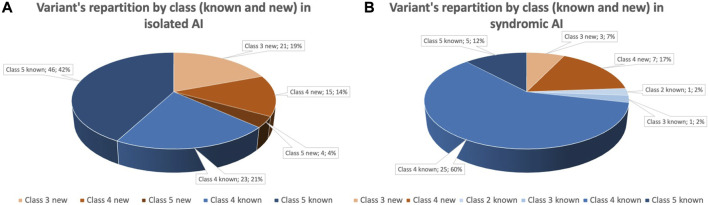
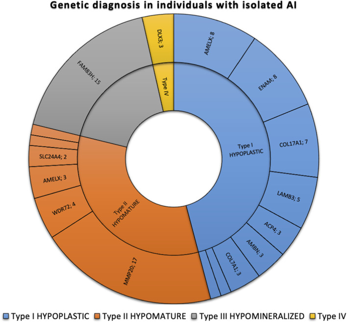
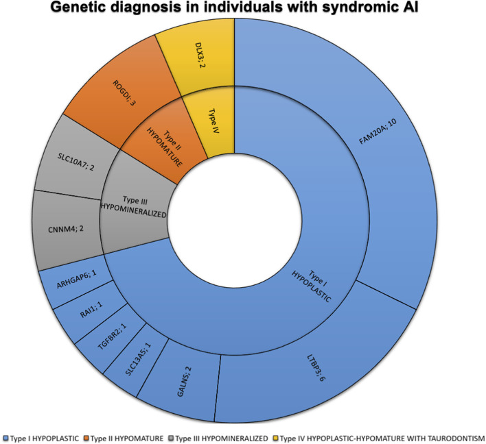
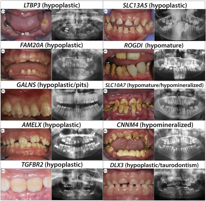
Amelogenesis imperfecta (AI) is a heterogeneous group of genetic rare diseases disrupting enamel development (Smith et al., Front Physiol, 2017a, 8, 333). The clinical enamel phenotypes can be described as hypoplastic, hypomineralized or hypomature and serve as a basis, together with the mode of inheritance, to Witkop’s classification (Witkop, J Oral Pathol, 1988, 17, 547–553). AI can be described in isolation or associated with others symptoms in syndromes. Its occurrence was estimated to range from 1/700 to 1/14,000. More than 70 genes have currently been identified as causative. Objectives: We analyzed using next-generation sequencing (NGS) a heterogeneous cohort of AI patients in order to determine the molecular etiology of AI and to improve diagnosis and disease management. Methods: Individuals presenting with so called “isolated” or syndromic AI were enrolled and examined at the Reference Centre for Rare Oral and Dental Diseases (O-Rares) using D4/phenodent protocol (www.phenodent.org). Families gave written informed consents for both phenotyping and molecular analysis and diagnosis using a dedicated NGS panel named GenoDENT. This panel explores currently simultaneously 567 genes. The study is registered under NCT01746121 and NCT02397824 (https://clinicaltrials.gov/). Results: GenoDENT obtained a 60% diagnostic rate. We reported genetics results for 221 persons divided between 115 AI index cases and their 106 associated relatives from a total of 111 families. From this index cohort, 73% were diagnosed with non-syndromic amelogenesis imperfecta and 27% with syndromic amelogenesis imperfecta. Each individual was classified according to the AI phenotype. Type I hypoplastic AI represented 61 individuals (53%), Type II hypomature AI affected 31 individuals (27%), Type III hypomineralized AI was diagnosed in 18 individuals (16%) and Type IV hypoplastic-hypomature AI with taurodontism concerned 5 individuals (4%). We validated the genetic diagnosis, with class 4 (likely pathogenic) or class 5 (pathogenic) variants, for 81% of the cohort, and identified candidate variants (variant of uncertain significance or VUS) for 19% of index cases. Among the 151 sequenced variants, 47 are newly reported and classified as class 4 or 5. The most frequently discovered genotypes were associated with MMP20 and FAM83H for isolated AI. FAM20A and LTBP3 genes were the most frequent genes identified for syndromic AI. Patients negative to the panel were resolved with exome sequencing elucidating for example the gene involved ie ACP4 or digenic inheritance. Conclusion: NGS GenoDENT panel is a validated and cost-efficient technique offering new perspectives to understand underlying molecular mechanisms of AI. Discovering variants in genes involved in syndromic AI (CNNM4, WDR72, FAM20A … ) transformed patient overall care. Unravelling the genetic basis of AI sheds light on Witkop’s AI classification.
Overview
The claims and statistics of this paper haven’t been checked yet.
Run the check — free, no account needed. It reads the open-access text and checks the sources it cites.
Integrity signals
Registry
Cites
Statistics
Figures
Verification
Claims the paper makes
The claims and statistics of this paper haven’t been checked yet.
Run the check — free, no account needed. It reads the open-access text and checks the sources it cites.
Self-check
Percentages, sums and reported differences re-computed from the paper’s own numbers.
The self-check needs the paper’s full text.
Citations
Cites
Richards, S., Aziz, N., Bale, S., Bick, D., Das, S., Gastier-Foster, J., Grody, W. W., Hegde, M., Lyon, E., Spector, E., et al. (2015) · Genetics in Medicine · doi:10.1038/gim.2015.30 · Cited by 32,594
ClinVar: improving access to variant interpretations and supporting evidence
Landrum, M. J., Lee, J. M., Benson, M., Brown, G. R., Chao, C., Chitipiralla, S., Gu, B., Hart, J., Hoffman, D., Jang, W., et al. (2018) · Nucleic Acids Research · doi:10.1093/nar/gkx1153 · Cited by 4,196
Zoncu, R., Bar-Peled, L., Efeyan, A., Wang, S., Sancak, Y., & Sabatini, D. M. (2011) · Science · doi:10.1126/science.1207056 · Cited by 1,673
Aïoub, M., Lézot, F., Molla, M., Castaneda, B., Robert, B., Goubin, G., Néfussi, J. R., & Berdal, A. (2007) · Bone · doi:10.1016/j.bone.2007.07.023 · Cited by 91
Elements of morphology: Standard terminology for the teeth and classifying genetic dental disorders
de La Dure‐Molla, M., Fournier, B. P., Manzanares, M. C., Acevedo, A. C., Hennekam, R. C., Friedlander, L., Boy‐Lefèvre, M., Kerner, S., Toupenay, S., Garrec, P., et al. (2019) · American Journal of Medical Genetics Part A · doi:10.1002/ajmg.a.61316 · Cited by 85
Self-citation
Research works printed but not linked: 253
- Aaltonen, 1997, High-resolution physical and transcriptional mapping of the autoimmune polyendocrinopathy–candidiasis–ectodermal dystrophy locus on chromosome 21q22.3 by FISH, Genome Res., 7, 820, 10.1101/gr.7.8.820 · DOI
- Acevedo, 2015, Variability of systemic and oro-dental phenotype in two families with non-lethal Raine syndrome with FAM20C mutations, BMC Med. Genet., 16, 8, 10.1186/s12881-015-0154-5 · DOI
- Akyol, 2019, Recommendations for the management of MPS IVA: Systematic evidence- and consensus-based guidance, Orphanet J. Rare Dis., 14, 137, 10.1186/s13023-019-1074-9 · DOI
- Aldred, 1995, Amelogenesis imperfecta-towards a new classification, Oral Dis., 1, 2, 10.1111/j.1601-0825.1995.tb00148.x · DOI
- Aldred, 2003, Amelogenesis imperfecta: A classification and catalogue for the 21st century, Oral Dis., 9, 19, 10.1034/j.1601-0825.2003.00843.x · DOI
- Alsharif, 2018, Unilateral focal dermal hypoplasia (goltz syndrome): Case report and literature review, Case Rep. Dermatol, 10, 101, 10.1159/000488521 · DOI
- Ashikov, 2018, Integrating glycomics and genomics uncovers SLC10A7 as essential factor for bone mineralization by regulating post-Golgi protein transport and glycosylation, Hum. Mol. Genet., 27, 3029, 10.1093/hmg/ddy213 · DOI
- Aswath, 2018, A novel ROGDI gene mutation is associated with Kohlschutter-Tonz syndrome, Oral Surg. Oral Med. Oral Pathol. Oral Radiol., 125, e8, 10.1016/j.oooo.2017.09.016 · DOI
- Bardet, 2016, Claudin-16 deficiency impairs tight junction function in ameloblasts, leading to abnormal enamel formation, J. Bone Min. Res., 31, 498, 10.1002/jbmr.2726 · DOI
- Björses, 1999, Localization of the APECED protein in distinct nuclear structures, Hum. Mol. Genet., 8, 259, 10.1093/hmg/8.2.259 · DOI
- Bloch-Zupan, 2012, Dento/oro/craniofacial anomalies and genetics
- Brookes, 2017, Amelogenesis imperfecta caused by N-terminal enamelin point mutations in mice and men is driven by endoplasmic reticulum stress, Hum. Mol. Genet., 26, 1863, 10.1093/hmg/ddx090 · DOI
- Burgeson, 1985, The structure of type VII collagen, Ann. N. Y. Acad. Sci., 460, 47, 10.1111/j.1749-6632.1985.tb51156.x · DOI
- Burzynski, 1973, Autosomal dominant smooth hypoplastic amelogenesis imperfecta. Report of a case, Oral Surg. Oral Med. Oral Pathol., 36, 818, 10.1016/0030-4220(73)90333-2 · DOI
- Caciotti, 2018, Mis-splicing of the GALNS gene resulting from deep intronic mutations as a cause of Morquio a disease, BMC Med. Genet., 19, 183, 10.1186/s12881-018-0694-6 · DOI
- Caricasole, 2002, Molecular cloning and initial characterization of the MG61/PORC gene, the human homologue of the Drosophila segment polarity gene Porcupine, Gene, 288, 147, 10.1016/S0378-1119(02)00467-5 · DOI
- Celli, 1999, Heterozygous germline mutations in the p53 homolog p63 are the cause of EEC syndrome, Cell., 99, 143, 10.1016/s0092-8674(00)81646-3 · DOI
- Collier, 1997, An amelogenin gene defect associated with human X-linked amelogenesis imperfecta, Arch. Oral Biol., 42, 235, 10.1016/s0003-9969(96)00099-4 · DOI
- Crawford, 2007, Amelogenesis imperfecta, Orphanet J. Rare Dis., 2, 17, 10.1186/1750-1172-2-17 · DOI
- Daneshmandpour, 2019, Features, genetics and their correlation in Jalili syndrome: A systematic review, J. Med. Genet., 56, 358, 10.1136/jmedgenet-2018-105716 · DOI
- Darling, 1956, Some observations on amelogenesis imperfecta and calcification of the dental enamel, Proc. R. Soc. Med., 49, 759, 10.1177/003591575604901007 · DOI
- Daubert, 2016, Human enamel thickness and ENAM polymorphism, Int. J. Oral Sci., 8, 93, 10.1038/ijos.2016.1 · DOI
- de la Dure-Molla, 2014, Pathognomonic oral profile of Enamel Renal Syndrome (ERS) caused by recessive FAM20A mutations, Orphanet J. Rare Dis., 9, 84, 10.1186/1750-1172-9-84 · DOI
- Deidrick, 2016, Cognitive and psychological functioning in focal dermal hypoplasia, Am. J. Med. Genet. C Semin. Med. Genet., 172, 34, 10.1002/ajmg.c.31471 · DOI
- Dellow, 1998, Amelogenesis imperfecta, nephrocalcinosis, and hypocalciuria syndrome in two siblings from a large family with consanguineous parents, Nephrol. Dial. Transpl., 13, 3193, 10.1093/ndt/13.12.3193 · DOI
- DePristo, 2011, A framework for variation discovery and genotyping using next-generation DNA sequencing data, Nat. Genet., 43, 491, 10.1038/ng.806 · DOI
- Dong, 2005, DLX3 mutation associated with autosomal dominant amelogenesis imperfecta with taurodontism, Am. J. Med. Genet. A, 138, 10.1002/ajmg.a.30521 · DOI
- Dourado, 2019, Enamel renal syndrome: A novel homozygous FAM20A founder mutation in 5 new Brazilian families, Eur. J. Med. Genet., 62, 103561, 10.1016/j.ejmg.2018.10.013 · DOI
- Dubail, 2018, SLC10A7 mutations cause a skeletal dysplasia with amelogenesis imperfecta mediated by GAG biosynthesis defects, Nat. Commun., 9, 3087, 10.1038/s41467-018-05191-8 · DOI
- Dugan, 2015, New recessive truncating mutation in LTBP3 in a family with oligodontia, short stature, and mitral valve prolapse, Am. J. Med. Genet. A, 167, 1396, 10.1002/ajmg.a.37049 · DOI
- Durmaz, 2018, A novel PORCN frameshift mutation leading to focal dermal hypoplasia: A case report, Cytogenet Genome Res., 154, 119, 10.1159/000487580 · DOI
- Duverger, 2017, DLX3-Dependent regulation of ion transporters and carbonic anhydrases is crucial for enamel mineralization, J. Bone Min. Res., 32, 641, 10.1002/jbmr.3022 · DOI
- El-Sayed, 2009, Mutations in the beta propeller WDR72 cause autosomal-recessive hypomaturation amelogenesis imperfecta, Am. J. Hum. Genet., 85, 699, 10.1016/j.ajhg.2009.09.014 · DOI
- Lek, 2016, Analysis of protein-coding genetic variation in 60,706 humans, Nature, 536, 285, 10.1038/nature19057 · DOI · Paper page
- Feldmeyer, 2006, Confirmation of the origin of NISCH syndrome, Hum. Mutat., 27, 408, 10.1002/humu.20333 · DOI
- Feske, 1996, Severe combined immunodeficiency due to defective binding of the nuclear factor of activated T cells in T lymphocytes of two male siblings, Eur. J. Immunol., 26, 2119, 10.1002/eji.1830260924 · DOI
- Feske, 2010, CRAC channelopathies, Pflugers Arch., 460, 417, 10.1007/s00424-009-0777-5 · DOI
- Flores, 2002, p63 and p73 are required for p53-dependent apoptosis in response to DNA damage, Nature, 416, 560, 10.1038/416560a · DOI
- Fouillen, 2017, Interactions of AMTN, ODAM and SCPPPQ1 proteins of a specialized basal lamina that attaches epithelial cells to tooth mineral, Sci. Rep., 7, 46683, 10.1038/srep46683 · DOI
- Frick, 2009, Metabolic acidosis increases intracellular calcium in bone cells through activation of the proton receptor OGR1, J. Bone Min. Res., 24, 305, 10.1359/jbmr.081015 · DOI
- Frisk, 2018, Goltz syndrome in males: A clinical report of a male patient carrying a novel PORCN variant and a review of the literature, Clin. Case Rep., 6, 2103, 10.1002/ccr3.1783 · DOI
- Furukawa, 2017, Stim1 regulates enamel mineralization and ameloblast modulation, J. Dent. Res., 96, 1422, 10.1177/0022034517719872 · DOI
- Gasse, 2013, Homozygous and compound heterozygous MMP20 mutations in amelogenesis imperfecta, J. Dent. Res., 92, 598, 10.1177/0022034513488393 · DOI
- Gasse, 2017, Evolutionary analysis predicts sensitive positions of MMP20 and validates newly- and previously-identified MMP20 mutations causing amelogenesis imperfecta, Front. Physiol., 8, 398, 10.3389/fphys.2017.00398 · DOI
- Geoffroy, 2018, AnnotSV: An integrated tool for structural variations annotation, Bioinformatics, 34, 3572, 10.1093/bioinformatics/bty304 · DOI
- Geoffroy, 2015, VaRank: A simple and powerful tool for ranking genetic variants, PeerJ, 3, e796, 10.7717/peerj.796 · DOI
- Gibson, 2001, Amelogenin-deficient mice display an amelogenesis imperfecta phenotype, J. Biol. Chem., 276, 31871, 10.1074/jbc.M104624200 · DOI
- Goodwin, 2014, Abnormal Ras signaling in Costello syndrome (CS) negatively regulates enamel formation, Hum. Mol. Genet., 23, 682, 10.1093/hmg/ddt455 · DOI
- Greene, 2002, A new frameshift mutation encoding a truncated amelogenin leads to X-linked amelogenesis imperfecta, Arch. Oral Biol., 47, 211, 10.1016/s0003-9969(01)00111-x · DOI
- Guerrini, 2022, Phenotypic and genetic spectrum of ATP6V1A encephalopathy: A disorder of lysosomal homeostasis, Brain, 145, 2687, 10.1093/brain/awac145 · DOI · Paper page
- Guo, 2005, Physical interaction and functional coupling between ACDP4 and the intracellular ion chaperone COX11, an implication of the role of ACDP4 in essential metal ion transport and homeostasis, Mol. Pain, 1, 15, 10.1186/1744-8069-1-15 · DOI
- Guo, 2018, LTBP3 pathogenic variants predispose individuals to thoracic aortic aneurysms and dissections, Am. J. Hum. Genet., 102, 706, 10.1016/j.ajhg.2018.03.002 · DOI
- Happle, 1977, Striation of bones in focal dermal hypoplasia: Manifestation of functional mosaicism?, Br. J. Dermatol, 96, 133, 10.1111/j.1365-2133.1977.tb12534.x · DOI
- Hardies, 2015, Recessive mutations in SLC13A5 result in a loss of citrate transport and cause neonatal epilepsy, developmental delay and teeth hypoplasia, Brain, 138, 3238, 10.1093/brain/awv263 · DOI
- Harrison, 2019, Overview of specifications to the ACMG/AMP variant interpretation guidelines, Curr. Protoc. Hum. Genet., 103, e93, 10.1002/cphg.93 · DOI
- Hart, Amelogenesis imperfecta phenotype-genotype correlations with two amelogenin gene mutations, Arch. Oral Biol., 47, 261, 10.1016/s0003-9969(02)00003-1 · DOI
- Hart, 2004, Mutation in kallikrein 4 causes autosomal recessive hypomaturation amelogenesis imperfecta, J. Med. Genet., 41, 545, 10.1136/jmg.2003.017657 · DOI
- Hart, A nomenclature for X-linked amelogenesis imperfecta, Arch. Oral Biol., 47, 255, 10.1016/s0003-9969(02)00005-5 · DOI
- Hart, 2003, Novel ENAM mutation responsible for autosomal recessive amelogenesis imperfecta and localised enamel defects, J. Med. Genet., 40, 900, 10.1136/jmg.40.12.900 · DOI
- Harutunian, 2011, Tuberous sclerosis complex with oral manifestations: A case report and literature review, Med. Oral Patol. Oral Cir. Bucal, 16, e478, 10.4317/medoral.16.e478 · DOI
- Hassan, 2004, Dlx3 transcriptional regulation of osteoblast differentiation: Temporal recruitment of Msx2, Dlx3, and Dlx5 homeodomain proteins to chromatin of the osteocalcin gene, Mol. Cell. Biol., 24, 9248, 10.1128/MCB.24.20.9248-9261.2004 · DOI
- Heimler, 1991, Sensorineural hearing loss, enamel hypoplasia, and nail abnormalities in sibs, Am. J. Med. Genet., 39, 192, 10.1002/ajmg.1320390214 · DOI
- Holcroft, 2011, Identification of amelotin- and ODAM-interacting enamel matrix proteins using the yeast two-hybrid system, Eur. J. Oral Sci., 119, 301, 10.1111/j.1600-0722.2011.00870.x · DOI
- Hollister, 1973, The lacrimo-auriculo-dento-digital syndrome, J. Pediatr., 83, 438, 10.1016/s0022-3476(73)80268-9 · DOI
- Hu, Amelogenesis imperfecta in two families with defined AMELX deletions in ARHGAP6, PLoS One, 7, e52052, 10.1371/journal.pone.0052052 · DOI
- Hu, 2003, Enamelin and autosomal-dominant amelogenesis imperfecta, Crit. Rev. Oral Biol. Med., 14, 387, 10.1177/154411130301400602 · DOI
- Hu, Expression of the sodium/calcium/potassium exchanger, NCKX4, in ameloblasts, Cells Tissues Organs, 196, 501, 10.1159/000337493 · DOI
- Huckert, 2014, A novel mutation in the ROGDI gene in a patient with kohlschütter-tönz syndrome, Mol. Syndromol., 5, 293, 10.1159/000366252 · DOI
- Huckert, 2015, Mutations in the latent TGF-beta binding protein 3 (LTBP3) gene cause brachyolmia with amelogenesis imperfecta, Hum. Mol. Genet., 24, 3038, 10.1093/hmg/ddv053 · DOI
- Inoki, 2002, TSC2 is phosphorylated and inhibited by Akt and suppresses mTOR signalling, Nat. Cell. Biol., 4, 648, 10.1038/ncb839 · DOI · Paper page
- Inoue, 2002, Human Na+-coupled citrate transporter: Primary structure, genomic organization, and transport function, Biochem. Biophys. Res. Commun., 299, 465, 10.1016/S0006-291X(02)02669-4 · DOI
- Intarak, 2019, Genotype-phenotype correlation and expansion of orodental anomalies in LTBP3-related disorders, Mol. Genet. Genomics, 294, 773, 10.1007/s00438-019-01547-x · DOI
- Iwase, 2007, Evolutionary history of sex-linked mammalian amelogenin genes, Cells Tissues Organs, 186, 49, 10.1159/000102680 · DOI
- Jabs, 1993, A mutation in the homeodomain of the human MSX2 gene in a family affected with autosomal dominant craniosynostosis, Cell., 75, 443, 10.1016/0092-8674(93)90379-5 · DOI
- Jagtap, 2019, Trichodentoosseous syndrome: A case report and review of literature, BJR Case Rep., 5, 20190039, 10.1259/bjrcr.20190039 · DOI
- Jain, 2017, Tricho-dento-osseous syndrome and precocious eruption, J. Clin. Exp. Dent., 9, e494, 10.4317/jced.53348 · DOI
- Jalili, 1988, A progressive cone-rod dystrophy and amelogenesis imperfecta: A new syndrome, J. Med. Genet., 25, 738, 10.1136/jmg.25.11.738 · DOI
- Jani, 2020, Severity of oro-dental anomalies in Loeys-Dietz syndrome segregates by gene mutation, J. Med. Genet., 57, 699, 10.1136/jmedgenet-2019-106678 · DOI
- Jaureguiberry, 2012, Nephrocalcinosis (enamel renal syndrome) caused by autosomal recessive FAM20A mutations, Nephron Physiol., 122, 1, 10.1159/000349989 · DOI
- Ji, 2021, Maturation stage enamel defects in Odontogenesis-associated phosphoprotein (Odaph) deficient mice, Dev. Dyn., 250, 1505, 10.1002/dvdy.336 · DOI
- Kantaputra, 2003, Heterozygous mutation in the SAM domain of p63 underlies rapp-hodgkin ectodermal dysplasia, J. Dent. Res., 82, 433, 10.1177/154405910308200606 · DOI
- Katsura, 2014, WDR72 models of structure and function: A stage-specific regulator of enamel mineralization, Matrix Biol., 38, 48, 10.1016/j.matbio.2014.06.005 · DOI
- Katsura, 2022, WDR72 regulates vesicle trafficking in ameloblasts, Sci. Rep., 12, 2820, 10.1038/s41598-022-06751-1 · DOI
- Kausalya, 2006, Disease-associated mutations affect intracellular traffic and paracellular Mg2+ transport function of Claudin-16, J. Clin. Investig., 116, 878, 10.1172/JCI26323 · DOI
- Khandelwal, 2021, Phenotypic variability in distal acidification defects associated with WDR72 mutations, Pediatr. Nephrol., 36, 881, 10.1007/s00467-020-04747-5 · DOI
- Kim, 2008, FAM83H mutations in families with autosomal-dominant hypocalcified amelogenesis imperfecta, Am. J. Hum. Genet., 82, 489, 10.1016/j.ajhg.2007.09.020 · DOI
- Kim, ENAM mutations in autosomal-dominant amelogenesis imperfecta, J. Dent. Res., 84, 278, 10.1177/154405910508400314 · DOI
- Kim, MMP-20 mutation in autosomal recessive pigmented hypomaturation amelogenesis imperfecta, J. Med. Genet., 42, 271, 10.1136/jmg.2004.024505 · DOI
- Kim, 2019, Mutations in RELT cause autosomal recessive amelogenesis imperfecta, Clin. Genet., 95, 375, 10.1111/cge.13487 · DOI
- Kim, 2004, Amelogenin p.M1T and p.W4S mutations underlying hypoplastic X-linked amelogenesis imperfecta, J. Dent. Res., 83, 378, 10.1177/154405910408300505 · DOI
- Kim, Identification of a homozygous PEX26 mutation in a heimler syndrome patient, Genes. (Basel)., 12, 646, 10.3390/genes12050646 · DOI
- Kim, 2017, Analyses of MMP20 missense mutations in two families with hypomaturation amelogenesis imperfecta, Front. Physiol., 8, 229, 10.3389/fphys.2017.00229 · DOI
- Kim, 2020, Alteration of exon definition causes amelogenesis imperfecta, J. Dent. Res., 99, 410, 10.1177/0022034520901708 · DOI
- Kim, A novel de novo SP6 mutation causes severe hypoplastic amelogenesis imperfecta, Genes. (Basel)., 12, 346, 10.3390/genes12030346 · DOI
- Kindelan, 2000, Detection of a novel mutation in X-linked amelogenesis imperfecta, J. Dent. Res., 79, 1978, 10.1177/00220345000790120901 · DOI
- Kiritsi, 2015, Junctional epidermolysis bullosa with LAMB3 splice-site mutations, Acta Derm. Venereol., 95, 849, 10.2340/00015555-2073 · DOI
- Kittler, 2004, An endoribonuclease-prepared siRNA screen in human cells identifies genes essential for cell division, Nature, 432, 1036, 10.1038/nature03159 · DOI
- Kohlschütter, 1974, Familial epilepsy and yellow teeth-a disease of the CNS associated with enamel hypoplasia, Helv. Paediatr. Acta, 29, 283
- Konrad, 2006, Mutations in the tight-junction gene claudin 19 (CLDN19) are associated with renal magnesium wasting, renal failure, and severe ocular involvement, Am. J. Hum. Genet., 79, 949, 10.1086/508617 · DOI
- Kosaki, 2008, Split hand foot malformation with whorl-like pigmentary pattern: Phenotypic expression of somatic mosaicism for the p63 mutation, Am. J. Med. Genet. A, 2574, 10.1002/ajmg.a.32415 · DOI
- Kuga, 2016, FAM83H and casein kinase I regulate the organization of the keratin cytoskeleton and formation of desmosomes, Sci. Rep., 6, 26557, 10.1038/srep26557 · DOI
- Lacruz, 2015, Diseases caused by mutations in ORAI1 and STIM1, Ann. N. Y. Acad. Sci., 1356, 45, 10.1111/nyas.12938 · DOI
- Lagerström, 1990, Mapping of the gene for X-linked amelogenesis imperfecta by linkage analysis, Am. J. Hum. Genet., 46, 120
- Lagerström, 1991, A deletion in the amelogenin gene (AMG) causes X-linked amelogenesis imperfecta (AIH1), Genomics, 10, 971, 10.1016/0888-7543(91)90187-j · DOI
- Larrègue, 1975, Letter: Striated osteopathy in focal dermal hypoplasia, Arch. Dermatol, 111, 1365, 10.1001/archderm.1975.01630220129019 · DOI
- Lau, 1989, Human and mouse amelogenin gene loci are on the sex chromosomes, Genomics, 4, 162, 10.1016/0888-7543(89)90295-4 · DOI
- Laugel-Haushalter, 2019, A new SLC10A7 homozygous missense mutation responsible for a milder phenotype of skeletal dysplasia with amelogenesis imperfecta, Front. Genet., 10, 504, 10.3389/fgene.2019.00504 · DOI
- Lee, 2015, Novel LAMB3 mutations cause non-syndromic amelogenesis imperfecta with variable expressivity, Clin. Genet., 87, 90, 10.1111/cge.12340 · DOI
- Lee, 2006, Kidney claudin-19: Localization in distal tubules and collecting ducts and dysregulation in polycystic renal disease, FEBS Lett., 580, 923, 10.1016/j.febslet.2006.01.019 · DOI
- Lee, 2010, MMP20 hemopexin domain mutation in amelogenesis imperfecta, J. Dent. Res., 89, 46, 10.1177/0022034509352844 · DOI
- Lee, 2011, FAM83H mutations cause ADHCAI and alter intracellular protein localization, J. Dent. Res., 90, 377, 10.1177/0022034510389177 · DOI
- Lench, 1995, Characterisation of molecular defects in X-linked amelogenesis imperfecta (AIH1), Hum. Mutat., 5, 251, 10.1002/humu.1380050310 · DOI
- Li, 1995, Mapping of human and murine genes for latent TGF-β binding protein-2 (LTBP2), Mamm. Genome, 6, 42, 10.1007/BF00350892 · DOI
- Liang, 2019, AMBN mutations causing hypoplastic amelogenesis imperfecta and Ambn knockout-NLS-lacZ knockin mice exhibiting failed amelogenesis and Ambn tissue-specificity, Mol. Genet. Genomic Med., 7, e929, 10.1002/mgg3.929 · DOI
- Lindemeyer, 2010, Amelogenesis imperfecta due to a mutation of the enamelin gene: Clinical case with genotype-phenotype correlations, Pediatr. Dent., 32, 56
- Lu, 2018, Whole exome sequencing identifies an AMBN missense mutation causing severe autosomal-dominant amelogenesis imperfecta and dentin disorders, Int. J. Oral Sci., 10, 26, 10.1038/s41368-018-0027-9 · DOI
- Ludwig, 2003, Proton-sensing G-protein-coupled receptors, Nature, 425, 93, 10.1038/nature01905 · DOI
- Maas, 1996, EEC syndrome and genitourinary anomalies: An update, Am. J. Med. Genet., 63, 472, 10.1002/(SICI)1096-8628(19960614)63:3<472::AID-AJMG11>3.0 · DOI
- Mårdh, 2002, A nonsense mutation in the enamelin gene causes local hypoplastic autosomal dominant amelogenesis imperfecta (AIH2), Hum. Mol. Genet., 11, 1069, 10.1093/hmg/11.9.1069 · DOI
- Martelli-Júnior, 2011, Amelogenesis imperfecta and nephrocalcinosis syndrome: A case report and review of the literature, Nephron Physiol., 118, 62, 10.1159/000322828 · DOI
- Martino, 1992, Child with manifestations of dermotrichic syndrome and ichthyosis follicularis-alopecia-photophobia (IFAP) syndrome, Am. J. Med. Genet., 44, 233, 10.1002/ajmg.1320440222 · DOI
- Masunaga, 2006, Epidermal basement membrane: Its molecular organization and blistering disorders, Connect. Tissue Res., 47, 55, 10.1080/03008200600584157 · DOI
- Mátyás, 2006, Identification and in silico analyses of novel TGFBR1 and TGFBR2 mutations in Marfan syndrome-related disorders, Hum. Mutat., 27, 760, 10.1002/humu.20353 · DOI
- McCarl, 2009, ORAI1 deficiency and lack of store-operated Ca2+ entry cause immunodeficiency, myopathy, and ectodermal dysplasia, J. Allergy Clin. Immunol., 124, 1311, 10.1016/j.jaci.2009.10.007 · DOI
- McGrath, 1996, Compound heterozygosity for a dominant Glycine substitution and a recessive internal duplication mutation in the type XVII collagen gene results in junctional epidermolysis bullosa and abnormal dentition, Am. J. Patho, 148, 1787
- McNally, 2012, Gated regulation of CRAC channel ion selectivity by STIM1, Nature, 482, 241, 10.1038/nature10752 · DOI
- Mechaussier, 2020, Heimler syndrome, Adv. Exp. Med. Biol., 1299, 81, 10.1007/978-3-030-60204-8_7 · DOI
- Mendoza, 2007, A new locus for autosomal dominant amelogenesis imperfecta on chromosome 8q24.3, Hum. Genet., 120, 653, 10.1007/s00439-006-0246-6 · DOI
- Molla, 2010, Enamel protein regulation and dental and periodontal physiopathology in Msx2 mutant mice, Am. J. Pathol., 177, 2516, 10.2353/ajpath.2010.091224 · DOI
- Montaño, 2008, Growth charts for patients affected with Morquio A disease, Am. J. Med. Genet. Part A, 1286, 10.1002/ajmg.a.32281 · DOI
- Mory, 2012, A nonsense mutation in the human homolog of Drosophila rogdi causes kohlschutter–tonz syndrome, Am. J. Hum. Genet., 90, 708, 10.1016/j.ajhg.2012.03.005 · DOI
- Müller, 2006, Familial hypomagnesemia with hypercalciuria and nephrocalcinosis: Blocking endocytosis restores surface expression of a novel claudin-16 mutant that lacks the entire C-terminal cytosolic tail, Hum. Mol. Genet., 15, 1049, 10.1093/hmg/ddl020 · DOI
- Muto, 2012, Dissection of morphological and metabolic differentiation of ameloblasts via ectopic SP6 expression, J. Med. Investig., 59, 59, 10.2152/jmi.59.59 · DOI
- Nagamine, 1997, Positional cloning of the APECED gene, Nat. Genet., 17, 393, 10.1038/ng1297-393 · DOI
- Nakamura, 2008, Transcription factor epiprofin is essential for tooth morphogenesis by regulating epithelial cell fate and tooth number, J. Biol. Chem., 283, 4825, 10.1074/jbc.M708388200 · DOI
- Nakamura, 2004, The Krüppel-like factor epiprofin is expressed by epithelium of developing teeth, hair follicles, and limb buds and promotes cell proliferation, J. Biol. Chem., 279, 626, 10.1074/jbc.M307502200 · DOI
- Nalbant, 2005, FAM20: An evolutionarily conserved family of secreted proteins expressed in hematopoietic cells, BMC Genomics, 6, 11, 10.1186/1471-2164-6-11 · DOI
- Neuhaus, 2017, Next-generation sequencing reveals the mutational landscape of clinically diagnosed usher syndrome: Copy number variations, phenocopies, a predominant target for translational read-through, and PEX26 mutated in heimler syndrome, Mol. Genet. Genomic Med., 5, 531, 10.1002/mgg3.312 · DOI
- Nikolopoulos, 2020, New missense variants in RELT causing hypomineralised amelogenesis imperfecta, Clin. Genet., 97, 688, 10.1111/cge.13721 · DOI
- Nikolopoulos, 2021, Spectrum of pathogenic variants and founder effects in amelogenesis imperfecta associated with MMP20, Hum. Mutat., 42, 567, 10.1002/humu.24187 · DOI
- Noor, 2009, Oligodontia is caused by mutation in LTBP3, the gene encoding latent TGF-beta binding protein 3, Am. J. Hum. Genet., 84, 519, 10.1016/j.ajhg.2009.03.007 · DOI
- Nurbaeva, 2015, Dental enamel cells express functional SOCE channels, Sci. Rep., 5, 15803, 10.1038/srep15803 · DOI
- Ogawa, 1995, Mucopolysaccharidosis IVA: Screening and identification of mutations of the N-acetylgalactosamine-6-sulfate sulfatase gene, Hum. Mol. Genet., 4, 341, 10.1093/hmg/4.3.341 · DOI
- O’Sullivan, 2011, Whole-Exome sequencing identifies FAM20A mutations as a cause of amelogenesis imperfecta and gingival hyperplasia syndrome, Am. J. Hum. Genet., 88, 616, 10.1016/j.ajhg.2011.04.005 · DOI
- Ozdemir, Phenotype of ENAM mutations is dosage-dependent, J. Dent. Res., 84, 1036, 10.1177/154405910508401113 · DOI
- Ozdemir, MMP20 active-site mutation in hypomaturation amelogenesis imperfecta, J. Dent. Res., 84, 1031, 10.1177/154405910508401112 · DOI
- Papagerakis, 2008, Premature stop codon in MMP20 causing amelogenesis imperfecta, J. Dent. Res., 87, 56, 10.1177/154405910808700109 · DOI
- Parker, 1996, Molecular cloning of a novel human gene (D11S4896E) at chromosomal region 11p15.5, Genomics, 37, 253, 10.1006/geno.1996.0553 · DOI
- Parry, 2012, Mutations in C4orf26, encoding a peptide with in vitro hydroxyapatite crystal nucleation and growth activity, cause amelogenesis imperfecta, Am. J. Hum. Genet., 91, 565, 10.1016/j.ajhg.2012.07.020 · DOI
- Parry, A homozygous STIM1 mutation impairs store-operated calcium entry and natural killer cell effector function without clinical immunodeficiency, J. Allergy Clin. Immunol., 137, 955, 10.1016/j.jaci.2015.08.051 · DOI
- Parry, 2009, Mutations in CNNM4 cause Jalili syndrome, consisting of autosomal-recessive cone-rod dystrophy and amelogenesis imperfecta, Am. J. Hum. Genet., 84, 266, 10.1016/j.ajhg.2009.01.009 · DOI
- Parry, 2013, Identification of mutations in SLC24A4, encoding a potassium-dependent sodium/calcium exchanger, as a cause of amelogenesis imperfecta, Am. J. Hum. Genet., 92, 307, 10.1016/j.ajhg.2013.01.003 · DOI
- Parry, Mutations in the pH-sensing G-protein-coupled receptor GPR68 cause amelogenesis imperfecta, Am. J. Hum. Genet., 99, 984, 10.1016/j.ajhg.2016.08.020 · DOI
- Pavlic, 2009, Clinical and microstructural aberrations of enamel of deciduous and permanent teeth in patients with autoimmune polyendocrinopathy-candidiasis-ectodermal dystrophy, Arch. Oral Biol., 54, 658, 10.1016/j.archoralbio.2009.03.009 · DOI
- Peracha, 2018, Molecular genetics and metabolism, special edition: Diagnosis, diagnosis and prognosis of Mucopolysaccharidosis IVA, Mol. Genet. Metab., 125, 18, 10.1016/j.ymgme.2018.05.004 · DOI
- Pereverzev, 2008, Extracellular acidification enhances osteoclast survival through an NFAT-independent, protein kinase C-dependent pathway, Bone, 42, 150, 10.1016/j.bone.2007.08.044 · DOI
- Perniola, 2018, Twenty years of AIRE, Front. Immunol., 9, 98, 10.3389/fimmu.2018.00098 · DOI
- Picard, 2009, STIM1 mutation associated with a syndrome of immunodeficiency and autoimmunity, N. Engl. J. Med., 360, 1971, 10.1056/NEJMoa0900082 · DOI
- Plaisancié, 2015, MSX2 gene duplication in a patient with eye development defects, Ophthalmic Genet., 36, 353, 10.3109/13816810.2014.886270 · DOI
- Pollak, 2003, Sensorineural hearing loss and enamel hypoplasia with subtle nail findings: Another family with heimler’s syndrome, Clin. Dysmorphol., 12, 55, 10.1097/00019605-200301000-00010 · DOI
- Polok, 2009, Mutations in CNNM4 cause recessive cone-rod dystrophy with amelogenesis imperfecta, Am. J. Hum. Genet., 84, 259, 10.1016/j.ajhg.2009.01.006 · DOI
- Portsteffen, 1997, Human PEX1 is mutated in complementation group 1 of the peroxisome biogenesis disorders, Nat. Genet., 17, 449, 10.1038/ng1297-449 · DOI
- Poulter, A missense mutation in ITGB6 causes pitted hypomineralized amelogenesis imperfecta, Hum. Mol. Genet., 23, 2189, 10.1093/hmg/ddt616 · DOI
- Poulter, Deletion of ameloblastin exon 6 is associated with amelogenesis imperfecta, Hum. Mol. Genet., 23, 5317, 10.1093/hmg/ddu247 · DOI
- Prasad, A targeted next-generation sequencing assay for the molecular diagnosis of genetic disorders with orodental involvement, J. Med. Genet., 53, 98, 10.1136/jmedgenet-2015-103302 · DOI
- Prasad, Amelogenesis imperfecta: 1 family, 2 phenotypes, and 2 mutated genes, J. Dent. Res., 95, 1457, 10.1177/0022034516663200 · DOI
- Price, 1998, Identification of a mutation in DLX3 associated with tricho-dento-osseous (TDO) syndrome, Hum. Mol. Genet., 7, 563, 10.1093/hmg/7.3.563 · DOI
- Raine, 1989, Unknown syndrome: Microcephaly, hypoplastic nose, exophthalmos, gum hyperplasia, cleft palate, low set ears, and osteosclerosis, J. Med. Genet., 26, 786, 10.1136/jmg.26.12.786 · DOI
- Rao, 1971, Inherited defects in tooth structure, Birth Defects Orig. Artic. Ser., 7, 153
- Rapp, 1968, Anhidrotic ectodermal dysplasia: Autosomal dominant inheritance with palate and lip anomalies, J. Med. Genet., 5, 269, 10.1136/jmg.5.4.269 · DOI
- Ratbi, 2015, Heimler syndrome is caused by hypomorphic mutations in the peroxisome-biogenesis genes PEX1 and PEX6, Am. J. Hum. Genet., 97, 535, 10.1016/j.ajhg.2015.08.011 · DOI
- Rathi, 2014, Extending the use of a diagnostic occlusal splint to overcome existing lacunae of vertical dimension transfer in full mouth rehabilitation cases, Am. J. Med. Case Rep., 2, 291, 10.12691/ajmcr-2-12-9 · DOI
- Reese, 1997, Improved splice site detection in Genie, J. Comput. Biol., 4, 311, 10.1089/cmb.1997.4.311 · DOI
- Reuber, 1997, Mutations in PEX1 are the most common cause of peroxisome biogenesis disorders, Nat. Genet., 17, 445, 10.1038/ng1297-445 · DOI
- Rey, 2019, Protocol GenoDENT: Implementation of a new NGS panel for molecular diagnosis of genetic disorders with orodental involvement, Methods Mol. Biol., 1922, 407, 10.1007/978-1-4939-9012-2_36 · DOI
- Rinne, 2006, Pattern of p63 mutations and their phenotypes-update, Am. J. Med. Genet. A, 140, 1396, 10.1002/ajmg.a.31271 · DOI
- Robinson, 2014, Enamel maturation: A brief background with implications for some enamel dysplasias, Front. Physiol., 5, 388, 10.3389/fphys.2014.00388 · DOI
- Rølling, 1999, Dental findings in three siblings with Morquio’s syndrome, Int. J. Paediatr. Dent., 9, 219, 10.1046/j.1365-263x.1999.00127.x · DOI
- Ruspita, 2020, An msx2-sp6-follistatin pathway operates during late stages of tooth development to control amelogenesis, Front. Physiol., 11, 582610, 10.3389/fphys.2020.582610 · DOI
- Sabbioni, 1999, Exon structure and promoter identification of STIM1 (alias GOK), a human gene causing growth arrest of the human tumor cell lines G401 and RD, Cytogenet Celll Genet., 86, 214, 10.1159/000015341 · DOI
- Sawamoto, 2020, Mucopolysaccharidosis IVA: Diagnosis, treatment, and management, Int. J. Mol. Sci., 21, 1517, 10.3390/ijms21041517 · DOI
- Scherer, 1995, Assignment of the human homolog of mouse Dlx3 to Chromosome 17q21.3-q22 by analysis of somatic cell hybrids and fluorescence in situ hybridization, Mamm. Genome, 6, 310, 10.1007/BF00352432 · DOI
- Schossig, 2017, SLC13A5 is the second gene associated with Kohlschütter-Tönz syndrome, J. Med. Genet., 54, 54, 10.1136/jmedgenet-2016-103988 · DOI
- Schossig, 2012, Mutations in ROGDI cause kohlschütter-tönz syndrome, Am. J. Hum. Genet., 90, 701, 10.1016/j.ajhg.2012.02.012 · DOI
- Schulze, 1970, Developmental abnormalities of the teeth and jaws, 112
- Sewerin, 2022, Defective claudin-10 causes a novel variation of HELIX syndrome through compromised tight junction strand assembly, Genes. Dis., 9, 1301, 10.1016/j.gendis.2021.06.006 · DOI
- Seymen, 2016, Recessive mutations in ACPT, encoding testicular acid phosphatase, cause hypoplastic amelogenesis imperfecta, Am. J. Hum. Genet., 99, 1199, 10.1016/j.ajhg.2016.09.018 · DOI · Paper page
- Seymen, ENAM mutations with incomplete penetrance, J. Dent. Res., 93, 988, 10.1177/0022034514548222 · DOI
- Seymen, Novel ITGB6 mutation in autosomal recessive amelogenesis imperfecta, Oral Dis., 21, 456, 10.1111/odi.12303 · DOI
- Seymen, Exonal deletion of SLC24A4 causes hypomaturation amelogenesis imperfecta, J. Dent. Res., 93, 366, 10.1177/0022034514523786 · DOI
- Seymen, Novel MMP20 and KLK4 mutations in amelogenesis imperfecta, J. Dent. Res., 94, 1063, 10.1177/0022034515590569 · DOI
- Seymen, 2021, Novel mutations in GPR68 and SLC24A4 cause hypomaturation amelogenesis imperfecta, J. Pers. Med., 12, 13, 10.3390/jpm12010013 · DOI
- Shaheen, 2013, A novel syndrome of hypohidrosis and intellectual disability is linked to COG6 deficiency, J. Med. Genet., 50, 431, 10.1136/jmedgenet-2013-101527 · DOI
- Shapiro, 1987, RNA splice junctions of different classes of eukaryotes: Sequence statistics and functional implications in gene expression, Nucleic Acids Res., 15, 7155, 10.1093/nar/15.17.7155 · DOI
- Shore, 2010, The structure and composition of deciduous enamel affected by local hypoplastic autosomal dominant amelogenesis imperfecta resulting from an ENAM mutation, Cells Tissues Organs, 191, 301, 10.1159/000258703 · DOI
- Simmer, 2021, A genetic model for the secretory stage of dental enamel formation, J. Struct. Biol., 213, 107805, 10.1016/j.jsb.2021.107805 · DOI
- Simmer, 2009, Hypomaturation enamel defects in Klk4 knockout/LacZ knockin mice, J. Biol. Chem., 284, 19110, 10.1074/jbc.M109.013623 · DOI
- Simon, 1999, Paracellin-1, a renal tight junction protein required for paracellular Mg2+ resorption, Science, 285, 103, 10.1126/science.285.5424.103 · DOI
- Simpson, 2007, Mutations in FAM20C are associated with lethal osteosclerotic bone dysplasia (Raine syndrome), highlighting a crucial molecule in bone development, Am. J. Hum. Genet., 81, 906, 10.1086/522240 · DOI
- Simpson, 2009, Mutations in FAM20C also identified in non-lethal osteosclerotic bone dysplasia, Clin. Genet., 75, 271, 10.1111/j.1399-0004.2008.01118.x · DOI
200 of 253 shown
Cited by
Sorted by citations
Dental Management of Genetic Dental Disorders: A Critical Review
Dujic, H., Bücher, K., Schüler, I. M., Schmidt, P., Hertel, S., Timpel, J., Jablonski-Momeni, A., Schilke, R., Kapferer-Seebacher, I., Zschocke, J., et al. (2025) · Journal of Dental Research · doi:10.1177/00220345241305330 · Cited by 9
Metadata
- Authors
- Bloch-Zupan, A., Rey, T., Jimenez-Armijo, A., Kawczynski, M., Kharouf, N., O-Rare consortium, Dure-Molla, M. D. L., Noirrit, E., Hernandez, M., Joseph-Beaudin, C., Lopez, S., Tardieu, C., Thivichon-Prince, B., ERN Cranio Consortium, Dostalova, T., Macek, M., International Consortium, Alloussi, M. E., Qebibo, L., Morkmued, S., Pungchanchaikul, P., Orellana, B. U., Manière, M.-C., Gérard, B., Bugueno, I. M., & Laugel-Haushalter, V.
- Venue
- Frontiers in Physiology
- Publisher
- Frontiers Media SA
- Published
- 2023-05-09
- Volume
- 14
- ISSN
- 1664-042X
- PMC
- PMC10205041
- Cited by
- 74
- References
- 5
About these checks
Registry, retraction, Cites and statistics results are computed, not judged, with one exception: a printed reference that no index lists is read once by a model before it counts as a citation error. Claims are extracted by a model and kept only when their quote appears verbatim in the text; each cited source is then checked, with a model’s help, for whether it says what the claim relies on. Those verdicts can be wrong — read the claim’s quote and the cited source before relying on one.
Statistics and arithmetic are recomputed, not judged.
Confidence bands are shown only for fields calibrated on our annotated test set. None is calibrated yet, so no band is shown.
What counts as a claim ›How we measure confidence ›How we check this ›
Are you an author, or do you see a problem with this page? Request a review
